Duodenum
Historical document, translated for reference. It reflects medical knowledge of the 1920s–30s and is not medical advice.
Summary
The duodenum is the initial part of the small intestine following the stomach, named by the anatomist Herophilus for its length of approximately 12 finger-widths (24 cm). This article covers its embryology, comparative anatomy, histology, form, topography, and relationship with surrounding organs.
Encyclopedia article (1928–1936)
DUODENUM. Contents: Embryology and comparative anatomy .... 400 Anatomy and histology.............401 Ulcer of the D. k.....................407 Pathogenesis and etiology...........408 Symptomatology and clinical forms . . . . . 411 Complications.................414 Differential diagnosis.........415 Treatment....................416 Prevention . .'..............420 Surgical treatment...........421 Parasites of the D. k..................428 Duodenitis.....................429 Tumors of the D. k...................4S1 Injuries, fistulas and other diseases of the D. k. 435 Roentgenodiagnosis of diseases of the D. k.....440 Duodenum, the initial part of the small intestine, following directly after the stomach, was named by the anatomist Herophilus due to its length, which was calculated by the ancients as equal to 12 transverse fingers, i.e. 24 cm, with the end of the D. k. considered to be the place where it intersects with the root of the mesentery of the small intestines. At present, the end of the D. k. is considered to be the flexura duodeno-jejunalis, and its length is taken as averaging 27-30 cm. Since the loop of the D. k. is closely connected with the head of the pancreas, Luschka proposed for the D. k. the name intestinum pancreaticum. The capacity of the intestine is so significant (150-250 cm3) that it gave French authors reason to call the D. k. the "second" or "additional stomach".
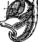
Embryology and comparative anatomy. Embryology. The D. k. separates very early. On the 6th week, the gastro-intestinal tract is located along the median line and is connected from beginning to end with the posterior wall of the abdomen by a dorsal mesentery, and the spindle-shaped stomach and D. k. additionally by a ventral one. At this stage, the D. k. represents a sagittally located loop, directed with its blind end toward the anterior abdominal wall; its lower branch, directed backward, is sharply separated (flex. duodeno-jejunalis) from the subsequent section (see figure 1). Even earlier, the entoderm of the intestine in the area of the D. k. gives rise to outgrowths of the liver and pancreas, with the hepatic outgrowth and the ventral part of the pancreas located in the ventral mesentery, and the dorsal outgrowth of the pancreas in the dorsal. When the stomach rotates around its axis with the anterior surface to the right, the loop of the D. k. follows it, with its convexity also turning to the right; at this time, the right side of the dorsal mesentery adheres to the posterior wall of the abdominal cavity and fuses with it. If for some reason this fusion does not occur, the D. k. retains its mesentery (mesoduodenum) for life. The remainder of the ventral mesentery after the development of the liver in it forms lig. hepato-duodenale.
Comparative anatomy. The upper part of the middle intestine, into which the pyloric appendages (in fish), liver and pancreas (in amphibians and reptiles) flow, does not differ from the rest of the intestine and has no special name. The D. k. separates only in birds and mammals, in which Brunner's glands develop. The attachment of the D. intestine to the wall appears only in primates, in others the mesentery is preserved to a greater or lesser extent. v. Karpov.

Anatomy and histology. Form. Due to embryological conditions, the loop of the D. intestine in the second half of intrauterine life acquires the form of an almost complete ring (Braune), the beginning and end of which are suspended: the first-by means of lig. hepato-duodenale, and the second-by means of m. suspensorius duodeni of Treitz. The latter originates from the circular musculature of the intestine and attaches at the root of a. mesenter. sup., a. coeliacae and the right crus of the diaphragm. Ring-shaped in the infant, the D. k. in adults rarely retains its original form (around 6%). In the vast majority of cases, due to the divergence of the ends of the intestine, the form of the D. k. acquires similarity with the Latin letter U (60%), or V (11%), or C (3%), or has other outlines (20%). Variations in the form of the D. k. are presented in figure 2 (V. Bobrov). Among the reasons determining the form of the D. k., besides peculiarities of intrauterine development, one must consider the type of structure of the chest cavity and the form of the abdomen, age, sex, degree of nutrition. With a narrow or excessively wide aperture of the chest cavity, in old age, in women who have given birth, with a hanging abdomen and severe weight loss, the D. k. has a tendency to take a folded form with bends and sharp angles.
It is customary to distinguish the following 4 parts of the D. k.: 1. Pars horizontalis super. (see figure 3); located directly behind the pylorus of the stomach, covered by peritoneum on almost 3/4 of its circumference, the most mobile of all parts, 3-4 cm long; it is separated from the next part by a rather sharp bend (angulus superior), due to which it is well demarcated, and in the living, due to filling with gases, it has the appearance of a ball and can be visible during fluoroscopy. Therefore it bears special names: bulbus duodeni, antrum duodenale, vestibule du duodenum. 2. Pars descendens, 9-12 cm long, the widest part, up to 18 cm in circumference or 47 mm in diameter. Here, piercing the posterior wall of the intestine and forming on the mucous membrane a special protrusion (papilla Vateri; see figure 4), the bile and pancreatic ducts open. The place of opening is most often located below the middle of this part (44%) or at its very middle (43%) and more rarely-above the middle (13%; in, g and e-transverse processes of III, IV and V lumbar vertebrae; 9 and 10-the descending and upper horizontal parts of duod.; 11 - XII rib. (From Zernov.) Suvorov). On another, barely noticeable elevation of the mucous membrane, which is not always present-papilla minor, Santorini-ends the accessory excretory duct of the pancreas. It is located somewhat above the Vaterian papilla. The second part of the D. k. passes into the next one under the angle (angulus inferior).
3. Pars horizontalis inferior varies greatly in length: from 1 to 9 cm, depending on the form of the D. k. (see figure 2). 4. Pars ascendens, 6-13 cm long, is separated from the jejunum by a sharp bend-flexura duodeno-jejunalis. Depending on the form of the D. k., its bends are expressed to a greater or lesser degree. This also changes the lumen of the intestine-narrowings appear on it, which in certain places are also caused by indentations of the intestinal wall from passing vascular-nervous bundles. Thus, the descending part is crossed by the vasa colica dexta running obliquely-transversely on its anterior surface, and the lower horizontal part-by the vasa mesenterica superiora running across it, forming a narrowing (isthmus duodeni). In pathological cases, arterio-mesenteric obstruction may form here.
Topography of the D. k. Almost the entire D. k. lies on the right lateral and anterior surfaces of the first three lumbar vertebrae, but its level varies within the limits from the XII thoracic to the V lumbar vertebra (see figure 3). The loop of the D. k. surrounds the head of the pancreas, which fuses with the D. k., being connected, among other things, by smooth muscle fibers, but most of all-by common blood vessels and excretory ducts. The head of the pancreas is covered by peritoneum only on its anterior and lateral surfaces, while the posterior surface is connected to the D. k. by connective tissue. The D. k. is covered by peritoneum on all sides except for the area of attachment to the head of the pancreas and the area of the flexura duodeno-jejunalis. The duodenal loop is fixed in place by the lig. hepatoduodenale, the lig. duodenocolicum, the lig. duodenojejunale, and the m. suspensorius duodeni of Treitz.
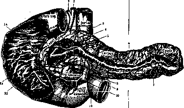
Figure 4. Duodenum and pancreas: 1-duct cysticus; 2-duct hepaticus; 3-duct choledochus; 4-ductus pancreaticus accessorius; 5-ductus pancreaticus; 6-margo superior; 7-margo inferior; 8-incisura pancreatica; 9-a. et v. mesentericae superiores; 10-a. pancreaticoduodenalis inferior; 11-flexura duodenojejunalis; 12-papilla Vateri; 13-plica longitudinalis duodeni; 14-papilla minor. (From Rauber's.)
with the ducts of the gland. This connection is especially intimate in the descending and lower horizontal parts of the duodenum, where the gland surrounds almost half of the circumference of the intestine. The duodenum together with the head of the pancreas is covered in front and behind by a special fascial plate, which further strengthens the connection of these organs. The walls of the stomach are separated from most of the duodenum by means of the mesocolon transversum. The pyloric part of the stomach, when filled, sags downward, presses on the mesocolon transversum, and covers the duodenum from the front. In such cases, the duodenum is very close to the stomach, but does not come into direct contact with it. The duodenum is connected to the liver by the lig. hepato-duodenale, and sometimes also by an accessory peritoneal ligament (lig. hepato-duodenale accessorium-21%). The liver comes into contact with its lower surface with the upper horizontal part of the duodenum, which forms a depression on the liver-impressio duodenalis. The gallbladder also touches the anterior surface of the initial part of the duodenum and is often (in 40%) connected to it by accessory peritoneal ligaments (lig. cystico-duodeno-epiploicum, lig. cysti-). The origin of these accessory ligaments is not always clear; some authors tend to consider them secondary, formed after chronic inflammations. The question remains unresolved.
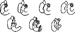

Figure 5. Relationship of the duodenum to mesocolon transv. and mesenterium.
The ascending colon with a short mesentery often comes into contact with the right lateral surface of the second part of the duodenum and when filled, partially displaces it to the left, while the transverse colon always has close contact with the duodenum. In some cases it is connected to the duodenum by means of its mesentery, which is fused with the anterior surface of the descending part of the duodenum (66%), in other cases the wall of the large intestine is fused with the anterior surface of the duodenum (see Figure 5) over a larger or smaller area (33%). The fusion band of mesocolon transversum divides the anterior duodenal area into two sections: the upper one, in contact with the liver and gallbladder, passing directly to the anterior surface of the stomach, and the lower one, covered by the overhanging colon transversum. Thus, for practical purposes, keeping in mind the spread of infection and exudates in the abdominal cavity, one can distinguish regio supramesocolica and regio submesocolica. This crossing of the mesentery is most often below the middle of partis descendentis duodeni (Suslov, Bobrov).
Figure 6. Relationship of the duodenum to the right kidney. Of the organs lying behind the duodenum and directly adjacent to it, it is necessary to especially note vena cava inf., which lies behind the descending part of the duodenum and angulus inferior, and the aorta, which more often lies behind the ascending part of the duodenum and less often behind the lower horizontal part (Bobrov). The bile duct lies behind the duodenum, between it and the head of the pancreas, going downward from lig. hepato-duodenale under pars horiz. sup. and then parallel to the descending part of the duodenum, which it gradually pierces, and opens together with the pancreatic duct on the Vaterian papilla either with a common opening with it or separately (which is important in case of blockage by gallstones). The right kidney has close contact with the descending part of the duodenum, being separated from it by its fascia praerenalis and a layer of loose retroperitoneal tissue. The area of contact of both organs varies depending on many reasons: 1) from the shape of the duodenum, from the height of its position on the posterior abdominal wall and from the degree of its filling; 2) from the height of the kidney position; 3) from the degree of filling of the ascending colon, which can push the duodenum toward the midline and itself occupy its place in front of the kidney (see Figure 6). Most often the right kidney comes into contact with its hilum. The left kidney rarely comes into contact with the ascending part of the duodenum. The root of the mesentery of the small intestine crosses the anterior surface of the lower horizontal part of the duodenum closer to the place where it passes into the ascending part of the duodenum (see Figure 5) and causes some narrowing of the lumen of the duodenum (see above). The described topographic relationships of the duodenum are subject to frequent and significant variations depending on the shape of the intestine and the position on the spine of both the duodenum itself and neighboring organs. In addition, the duodenum changes its topography due to its (relatively small) mobility. The pars horiz. sup. is most subject to displacement, as it follows the pylorus of the stomach, which moves in different directions by 7-11 cm. At the same time, pars horiz. sup. also changes its direction. The ascending part is displaced less (amplitude = 8 cm); even less - the lower horizontal part (6 cm), and least of all - the descending part (2-4 cm).
Lining membrane; B-submucous; C-muscular layer of the duodenum, common with the small intestine. (According to Lavdovskiy.)
with the head of the pancreas, is carried out from two sources: 1) from a. coeliaca, from which two arteries originate: a. pancr.-duod. superior and a. pancr.-duod. poster., and 2) from a. mesent. superior, which gives a. pancr.-duodenalis infer. Both systems anastomose with each other, forming two arches-anterior and posterior (Suslov). The same scheme applies to the veins flowing into vena porta. The lymphatic system of the duodenum in its topography is similar to the blood supply and accompanies it. There is a close connection between the lymphatic vessels of the duodenum and the pancreas, both direct and indirect (Bartels). Lymph from the duodenum drains into the lymphatic glands in front of and behind the head of the pancreas and then into lgl. coeliacae. Innervation of the duodenum: sympathetic system-from plexus coeliacus and plex. mesenter. super., parasympathetic-from vagus dexter (rami coeliaci).
v. Bobrov. Histology. The wall of the duodenum, like that of other intestines, consists of three layers: mucous (tunica mucosa), muscular (tunica muscularis) and serous (tunica serosa); the latter only in parts covered by peritoneum (see Figure 7); the mucous membrane is divided by a layer of smooth muscles into 2 floors-the proper mucous and submucous (submucosa). As in other parts, the mucous membrane forms circular, unclosed folds of Kerkring (plica circulares Kerkringi), which appear only in the descending part and increase in number and size in the ascending part; in the descending part, the bile and pancreatic ducts raise the mucous membrane into a fold (plica longitudinalis) about 11 mm long. The entire surface of the mucous membrane is densely covered with intestinal villi (22-40 per 1 mm³); in the duodenum they are lower and wider than in the small intestine, resembling leaflets; between the bases of the villi in the mucous membrane are located the tubular Lieberkühn glands, or crypts; in some places there are lymphatic nodules, so-called solitary follicles. Peyer's patches are found relatively rarely and in small numbers. A special feature of the duodenum is the tubulo-acinar Brunner's glands (see below). The muscular layer consists of an inner circular and an outer longitudinal layer of smooth muscles, somewhat thicker than in the rest of the intestine (0.3-0.5 mm). Nerve plexuses: Meissner's in the submucosa and Auerbach's in the muscular layer, as in other parts.
v. Karpov. Brunner's glands are located in the submucosal layer of the initial part of the duodenum. In structure and function they are similar to the pyloric glands and consist of branching, tortuous tubules lined with cylindrical epithelium. A series of tubules form lobules. To study the working conditions of Brunner's glands and the composition of the juice they secrete, in I. P. Pavlov's laboratory, the operation of isolating the 'Brunner's section' of the intestine was proposed. The secretion of juice from Brunner's glands occurs continuously, regardless of the hungry or fed state of the animal. Mechanical irritation, which does not cause secretion of the gastric glands, enhances the secretion of the 'Brunner's section'. The following substances, introduced directly into the isolated segment of the 'Brunner's section', also enhance the secretion of Brunner's glands: 1) weak solutions of HCl, products of digestion of fibrin by fundal juice, physiological solution, etc. The direct introduction of fatty substances into the isolated segment of the 'Brunner's section' gives only a slight and short-lived increase in secretion. The enhancing effect of fatty substances is much more energetic if the latter are in other parts of the digestive tract. The juice of Brunner's glands is a colorless liquid of weakly alkaline reaction. The main enzyme of the juice of Brunner's glands is a proteolytic enzyme that shows its action in an acidic environment. When acidified with 0.5% HCl, the juice of Brunner's glands curdles milk. In addition, it has some digestive effect on various fats and cane sugar. The enzymes of pancreatic juice, mainly protein, are activated by the juice of Brunner's glands. The physiological role of the 'Brunner's section', similar to the pyloric part of the stomach, apparently consists in digesting the connective tissue base of adipose tissue of meat, whereby fat becomes accessible to further processing by the enzymes of pancreatic juice and intestinal juice.
Duodenum. "Italics. Ulcer of the duodenum. History of the question. Description of individual symptoms of ulcer of the duodenum belongs to the beginning of the 19th century. However, the complete clinical symptom complex of this disease was first described in 1887 by the French author Bucquoy. This model description of the disease was forgotten, and even such clinicians as Nothnagel and Dieu-lafoy considered ulcer of the duodenum a disease that occurs without symptoms and is inaccessible to diagnosis. Only from 1900 onwards does a whole series of works by English and American therapists and surgeons (Moynihan, Mayo, Mayo-Robson and others) appear with a detailed description of the clinical picture of ulcer of the duodenum and indications of the frequency of this disease and the possibility of diagnosing it during life. These indications by American and English authors on the high frequency of ulcer of the duodenum stood in complete contradiction to the then observations of European therapists and surgeons and to patho-anatomical statistics. Perry and Shaw on 17,652 autopsies and Krug on 12,020 autopsies found only 0.4% of ulcer of the duodenum; Nekrasov and Pevzner on 1,627 autopsies of the Institute of Pathological Anatomy of 1st Moscow State University (over 11 years) - 0.64%. This discrepancy needs to be explained by the following reasons: 1) insufficiently thorough examination of the duodenum and differentiation of gastric ulcer from ulcer of the duodenum only at autopsy; 2) very delicate and insignificant scars after healing of ulcer of the duodenum, which in some cases can only be determined by microscopic examination. A more thorough examination by Carl Hart and his students during autopsies of the duodenum already gave on 3,058 autopsies - 163 cases of its ulcer, i.e. 5.3%. The fact of discrepancy between the huge number of operated cases in England and America and the relatively small number of operated cases in European countries should be explained not by the fact that this disease occurs more frequently in England and America, but by the fact that 1) despite the sufficiently developed symptomatology of this disease, in cases of ulcer of the duodenum the diagnosis of functional gastric disorder is often made, 2) in the operation of complicated ulcer of the duodenum the diagnosis of gastric outlet ulcer is made, 3) ulcer of the duodenum very often occurs simultaneously with gastric ulcer and passes under the diagnosis of the latter (Mayo), and 4) in the case of the location of ulcer of the duodenum in the upper part near the pylorus it is often taken for gastric outlet ulcer, since the pyloric vein separating the stomach from the duodenum is an unreliable landmark.-Ulcer of the duodenum occurs more frequently in men than in women. A summary of statistics by various authors, compiled by Melchior and covering 1,560 operated cases, gives the ratio: 3-4 ulcers in men to 1 in women. Approximately the same ratio was also obtained by Voznesensky in his 42 cases. The largest number of cases falls on the age of 20-40 years. However, ulcer of the duodenum also occurs in more mature age (according to Gruber, the largest number of diseases falls even on the age of 51-70 years), as well as in children, starting from the first days of life, more often in the first year of life. Pathogenesis and etiology. To explain the pathogenesis of ulcer of the duodenum (as well as of gastric ulcer) a whole series of theories have been proposed, of which the most acceptable should be considered: 1. The vascular theory, proposed by Virchow as early as 1853: violation of blood supply to the wall of the duodenum (stomach) is the primary cause of necrosis with subsequent digestion of this place by gastric juice. Such a violation of blood circulation can be: a) on the basis of embolism, which can occur in diseases of abdominal organs (retrograde embolisms; Mintz and others), in endocarditis, in operations on abdominal organs, especially on the omentum; such embolisms can also be of an infectious nature; b) on the basis of thrombosis, e.g. in arteriosclerosis (Openchovsky, Hauser and others); c) on the basis of changes in the vascular wall in various blood diseases and in intoxications and d) on the basis of spasm of blood vessels or the muscular wall (Talma, v. Bergmann, Ruhmann and others). Opponents of this theory point out on the basis of experiments on animals that violation of blood circulation does not always lead to the formation of an ulcer (Miller, Pavy and others) and that ulcers of the duodenal mucosa can form without any preceding violation of blood circulation in this place (Asehofi, Biichner).-2. The theory of digestion (peptic theory), put forward by Riegel and Bouveret, indicating that the acidity of gastric juice and its digesting ability are the primary cause of the formation of gastric and duodenal ulcer. A whole series of outstanding clinicians supported this theory (Hayem, Mathieu, Ewald and others), considering predisposing moments for the digestion of the mucosa the violation of the proper function of the organ (motor and secretory); Aschoff and his students insist that acute digestion of the duodenal mucosa (akute peptische Schadigung) can occur without any changes in the mucosa and without disturbance of blood circulation of the duodenum; this 'fresh peptic affect' is the cause of superficial erosion, and all chronic ulcers form, in Aschoff's opinion, from superficial lesions of the mucosa due mainly to mechanical causes. For the transition to a chronic ulcer the primary peptic affect must be so great and located in such a place ['sensitive zones' of Aschoff, not specific in their histological structure, but accessible to the influence of acidic gastric juice,-the area of Brunner's glands, the pyloric part, the area of the lesser curvature (Magenstrasse) and the cardial part of the esophagus], that the protective forces of the mucosa prove ineffective against further peptic influence of gastric juice on this place. In this sense, the insufficient secretion of bile and alkaline pancreatic juice into the duodenum or the lack of anti-peptic enzyme in the blood plays an important role in the formation of duodenal ulcer (Weinland, Katzenstein). Opponents of this theory point out that ulcers of the duodenum are often found with complete achylia and that in cases of a sharp increase in acidity in duodenal ulcer this increase should be considered a consequence of the already formed ulcerative process, a consequence that can hinder the healing of the ulcer (Fedorov, Zweig and others).-3. The theory of protoplasmic weakness. Different authors approach this theory from different points of view: some consider the decrease in resistance of the mucosa and the weakening of the regenerative capacity of tissues a manifestation of congenital (constitutional) weakness of the entire organism (status lymphaticus-Stoerk, asthenia universalis-Stiller), others-a manifestation of chronic infection or chronic intoxication [tuberculous toxemia or blood diseases (chlorosis, anemia) and others]. To this group can also be attributed the theory put forward recently by Balint and Zimnitsky about the insufficient alkalinity of tissue as a cause of ulcer formation. Opponents of this theory point out the enormous number of cases of status lymphaticus, asthenia universalis, chronic intoxications and infections without ulcer formation.-4. The theory of chronic inflammation of the mucosa. Inflammation of the mucous membrane and submucosal tissue is, in the opinion of many authors, if not the cause, then in any case a very sharp predisposing factor for the formation of an ulcer; duodenal ulcer (duodenitis ulcerosa) is, in the opinion of some, the first stage of peptic ulcer of the duodenum (Dieulafoy). This theory has gained particular prevalence in the sense of explaining the chronic course of ulcers (especially gastric ulcers). Some consider the lymphangitis developing in inflammation of the mucosa the cause of the progressive development of the ulcer; others assume that focal changes of the mucosa itself on the basis of inflammation (hyperplasia, metaplasia, atrophy) determine the development and chronic course of the ulcer (the Vienna school headed by Stoerk, Kopetzny). Opponents of this theory cite cases of chronic acidic catarrh of the stomach without ulcer formation and cases of so-called gastritis and duodenitis ulcerosa, which in their course are allegedly not similar to true gastric ulcer or duodenal ulcer.-5. The infectious theory, according to which the cause of the ulcer is infection; general infection, infected infarctions, infected emboli from different organs and cavities (e.g. the mouth) serve as the primary cause of the ulcerative process in the duodenum or stomach (Dieulafoy, Moutier, Neumann and others). Recently this theory has been supported by the French school headed by Rou and Duval (J. Ch. Roux, Duval); however, they express the view that infection only contributes to the chronic course of the ulcer, its spread, recurrences; thanks to infection insignificant injuries to the duodenal or gastric mucosa turn into a true chronic ulcer. Opponents of this theory point out that infection is a secondary, not primary factor.-6. Theories of nervous origin of ulcer. To this group of theories should be attributed the theories of ulcer formation a) on the basis of lesions of the brain (Hart, Burdenko, Mogilnitsky and others); b) on the basis of lesions of the spinal cord (Hart found organic lesions of the spinal cord in 17% of cases of ulcus duodeni); c) on the basis of pathological conditions of the autonomic nervous system. The latter theory has particularly gained many supporters recently.
Numerous experimental studies have shown that the severing of the vagus nerve or the splanchnic nerve, or the extirpation of the celiac plexus, or the injection of adrenaline can cause ulcers of the duodenum or stomach in animals. According to the opinion of most authors, in these cases, a spasm of the stomach musculature and a spasm of the blood vessels occurs, followed by ischemia and necrosis (Talma, Westphal, Ruhmann, etc.); since the lesion of the vagus nerve, as well as the splanchnic nerve, can cause the same phenomena, some (v. Bergmann and his school) believe that the cause of duodenal ulcer (or stomach) is disharmony in the autonomic nervous system. This disharmony is more often observed in people constitutionally predisposed to Basedow's disease or tetany or to other endocrine diseases. Close to this theory are those authors who believe that the cause of ulcers in constitutionally predisposed people is a reflex going to the duodenum or stomach from some other diseased organ of the abdominal cavity or abdominal wall (hernia of the linea alba, appendicitis, cholecystitis) (Kalk, Roessle, Grekov, Strazhesko, Breitman, etc.); c) the theory of the development of duodenal ulcer on the basis of trophoneurosis or trooneuritis: a lesion (anatomical or functional) of the trophic nerves supplying a certain part of the stomach wall or duodenum leads to the formation of an ulcer (Gubergrits). All theories of the neurogenic origin of duodenal ulcer, however, also give rise to a number of objections. Many clinicians point out that disharmony in the autonomic nervous system and in constitutionally predisposed people develops secondarily on the basis of the developed duodenal ulcer or stomach ulcer (Fedorov, Oppel, Konjetzny). Moreover, if most neurogenic theories are accepted, then the ulcer is formed as a result of a violation of vascular function; thus, all objections raised against the vascular theory remain valid.-It should also be mentioned about Stuber's hypothesis, according to which duodenal ulcer is the result of tryptic digestion of the mucous membrane; but the fact that in the small intestine, constantly subjected to the action of the tryptic enzyme, an ulcer is formed when gastric juice directly affects the mucous membrane of the small intestine, causes most authors to express the opinion that in duodenal ulcer we are dealing with peptic, not tryptic effects. Summarizing all the above about the pathogenesis of duodenal ulcer, it must be stated that many of the theories presented are quite reasonable and confirmed by experimental and clinical data. It should therefore be recognized that the pathogenesis of duodenal ulcer (and stomach) can be different (Hart, v. Bergmann, etc.); moreover, in many cases, several pathogenetic factors undoubtedly act together; individual of these factors can be different in each case in the strength of their action, and each of these factors can in different cases act primarily or secondarily, being either the first stimulus for the formation of an ulcer or only a factor maintaining the chronic course of it. Clarification of the etiology in each individual case of duodenal ulcer presents great difficulties. Clinical practice shows that duodenal ulcer often develops: 1. With severe abdominal injuries, with contusions; these injuries may not be sharp, but frequent and prolonged; to this group can be attributed duodenal ulcers in cobblers, saddlers (constant pressure on the abdominal wall with the end of a board on which the skin is stretched) and other workers. 2. With severe and frequent chemical or thermal injuries: ulcer in alcoholics, in cooks tasting very hot food, in people consuming very spicy and sharp dishes. It should be noted that in this group, usually suffering in the initial stage of the disease from superacidity or hypersecretion, one of the predisposing factors is improper distribution of food intake time (eating not on time, prolonged fasting). 3. With chronic intoxications: lead poisoning in printers, painters, potters, according to recent research-in drivers (a large amount of lead in the air in garages) and many others, nicotine poisoning, poisoning in uremia. 4. With tuberculosis and syphilis. The frequent development of duodenal ulcer in syphilitics should be considered recently proven (Oppenheimer, Hausman, Pevzner). 5. After amputation of limbs, after surgery on the omentum, after appendectomy, in which case often with severe bleeding (ulcus duodeni sanguinolentum). 6. With chronic inflammatory processes in the abdominal cavity, especially often with chronic appendicitis; Delafoy therefore calls the true duodenal ulcer, in contrast to the superficial one, appendicular ulcer (ulcere duodenal appendiculaire, ulcere consecutif a l'appendicite). 7. With septic processes (acute and chronic)-erysipelas, pemphigus, septicemia, etc. 8. With large burns or large frostbites of the body. 9. With heart diseases (endocarditis) and blood vessels, with stagnation in the portal vein. 10. With lesions of the brain and spinal cord. 11. With blood diseases (chlorosis, anemia). Symptomatology and clinical forms of duodenal ulcer. Duodenal ulcer without any complications (see below) can be diagnosed in cases where it proceeds with a certain clinical symptom complex. Great difficulties for diagnosis are presented: I. So-called latent cases, in which the ulcerative process proceeds covertly until a complication appears in the form of profuse bleeding or perforation; a careful anamnesis often gives in these cases complaints of general weakness, loss of working capacity, in children poor physical development; sometimes there are indications of periodically occurring dizziness or semi-fainting states or of pains in the abdomen of an indefinite nature, not frequent, periodic; these minor, sometimes not sharply expressed symptoms should be the reason for a systematic analysis of feces for the so-called "hidden" blood, a positive result of which confirms the suspicion of duodenal ulcer.-II. Cases of duodenal ulcer proceeding with so-called duodenal crises, i.e., with acute attacks of pain, occurring suddenly in the midst of complete health, localized in the upper part of the abdomen, more often on the right; these attacks last from several hours to several days, accompanied by sour regurgitation, occasionally vomiting small amounts of acidic fluid or food. The attacks disappear as suddenly as they appeared; the patient immediately feels well and can eat and drink without punishment; such a favorable state continues until a new attack.-Duodenal crises can be accompanied by severe bleeding per os or per anum. The intervals between such crises can be several months, sometimes more (a year, two). In the pain-free period, sometimes (far from always) it is possible to establish the presence of some objective symptoms of duodenal ulcer.--III. Usually do not cause difficulties for diagnosis cases with a very characteristic anamnesis and a characteristic clinical symptom complex. a) Cases with a characteristic anamnesis, subjective symptoms, but without objective data. Patients have long complained of periods of pain that occur from time to time; these pains appear in the upper part of the abdomen, under the xiphoid process, in the right side, occasionally in the left hypochondrium, often several (5-6) hours after eating-"late pains" of German authors; these pains also often occur at night, and patients in many cases do not associate them with food intake. After liquid food, pains appear earlier, after solid and abundant food-later; the interval between the appearance of pains and food, with the same food, for a given patient is strictly defined. Most immediately after food intake, the pains completely disappear for a certain number of hours (hunger pains-hunger pain-of American and English authors); pains disappear regardless of the quality of food, which is characteristic of duodenal ulcer, unlike stomach ulcer. Before the onset of pains, sometimes salivation appears, sometimes an unpleasant sensation under the xiphoid process or behind the lower end of the sternum, turning into sharp pain, if the patient does not immediately take food. When lying on the right side, pains usually intensify. Periods of pain with the above-described symptoms last from several weeks to several months and are replaced by light intervals without pain, when the patient can eat and drink without punishment and feels healthy. In some cases, besides this very characteristic anamnesis and these subjective symptoms, no objective signs (even with the help of X-ray series of the duodenum) are found, b) Cases where there are characteristic objective signs: 1) A painful point on the right, at the level of the navel or slightly higher along the parasternal line, determined by deep pressure; soreness is more pronounced when examined in a standing position. Mendel's symptom-pain on tapping with a hammer in this place (obtained more often with local irritation of the peritoneum).
Tension mainly of the right rectus muscle (defense musculaire) is observed during the actual attack of pain or in relatively advanced cases. 2) A tender point on pressure from behind on the right, at the level of the X-XII thoracic vertebrae (Boas), more often at the level of the XII thoracic and I lumbar (Pevzner). The tender points in front and behind acquire special diagnostic significance when they are determined in the area of the duodenum during X-ray examination. 3) The presence of 'hidden' blood in the stool, i.e., a repeatedly positive chemical reaction for blood in the stool. The chemical or spectroscopic reaction for blood in duodenal (and gastric) ulcers is not always obtained, and apparently only during periods of exacerbation of the ulcerative process, therefore a negative result of these tests does not give the right to reject the diagnosis of duodenal ulcer in the presence of a characteristic history and other objective symptoms. 4) Gastric secretion: increased acidity (superaciditas) is found only in 40-50% of all cases of duodenal ulcer, in the others acidity is normal or reduced, sometimes there is even complete achylia (Moynihan, Singer). Nevertheless, the presence of supersecretio continua or alimentaria, repeatedly confirmed together with high acidity of gastric secretion, is an important diagnostic sign of duodenal ulcer or gastric ulcer. 5) Data from X-ray examination-see below (X-ray diagnosis of diseases of the duodenum). The course of duodenal ulcer is mostly chronic; but there are undoubtedly cases where the ulcer, due to very favorable conditions, heals very quickly, leaving a delicate scar on the mucous membrane. The periodic manifestation of symptoms is the most characteristic feature of the course of this disease. In latent cases, when the only symptoms are general weakness, loss of working capacity, anemia, in cases with duodenal crises, in cases where all characteristic subjective and objective symptoms are present, there are always more or less prolonged intervals (for several weeks, months, sometimes even several years), during which the patient feels healthy, eats and drinks without any consequences, and is fully capable of work. Painful periods often occur in spring or autumn, sometimes under the influence of excessive physical exertion (Singer, Pevzner) or nervous shocks. The absence of all symptoms during this free period can be explained by either complete healing of the ulcer with subsequent relapse (Melchior), or the formation of a new ulcer (Hauser), or the development of granulation tissue, which undergoes necrosis during periods of increased digestive power of gastric juice (Askanazy), or a decrease in the sensitivity of the autonomic nervous system without healing of the ulcer (v. Bergmann). Complications of duodenal ulcer. 1. Profuse bleeding. The number of cases with profuse bleeding reaches according to statistics of some authors 40% (Fenwick, Moynihan, Kraus). Mortality in such bleeding reaches 11.6% (Miiller), and according to some authors-even more. Sometimes death occurs so quickly that it is not possible to resort to surgical intervention (according to statistics of Savariaud-18.5%). Fatal bleeding is mostly caused by ulcers of the posterior wall, penetrating into the pancreas and adherent to it (proximity of large arteries). Most often melaena is observed with enormous discharge through the intestine of dark-colored blood with clots, often with an admixture of red blood; at the same time sometimes bloody vomiting with the discharge of small amounts of blood is observed; very rarely blood is discharged only with vomiting. To the patient after cessation of bleeding appetite quickly returns, and sometimes working capacity. However, bleeding often recurs despite the patient's good condition.-2. Perforation is a more frequent complication of duodenal ulcer than of gastric ulcer. According to Gart's data, perforation in duodenal ulcer is observed in approximately 10% of all cases, more often in ulcers of the anterior wall of the upper part of the duodenum, and both in cases running latently and in cases with a characteristic clinical symptom complex. Perforation occurs more often after sudden physical exertion or after a plentiful meal. In the midst of complete health without clearly visible causes in the patient there appears a sharp attack of pain in the right half of the abdomen (the dagger blow of French authors-coup de poignard)-the abdominal wall becomes hard as a board and painful on palpation (especially on the right), Hippocratic facies appears, cold sweat and frequent pulse. If the patient survives, usually a purulent peritonitis develops, with the pus often accumulating mainly in the area of the cecum or laterally from colon ascendens and thus simulating appendicitis; in rarer cases the abscess forms in the retroperitoneal tissue, under the diaphragm or under the lower surface of the liver. - 3. Periduodenitis with more or less pronounced symptoms of narrowing of the duodenum, sometimes with subsequent dilatation of the stomach and with all symptoms resembling narrowing of the pylorus. If the narrowing is localized on the other side of papillae Vateri, then a large amount of bile and pancreatic juice is found in the stomach, and 111!

Ulcer of the duodenum and the gallbladder. (From Moynihan.)
is profuse vomiting of almost pure bile. -- 4. Sometimes motor insufficiency of the stomach in duodenal ulcer is a consequence of reflex pyloric spasm; in these cases, there is sometimes periodic motor insufficiency with attacks of pain and vomiting of large amounts of acidic fluid (Reichmann's symptom complex). -- 5. Adhesions of the duodenum to the gallbladder (see Figure 8) as a consequence of periduodenitis can give a clinical picture of chronic cholecystitis; in these cases, the presence of a duodenal ulcer and inflammation of the gallbladder often give a complex clinical picture with the periodic predominance of either ulcer or cholecystitis symptoms. Differential diagnosis. 1. Differential diagnosis between duodenal ulcer and pyloric ulcer, according to some authors, is impossible; therefore, they propose calling duodenal ulcer and pyloric ulcer parapyloric ulcers. However, strict periodicity, sudden onset and sudden disappearance of the painful period independently of treatment, complete relief of pain even by the coarsest, harshest food, comparatively rare appearance of vomiting of large amounts of acidic fluid and the presence of supersecretion, as well as radiological data, make it fully possible to differentiate a typical duodenal ulcer from a pyloric ulcer. The clinical picture is obscured when the duodenal ulcer has also spread to the outlet part of the stomach or if, in addition to the duodenal ulcer, there is an ulcer in the stomach itself, which is observed relatively frequently. 2. Differential diagnosis between cholelithiasis and duodenal ulcer often presents difficulties. The onset of painful periods more often in autumn and spring, relief of pain by taking any food, a painful point without radiation of pain upwards, the absence of bilirubin, urobilin, and urobilinogen in the urine and a marked increase in bile pigments in the blood during an attack, the normal state of the liver and gallbladder upon objective examination, and radiological data in most cases make it possible to recognize a duodenal ulcer. Diagnosis presents great difficulties in the presence of periduodenitis in addition to cholecystitis, since the latter can produce the symptom complex typical of duodenal ulcer. Differential diagnosis is facilitated if objective data typical of cholecystitis are present (painful points, corresponding data from the examination of duodenal contents, etc.), constriction (on the radiological picture) of the pyloric part of the stomach to the right, and a sharp dependence of pain on movements, jolting, and bending the body backward. 3. Differential diagnosis between gastric neurosis and duodenal ulcer. In neurosis, 1) individual attacks or painful periods rarely resemble one another, 2) pain is not relieved by food intake, 3) there are no objective data characteristic of duodenal ulcer (painful points, occult bleeding, radiological data). 4. Differential diagnosis between tabetic crises and duodenal crises presents great difficulties. The appearance of Romberg's and Förster's symptoms and pupillary inequality specifically during an attack speaks in favor of the presence of tabes dorsalis; however, ulcus duodeni also frequently develops in tabes dorsalis. 5. Differential diagnosis between appendicitis and duodenal ulcer is sometimes difficult, because subjective pain in appendicitis is sometimes localized in the epigastrium and because, on the other hand, tenderness at McBurney's point coincides with the painful point of a descended duodenal bulb. In the first case, a detailed anamnesis, and in the second, careful roentgenoscopy help make the correct diagnosis. It should, however, be remembered that duodenal ulcer often develops on the basis of appendicitis, and symptoms remaining after appendectomy may depend not on adhesions and not on typhlitis, but on a developed duodenal ulcer. Treatment. Treatment of duodenal ulcer in uncomplicated cases is conservative, since duodenal ulcer under favorable conditions heals relatively easily (pathological-anatomical statistics show a large percentage of scarred ulcers). The goal of therapy is first of all to give maximum rest to the diseased organ, reduce irritation of the duodenal mucosa, lower the excitability of the nervous system, and raise the body's strength; in cases where the immediate etiological moment is clear (chronic intoxications, chronic inflammations, etc.), it must of course be eliminated as far as possible. According to intensity, one can distinguish 1) ambulatory (Boas) or mild treatment (v. Bergmann) without bed rest, 2) "half" or "incomplete" treatment with bed rest after daily 8-hour work (without sharp physical exertions; Pevzner), 3) "typical" anti-ulcer treatment with bed rest for 3-4 weeks. -- Typical treatment of duodenal ulcer consists of 1) therapeutic nutrition sparing the secretory and motor functions of the stomach and duodenum, 2) bed rest, which usually calms the nervous system and gives greater rest to the diseased organ, 3) application of heat, 4) drug therapy, 5) treatment with mineral waters. Therapeutic nutrition. In an effort to give complete rest to the ulcerated mucosa, some begin treatment of any ulcer by prescribing complete starvation to the patient for several days. However, the latter does not give complete rest to the duodenum and stomach (hunger peristalsis), and in duodenal ulcer, it usually even intensifies pain. Starvation days may be prescribed only when duodenal ulcer is complicated by profuse hemorrhage, perforation, or during attacks of sharp motor insufficiency of the stomach with vomiting. Conversely, frequent introduction of food (every 2-3 hours) in small portions is most appropriate. The dietary regimen proposed by Leube, the founder of typical ulcer treatment, has now been abandoned because it contains sharply secretagogue nutrients (broth, meat extracts, beefsteak) and provides an insufficient number of calories. -- The diet proposed by Lenhartz, with the supply of a large amount of food to preserve the patient's strength (on the 7th day after hemorrhage, raw scraped meat, rice porridge, and 8 eggs), has found few supporters, since statistics showed an increase in mortality from hemorrhages with this diet up to 10.5% compared to the previous 3% (Bamberger). -- Yarotsky's diet is built on the idea of introducing into the stomach food that causes the least secretion and leaves the stomach quickly: for the first 10-12 days, the patient is given only 8 raw egg whites and 160 g of butter daily, but at different times; the number of food intakes during the day is limited as much as possible (to avoid the secretion of appetitive juice), the amount of introduced liquid is limited to reduce the secretion of gastric juice; milk and cream are excluded from the diet. This dietary regimen is poorly tolerated by the majority of patients, causing nausea, vomiting, and a long-term aversion to butter and eggs; in some, it causes phenomena of acidosis; the gastric secretion, which drops with this nutrition, reaches previous figures as soon as the patient begins to receive ordinary (non-irritating) food. -- Sippy's diet is built on the idea of introducing a sufficient amount of food in small portions with simultaneous neutralization of acidic gastric juice. Food is given from 7 a.m. to 7 p.m. every hour, and 30 minutes after each meal an alkali is given (Magnes. usta + Natr. bicarbon. aa 0.65, or Bismut. subcarbonic. 0.65 + Natr. bicarbon. 1.3-1.95); from 7 p.m. to 10 p.m., alkali is given every half hour; in cases complicated by motor insufficiency, 6.5 of Natr. bicarbon. is given every half hour between meals. The amount of alkali can be increased if daily examination of gastric juice (twice during the day, once in the evening) shows insufficient neutralization of the gastric contents. Sippy's actual food ration is as follows: for the first 2-3 days, 85.0 of milk mixed half-and-half with cream is given 12 times a day, then boiled eggs and flour dishes are added; as a result, on the 10th day, the patient, in addition to milk with cream, receives 3 eggs and about 270 g of porridge or grain soup. In the third week, russets, mashed potatoes, and purée soups are added; in the fourth week, food is given every 3 hours. During the year, only "soft, non-irritating food" (puree of greens, fruits, jellies, etc.) with intakes of alkalis for several months is permitted.
Sippy's method, widespread in England and America, has not gained much ground in Europe; the main objections against it boil down to the following: 1) irritation of the gastric mucosa by the resulting carbonic acid and subsequent increase in secretion in people with an irritated mucosa when given large amounts of alkali (Kalk, Lange); 2) sometimes developing alkalosis with symptoms of intoxication (headaches, dizziness, nausea, muscle and joint pain); 3) the difficulty of carrying out the treatment for a whole year (as Sippy requires), when in addition to breakfast, lunch, and dinner, the patient must also drink milk with cream 5-7 times during the day and take alkalis many times. The dietary regimen, based on Leube's principles but with various changes according to modern concepts, is as follows: the patient receives food 5 times a day (every 3 hours), consisting of 2-3 glasses of milk (rich) or milk with cream, 2-3 eggs, mucous soup (from oatmeal, barley groats) with butter or liquid jelly, kissel (not cranberry) or cream (total 1,600-1,800 calories on bed rest); after 2 weeks, up to 100 g of meat or fish in the form of quenelles (2 times 50 g), pureed, whipped milk porridges are added; on the 4th week, rusks (100 g per day), mashed potatoes with butter and cream (total 2,000-2,500 calories on bed rest) are added; from the 5th week, pureed greens and vegetables (except spinach, legumes, and Antonovka apples) and stale white bread, steamed puddings, noodles, macaroni, egg dishes (3,000-3,500 calories without bed rest) are added. For patients who tolerate sugar, 20-30 g of sugar per day is added to milk or porridges (in addition to the sugar in kissels); in cases where milk is poorly tolerated, very weak tea with cream, or almond milk, or instead of milk, an extra portion of mucous soup with butter, or liquid jelly is given. From the 2nd week of treatment, due to the predominantly carbohydrate diet and insufficient supply of vitamins, the patient is given daily about 50 g of carrot juice, or 20 g of orange juice with water and sugar, or 20 g of freshly squeezed grape juice, or 5 g of lemon juice (in 3/4 glass of water with sugar), or one teaspoon of raw potato juice per plate of mucous soup (Pevzner). Those who have suffered an ulcer must follow a special dietary regimen for a very long time: eat at regular intervals, avoid sharp errors in diet (overeating, starvation, eating dry food, alcohol abuse, consumption of spicy, savory dishes); during the first year the following are not allowed: raw greens (cabbage, cucumbers, salads), raw fruits (except sweet grapes or sweet orange juice), appetizers, spicy or very salty food, strong coffee, strong tea. Bed rest is a very important element of typical ulcer treatment and is carried out for 3-4 weeks, and in complicated cases (especially with bleeding) for 5-6 weeks. Anti-ulcer treatment without bed rest does not in all cases relieve patients of pain and other subjective symptoms and gives fewer guarantees of complete cure (more often the ulcer passes into a latent state). Only typical treatment with bed rest gives an answer to the question of whether conservative treatment can help (von Bergmann, Kalk). It is precisely in cases with mildly expressed, but strictly defined ulcer symptoms that typical treatment with bed rest is indicated, because it is precisely in cases of recently onset, unneglected cases that a more stable treatment result is obtained, preventing the formation of a callous ulcer (v. Bergmann). If it is impossible (due to socio-economic conditions) to carry out treatment with bed rest, one should at least carry out "incomplete" or "half" (Pevzner) typical treatment: after the end of the eight-hour working day, sharp physical exertion is forbidden; the patient goes to bed at 5-6 p.m. until the next day (complete rest for 12-15 hours a day). Outpatient treatment of an ulcer (Boas) without any bed rest in many cases either brings no benefit or helps for a short time, giving rapid relapses. With "incomplete" typical treatment, when the patient is allowed to work, the diet from the very beginning must correspond to the 3rd week of complete typical treatment, i.e., provide a supply of at least 2,500 calories. The application of heat is also necessary in any treatment of a duodenal ulcer. In typical treatment, it is necessary to use not simple heat (in the form of a heating pad), but poultices (cataplasms) of flaxseed or oats (Leube) for at least 6 hours a day (consecutively for at least two hours). The latter cause leukocytosis, lower stomach secretion (Berlin, Levin) and act as protein therapy (Kalk). Cataplasms are contraindicated in the first three months after profuse bleeding, but ordinary heat (compresses, heating pads) is allowed after 3-4 weeks. Drug therapy. Alkalis in the treatment of ulcers are necessary and contribute to the relief of pain and other symptoms (heaviness, heartburn, belching). Large quantities of alkalis, up to 100.0 g per day (Sippy), are not always tolerated. It is advisable to prescribe alkalis in a moderate amount half an hour before meals and before bedtime (Magist. bismut. 1.5 + Magn. usta or Perhydrol 0.5 4 times a day). In the presence of superacidity, alkalis must be given for a long time and after the end of typical treatment. Atropine acts antispasmodically in ulcers, calms the parasympathetic nervous system, reduces the tone and peristalsis of the stomach (von Tabora, von Bergmann); it is prescribed at the beginning of treatment while pain and symptoms of stomach spasm are present (internally up to 3 times a day, 8 drops of a 1:1,000 solution; subcutaneously from 1/2 to 1 mg). In cases where typical treatment does not relieve the patient of pain, protein therapy (acting in a pain-relieving manner - v. Bergmann, Kalk) or X-ray therapy sometimes helps; these methods of treatment should be considered only as auxiliary when carrying out typical treatment. Mud therapy also belongs to such methods, which acts both as a thermal factor and as a specific one, similarly to protein therapy (Berlin). In order to influence the catarrh of the mucosa accompanying the ulcer, a solution of Arg. nitr. (Boas, Gerhardt) is prescribed (better after the end of typical treatment) (Arg. nitrici 0.1-0.15 : 180.0, one tablespoon 2 times a day half an hour before meals). Mineral waters and salts. Waters of Glauber's salts (especially Karlsbad-Mühlbrunnen) and waters of bitter salts (Apenta, Batalinskaya) lower stomach secretion and act against mucosal catarrh (Schade); they are prescribed hot on an empty stomach 100.0-200.0; instead of mineral waters, mineral salts can be successfully used (Natrii sulfurici or Sal Carolin. 1/2 teaspoon per half glass of hot water). The use of mineral waters, salts, and drug therapy is indicated for both typical and incomplete ulcer treatment. Treatment of a complicated duodenal ulcer depends on the nature of the complication. In the absence of contraindications, typical anti-ulcer treatment is carried out, and in case of a negative result, other measures are used (surgical assistance; see below). In case of profuse bleeding, absolute rest is prescribed (preferably complete isolation); 1-3 days of absolute starvation, prescription of hemostatic agents, intravenous infusion of a 25-30% glucose solution (20 cm3 3 times) (Kalk). On the third day - drip enemas (10% inverted sugar solution), frozen milk (one tablespoon up to 6-7 times a day); subsequently, the amount of milk is gradually increased, mucous soup from oatmeal, a 5% sugar solution, soft-boiled egg are added, reaching within a week, if the profuse bleeding has stopped, the diet of typical treatment of an uncomplicated ulcer. (For indications for surgical assistance in profuse bleeding, see below.) The absence of a positive result during typical anti-ulcer treatment indicates complications requiring surgical assistance, or an erroneous diagnosis. With a positive result of treatment, to prevent relapses, in addition to the preventive measures indicated below, it is recommended to repeat a two-week anti-ulcer treatment 2 times a year (for 3 years): one week with absolute bed rest with the therapeutic nutrition of the first week of typical treatment, the second week - transition to work with the diet of the fourth week of typical treatment. Prevention.
In cases of anemia without a clarified etiological moment, poor development of children for no apparent reason, sudden and periodically occurring sharp drops in working capacity or fainting states during intensive work among persons engaged in professions predisposing to ulcers (professions associated with lead poisoning, as well as shoemakers, rollers, turners, cooks, etc.), in persons suffering from sharp superaciditas and hypersecretion, a specific dietary regimen must be carried out to prevent the development of a duodenal ulcer: first of all, they should receive food on time and as often as possible (every 3-4 hours), since very large intervals between meals and receiving food at different times are one of the most frequent predisposing moments for the formation of a duodenal ulcer. In addition, in these cases it is necessary to avoid 1) food substances acting as strong sialagogues (meat and fish broths, all salty dishes, food substances containing caffeine, alcoholic beverages, etc.), 2) food substances that strongly irritate the mucous membrane mechanically and chemically, 3) very hot food and drink. The diet of such persons should include a sufficient amount of milk, butter, and other food substances rich in vitamins. Persons engaged in such professions should also periodically undergo a thorough medical examination. Persons engaged in heavy physical labor and suffering from a duodenal ulcer should be transferred for a long time to lighter work after anti-ulcer treatment, even if they completely lack any symptoms of the ulcer. Those who have suffered a duodenal ulcer must lead a regular lifestyle, eat properly, and avoid frequent and sharp errors in diet and very sharp physical exertions throughout their lives, as all this can serve as the cause of a relapse or the new formation of an ulcer to which they are predisposed.
.
M. Pevzner. Surgical treatment of duodenal ulcers. Operative intervention for duodenal ulcers is used either 1) as a method of treatment for chronic ulcers that do not respond to internal therapy or are complicated, or 2) as an emergency operation for profuse hemorrhages and perforations. I. Indications for surgical intervention in chronic ulcers have two purposes in mind: to prevent perforations and hemorrhages, which complicate duodenal ulcers significantly more often than gastric ulcers, and to relieve the patient from the severe manifestations of the ulcer itself, i.e., mainly from pain and progressive exhaustion. Therapeutic treatment can provide a complete cure in a number of cases, in others it eliminates or alleviates periods of exacerbations ("crisis"), but it is not always able to prevent their recurrence, or requires prolonged and even constant adherence to a certain regimen in terms of nutrition, facilitated working conditions, which is unfeasible for many patients due to domestic conditions. Surgical treatment should be considered indicated when persistently and correctly performed conservative therapy is unsuccessful, when there are frequent recurrences or it is impossible for the patient to observe the necessary norm of nutrition and treatment, and in progressive anemia due to chronic blood loss and exhaustion from malnutrition. In addition, one should always keep in mind that chronic duodenal ulcers often lead to its organic narrowing or the ulcer takes on

a callous form and loses the ability to heal; for such cases, surgical intervention sooner or later becomes necessary. Moynihan, one of the great authorities on the subject of duodenal ulcers, pointed out that "the treatment of chronic duodenal ulcers must always be surgical; the only question is the time when to offer the patient an operation." At a young age, conservative treatment promises greater success than at a middle or elderly age. In the Mayo Clinic, out of the total mass of cases referred to it, the greater half (65%) are subjected to operation.
Methods of surgical treatment of duodenal ulcers have much in common with methods used for gastric ulcers, and in many cases these two areas merge. The boundary separating gastric ulcers from duodenal ulcers is, according to Mayo, the pyloric vein (see figure 9), in most cases clearly distinguishable from the outside, but this is only an external landmark-Figure 9. The pyloric vein- the boundary is the pyloric sphincter. Strictly duodenal ulcers are almost always (92-98%) located in the upper horizontal part of the intestine (see figure 10) and furthermore, according to Mayo, at a distance of 1-1.5 cm from the pylorus; ulcers of the vertical and lower horizontal part occur in no more than 8%. But at the same time, one must keep in mind a very important fact from a surgical point of view that in some cases the ulcer occupies simultaneously the very pylorus, sometimes spreading to the stomach and further, then in some cases (5-10%) there are simultaneously ulcers in the stomach and in the duodenum, and finally in 16-20% pure duodenal ulcers turn out to be multiple. Depending on this, the methods of operations vary in individual cases, and they are divided into radical and palliative. With small ulcers of the anterior wall of the duodenum, in the absence of extensive adhesions, the method of excision of the ulcer (excisio ulceris) with two half-moon incisions can be used, after which the defect must be sutured in a direction transverse to the axis of the intestine. Excision of the ulcer can also be done in a longitudinal (horizontal) direction, but the defect must also be closed transversely here to avoid narrowing of the intestine. For ulcers extending to the pylorus, excision is done together with

^#*7
Figure 10. Ulcer of the duodenum. (From Moynihan.) 14

Figure 11. Excision of a duodenal ulcer with a cylindrical segment of the intestine. (From Moynihan.)
its anterior half (Mayo, Finney). Ulcers sitting on the posterior wall of the duodenum are excised by some surgeons transduodenally from the side of the mucous membrane (Judd) or burned out with a cautery (Rovsing). In other similar cases, or with more extensive or multiple ulcers, circular resection of the duodenum as a whole within the intestine itself is used, with the ends connected by sutures (end-to-end) (see figures 11 and 12) or the distal end of the duodenum is sutured tightly, and the proximal end is sutured (end-to-side) into the vertical section of the duodenum, while the pylorus remains untouched and continues to function (Moynihan). Such an excision is technically easier to finish by suturing both ends tightly with subsequent gastroenterostomy (Billroth II method). For ulcers extending to the pylorus and further towards the stomach, and for multiple duodenal ulcers, a radical operation can be done only by circular resection (by the type of Billroth I or II operation). Finsterer also uses his method of expanded excisions in duodenal ulcers, capturing the greater part of the stomach (the pyloric part and part of the body of the stomach), in order to remove the very source of acid formation. Such expanded operations, however, rightfully find no followers. The methods of radical operation are used by the majority of surgeons only in the presence of certain favorable local relations indicated above; they also require proper mobilization of the duodenum. If the ulcer is located further from the pylorus or is extensive, grows into the pancreas, or has extensive adhesions to surrounding organs, the radical operation becomes a difficult task and a heavy intervention, often unfeasible. In such cases, even supporters of resections are limited to palliative operations. 1. Excisions and resections find supporters mainly among American and some German surgeons (Finsterer, Haberer, Eiselsberg, etc.), among whom the number of resections reaches 2/3 of all cases, whereas in the Mayo Clinic excisions and resections even in recent years do not exceed 15%. 2. The majority of modern surgeons in pure duodenal ulcers predominantly or even exclusively use a palliative operation-gastroenterostomy (Beer, Mayo, Moynihan, among Russians-Oppel, Halpern, Savkov, Shelkov). Almost exclusively

Figure 12. Circular resection of the duodenum with an ulcer. (From Moynihan.)

Posterior gastro-enterostomy according to Petersen finds application, but in some cases, due to the peculiarities of duodenal ulcers, certain additional techniques are employed. Moynihan and Wilms, considering the greater tendency of duodenal ulcers to perforation, use suture of the ulcers (see Figure 13) or sutures on the walls of the duodenum itself or sutures connecting the lig. hepatoduodenale and lig. gastrocolicum or the greater omentum above the base of the ulcer. Gastro-duodenostomy, i.e., anastomosis between the stomach and the vertical part of the duodenum, bypassing the ulcer, resp. narrowing, is applied only in individual cases (Mayo). It seemed a completely rational procedure to supplement gastro-enterostomy in duodenal ulcers with exclusion of the pylorus by one method or another, since according to firmly established observations, the anastomosis functions only when the emptying of food masses through the pylorus is impossible or greatly hindered; with a patent pylorus, food masses bypass the anastomosis. The therapeutic value of gastro-enterostomy in duodenal ulcers is explained either by the fact that the duodenum is narrowed (by scarring of the ulcer or by suturing it during operation), or by the presence of pylorospasm. It is therefore possible to assume that after the ulcer heals and the pylorospasm resolves, food masses will again begin to pass through the duodenum, and perhaps this is the reason for recurrences after gastro-enterostomies. Moynihan, therefore, in recommending suturing of ulcers, had in mind not only the possibility of their perforation, but also the inevitable narrowing of the duodenum when suturing. However, the exclusion of the pylorus, widely used at first, proved dangerous in another respect-it promotes the development of secondary peptic ulcers in the jejunum or in the anastomosis itself. This complication is so formidable in itself that it has forced almost everyone to abandon exclusion, at least complete exclusion. Some surgeons allow themselves only to narrow the pylorus by one of the 'auto-plastic' methods, which makes the exclusion either incomplete or temporary, but at the same time does not destroy the normal reflex to the pancreas.
The results of surgical treatment of duodenal ulcers would seem to be better after radical operations than after palliative operations; however, the large statistics of individual authors, as well as combined statistics, show that even after gastro-enterostomies the results are not inferior to those after resections; recurrences are observed in a certain percentage of cases, but they have also been observed after radical operations. According to Moynihan's data (on 164 cases), complete healing after gastro-enterostomies occurred in 79% and in addition improvement in 9.6%, while failure was noted only in 1%. Subsequent statistics give figures close to these: in Mayo's cases, healing occurred in 82% and improvement in 9.5%; in Hochenegg's cases, recovery in 89.3%. From Russian summaries, one can note Savkov's data-70% and Shelkov's-85% recovery. Balfour presented very instructive data on the long-term results of operations for duodenal ulcers in 100 doctors operated on in Mayo's clinic: out of 92 gastro-enterostomies, complete recovery, followed up to 81/2 years, was noted in 87%, significant improvement in 5%. In 6.7%, a return of symptoms occurred, requiring either secondary intervention or a course of conservative therapy. As for the immediate mortality after gastro-enterostomies, it in the hands of individual surgeons currently ranges from 3-2% to 0. Mortality after radical operations in the hands of surgeons with exclusively large experience in this field gives a higher percentage (Finsterer on 389 cases 2.8%, Gaberer on 215 cases 4%), but these figures by no means should serve as an expression of the small danger of resections in the hands of any surgeon; at present, resections of duodenal ulcers cannot be placed on the same level as resections of gastric ulcers, and the usual method of operation here remains gastro-enterostomy. II. Emergency surgical intervention is indicated in some cases in profuse hemorrhages, but intervention on the source of bleeding itself-branches of the a. pancreatoduodenalis-presents an extremely difficult and in most cases unfeasible task. Infiltration of the bleeding vessel at the base of the ulcer usually fails due to the ligatures cutting through; ligation of the a. pancreatoduodenalis is extremely technically difficult; the most radical procedure is excision of the ulcer itself, but since hemorrhages almost always come from ulcers of the posterior wall-not at their classical location-but from ulcers of the posterior wall, eroding into the pancreas, the resection of such ulcers, very difficult even in the absence of acute hemorrhage, becomes an even more difficult operation during acute hemorrhage. However, some surgeons consider resection here the only rational method (Eiselsberg, Gaberer, Hesse). Usually, however, in such cases they are limited to simple gastro-enterostomy or supplement it with exclusion of the pylorus or perform transduodenal cauterization of the ulcer. Most surgeons prefer generally to refrain from intervention during major hemorrhages, waiting for an interval and applying all measures to stop the bleeding, up to blood transfusion. III. Perforation of ulcers. Duodenal ulcers have a greater tendency to perforation than gastric ulcers: according to surgical material almost 1/3 of duodenal

Figure 14. Perforated ulcer of the duodenum. (According to Codman.)
ulcers are operated on for perforations (Melchior), in which case the majority of cases involve ulcers of the anterior wall of the duodenum at their classical location. The perforation opening, usually round in shape, up to the size of a pea and rarely larger, gapes due to the induration of the surrounding tissue (see Figure 14). The symptoms of complete perforation are sometimes preceded by an exacerbation of ulcer symptoms, especially pain, but usually perforation occurs suddenly under the influence of slight strain or a gross violation of diet. In the vast majority of cases, the condition develops extremely rapidly: immediately the most severe pains in the abdomen appear, leading to shock: general prostration, cold sweat on the face, wide-open eyes, shallow breathing; the muscles of the abdominal wall are tense, hard as a board; vomiting is observed in this stage only in */4 of cases. Only in exceptional cases do patients die in this stage from shock, usually followed by the rapid development of general peritonitis, from which patients perish—in half of cases within the first day, and only in exceptional cases does the condition drag on to the 4th and even 7th day. The diagnosis of acute perforating peritonitis is usually easily established from the course of the disease and the data of objective examination. Differential diagnosis from perforating gastric ulcers can be made based on characteristic anamnestic data, but this has no practical significance here. It is much more difficult to exclude perforation of the gallbladder and the vermiform appendix; the latter circumstance has great practical importance, as it decides the question of the choice of location for opening the abdominal cavity. Meanwhile, reasons for incorrect diagnosis are often present because in perforations of duodenal ulcers and the gallbladder, the contents that have flowed into the abdominal cavity are directed from under the liver downward along the outer side of the ascending colon (canalis, lateralis, dextr.) into the right iliac fossa and then into the pelvic cavity; therefore, pains and all local phenomena are expressed predominantly in the right half of the abdomen and in the region of the cecum. This circumstance often serves as a reason for the source of perforation remaining unrecognized even during operation. In relatively rare cases, perforation takes a subacute character due to the presence of a number of favorable conditions for this: an empty stomach at the moment of perforation, the presence of adhesions in the surrounding area, or the covering of the perforation opening by the omentum or a neighboring organ. In such cases, peritonitis may remain limited; subsequently this leads to the formation of an abscess, sometimes with subsequent perforation into the free abdominal cavity and diffuse peritonitis, or pyemia develops. Perforation with subsequent formation of subdiaphragmatic or subhepatic abscesses occurs in 1% (Melchior)-4% (Mayo) of cases. In rare cases, perforation of a duodenal ulcer occurs not into the free abdominal cavity but into the retroduodenal cellular tissue; this almost always refers to ulcers of the posterior wall of the vertical part and less frequently to ulcers of the upper horizontal part of the duodenum. Such perforations give rise to retroduodenal and then retroperitoneal phlegmon, mostly with a fatal outcome, but in favorable cases an isolated abscess forms, which in the further course may descend downward in the type of a descending abscess into the lumbar or iliac region and after opening gives rise to a duodenal fistula. The treatment of acute perforations is always surgical, without the slightest delay and in any case without waiting for the end of the shock period. The operation becomes useless in advanced cases, in stages close to the agonal. The surgical techniques for perforating duodenal ulcers include the following three moments: 1) closure of the perforation site by one method or another; 2) cleansing of the abdominal cavity from gastric contents and pus, drainage; 3) creation of a gastroenteroanastomosis. On each of these questions there is a difference of opinion regarding the advantages and benefits of various techniques. 1. Regarding the suturing of the perforation, it should be noted as a fact favorable for surgical intervention that the perforation opening is almost always located on the anterior wall of the duodenum and finding the site of perforation and suturing it usually does not present great difficulty. Sutures are applied directly to the edges of the perforation or the edges are first freshened by excision of the intestinal wall to the limits of healthy tissue, in other words—preliminary excision of the ulcer is performed. Sutures, if possible, are applied in two layers, and to reinforce them the omentum is drawn over. In some cases, however, applying sutures encounters almost insurmountable difficulties, and then either omentoplasty or simply closing the defect with a tampon brought out externally (Petrov) is recommended. As a very important practical technique, emptying the stomach by introducing a probe before the operation or already on the operating table is recommended (Grekov, Petrashevskaya). 2. After suturing the ulcer, cleansing of the abdominal cavity follows, which is performed either by the "dry" method (wiping with gauze sponges) or by washing with physiological solution. Both methods have their proponents and opponents. In relation to perforating duodenal ulcers, a second favorable moment can be noted, consisting in the fact that the contents of the duodenum contain little virulent infection or even turn out to be sterile due to high acidity. This makes it possible in fresh cases to limit oneself (in the absence of food masses) to wiping and tightly closing the abdominal cavity. If there is a purulent exudate

Figure 15. Duodenal ulcers: 1—perforated ulcer; 2—penetrating into the pancreas. (After Singer.)
if already in the iliac fossa, then after suturing the upper wound, drainage is made above the pubis (Moynihan). 3. The question of an additional gastroenterostomy for perforated ulcers is resolved in the affirmative by most surgeons for the following reasons: the suture is not always reliable, suturing the perforation may cause narrowing of the duodenum, while the presence of a gastroenteroanastomosis protects the sutures from divergence; moreover, when suturing the perforation, one does not have to consider the possibility of stenosis and can apply the suture more firmly; one should also keep in mind the multiplicity of ulcers (see Figure 15).-If it is necessary to complete the operation as quickly as possible, one should refrain from a gastroenteroanastomosis, but some surgeons generally consider a gastroenteroanastomosis an unnecessary complication. The outcomes of surgical treatment for perforated ulcers have significantly improved in recent years; while according to statistics before 1900, recovery was noted only in 15-30% (Weir, Brunner), according to more recent data, recovery occurred in 50% (Melchior), 60% (Fowler), 81% (Mayo), 94% (Deaver) and even 100% (Mitchell). The success of surgical intervention mainly depends on the time: the sooner the operation is started, the more favorable the prognosis. Operated on within 12 hours from the moment of perforation, all recovered (Citronblatt); of those operated on within 24 hours, only half recovered; after a day, the operation gives 80% mortality, and those operated on after 48 hours all died. Without being misled by the good results of operations performed soon after perforation, one should nevertheless consider emergency intervention indicated not only when perforation has already occurred, but also for ulcers threatening perforation (ulcus perforaturus). For retroduodenal perforations, if the diagnosis can be established, the phlegmon is opened by a right-sided lumbar incision (Berg); if retroduodenal perforation is established only during laparotomy, the abscess is opened from the front after mobilization of the duodenum and the placement of separating tampons. For descended abscesses, the opening is made at their location, V. Dobrotvorsky. Parasites of the duodenum.-The duodenum is a habitat for various animal parasites- protozoa and worms. However, their localization in the duodenum is not obligatory, and most of these parasites can also be found in other parts of the digestive tract, mainly the small intestines. Of the protozoa, Giardia (Lamblia) intestinalis, which is widespread in all countries of the world, can parasitize in the duodenum. The presence of Entamoeba histolytica in the duodenum has been described. Parasitic worms can be represented in the duodenum by a number of species. Of the trematodes, Fasciolopsis Buski (South and East Asia), Watsonius (Cladorchis) Watsoni (South Africa) are encountered. Of the cestodes-the large tapeworms found in all countries-Taenia solium, Taeniarhynchus saginatus (Taenia saginata) and Diphyllobothrium latum (Bothriocephalus latum)-often settle in the duodenum. Of the nematodes, the following are found here: Strongyloides stercoralis, whose larval form penetrates into the thickness of the mucous membrane, Ankylostoma duodeniale and Necator americanus. These three forms are widespread in all tropical countries, and Strongyloides stercoralis-also in some areas with a temperate climate; Ankylostoma and Necator are found in many countries among underground miners. In addition, Ascaris lumbricoides (a parasite widespread everywhere) and various species of the genus Trichostrongylus, found both on the territory of the USSR and in a number of countries in Africa and Asia, are found in the duodenum. The presence of duodenal parasites is recognized during life by means of duodenal probing. In the contents of the duodenum, protozoa (vegetative or encysted forms) and worm eggs are found. For more details, see the articles on individual Parasites.
E. Pavlovsky. Duodenitis (catarrh of the D. k.). The disease was first described by Broussais in 1825, and among modern authors, Gaultier has provided the most comprehensive account. The identification and recognition of duodenitis as an independent nosological unit is difficult because in most cases, duodenitis is observed simultaneously with other diseases (gastritis, ulcus duodeni, angiocholitis, enteritis). In the form of an isolated disease, duodenitis apparently occurs rarely. Etiology. 1. Infection. With normal secretory function of the stomach, the contents of the D. k. are sterile, but with reduced secretion and especially with achylia of the stomach, the D. k. contains intestinal bacilli and sometimes other (conditionally pathogenic) bacteria, which can cause the development of duodenitis. Duodenitis has been described in various acute and chronic infectious diseases. Among helminthiases, ankylostomiasis and ascariasis can be the cause of duodenitis development, and from Protozoa—Lamblia intestinalis. 2. Food irritants, especially in the absence of hydrochloric acid in the stomach and with gaping of the pylorus, can also cause duodenitis. 3. Stagnation of chyme and secretions in the D. k. due to compression by tumors of neighboring organs, kinking on the basis of perivisceritis, scars, postoperative adhesions, or developmental and positional anomalies of the duodenum also contributes to the development of duodenitis; it is possible that purely functional disorders of the motor function of the duodenum can also contribute to stagnation and thus the development of duodenitis. Stagnation in the D. k. promotes the development of duodenitis, partly by creating favorable conditions for infection. 4. External mechanical factors that disrupt the nutrition of the mucous membrane, such as professional injuries (especially among tailors, cobblers, carpenters) or compression by tight clothing, are also given importance in the origin of duodenitis. 5. Chronic gastritis with reduced secretory function, primary ulcer of the duodenum, diseases of the bile ducts, venous stagnation due to cardiac decompensation, liver diseases, and all processes that impede blood circulation in the area of the inferior vena cava and portal vein, blood diseases, and general nutritional disorders in which the resistance of the mucous membrane to accidental infections is impaired can also contribute to the development of duodenitis. The symptomatology varies depending on the form of the disease: in simple catarrhal duodenitis, all symptoms are expressed less sharply than in ulcerative duodenitis, where the clinical picture often resembles that of ulcus duod. (sharp pains, melaena). Subjective symptoms. 1. Appetite is reduced only in chronic colibacillary duodenitis or with accompanying gastritis. 2. Pains 2-3 hours after eating (sometimes after 4-5 hours), of colicky or aching nature, usually above the navel or in the right hypochondrium; with localization in the right hypochondrium, the pains can simulate biliary colic. 3. A feeling of fullness and heaviness in the epigastric region. 4. Regurgitation several hours after eating, occasionally vomiting of bile and mucus without food admixtures. 5. Intestinal symptoms: bloating, delay of gases with their subsequent abundant discharge. 6. Nausea, general malaise, depressed mood. Objective symptoms. 1. Diffuse tenderness in the epigastric region or along the right parasternal line, without sharply expressed tender points, as in ulcus duodeni. 2. Jaundice in varying degrees, more often subicterus due to stagnant phenomena in the area of pap. Vateri (icterus duodenalis of Chvostek) or accompanying ampulitis (Loeper). In the absence of visible jaundice, an increased content of bilirubin in the blood is often still observed. 3. Changes in duodenal contents: increase in mucus, leukocytes, intestinal epithelium, bacteria. Unlike cholecystitis, the content of leukocytes, bacteria, and parasites (lambliae) is especially high in the first portions of duodenal contents (in 'bile' A), while the gallbladder bile may contain almost none of these. 4. The coprological picture depends on the degree of violation of bile secretion (hypo-cholia, soaps of fatty acids) or pancreatic secretion (steatorrhea, creatorea). 5. Radiological changes in uncomplicated duodenitis are usually absent. In duodenitis due to adhesions, compression, or stasis, radioscopic signs of these changes are sometimes determined. The course and prognosis depend on the severity of the process and the general condition of the body. Purulent and ulcerative duodenitis run acutely, violently, often with a fatal outcome due to the resulting peritonitis. Catarrhal duodenitis easily passes into a chronic form and may be a predisposing factor for the formation of ulcus duodeni. In differential diagnosis, the presence of an ulcer of the D. k. must first be excluded based on the absence of signs typical for the latter (characteristic painful phenomena, typical tender points, bleeding, typical radiological changes) and the absence of the periodicity of course characteristic of an ulcer of the D. k. In periduodenitis, the anamnesis (ulcer or cholecystitis) and the radiological picture are characteristic. In contrast to gastritis, the gastric contents in duodenitis are usually normal. In cholecystitis, the pains are not related to food intake, radiate to the shoulder and scapula, and are usually more intense than in duodenitis. Treatment. In acute phlegmonous duodenitis with rapidly developing signs of peritonitis, surgical help is indicated. Treatment of chronic duodenitis should, as far as possible, be etiological (chronic intoxication, gastritis, cholecystitis, colibacillosis, ascarids, lambliae, etc.). For direct action on the mucous membrane in chronic duodenitis, duodenal lavages (tubage) with mineral waters, physiological or slightly hypertonic solution of NaCl (0.7-1%), 5-10% solutions of MgSO4, as well as instillations of 50-100 cm3 of 5% calcium chloride (Gaultier) are used. Good effect is obtained from washing with weak chamomile solutions and solutions of Argent, nitr. 1:10,000 (Pevzner). In colibacillary duodenitis, tubage with a 15% solution of MgSO4 (20-30 cm3) or a 1% solution of ipecacuanha (200 cm3; according to Oliver) is indicated. Dietotherapy for duodenitis comes down to prescribing fractional nutrition and excluding all sharp food irritants. In the presence of dense adhesions and scars that maintain stagnation in the D. k., surgical intervention (division of adhesions, gastroenterostomy, duodenojejunostomy) is indicated.
i. Lorie. Tumors of the D. Benign forms of duodenal tumors occur very rarely and are of little clinical interest, as they sometimes do not manifest themselves at all; they are almost inaccessible to diagnosis, and the true nature of the disease is only clarified during laparotomy. Individual cases of surgical intervention for narrowing of the duodenum due to fibromyoma, congenital cyst, and finally several cases of papillary adenomas have been described; but since the true nature of such papillae was not always verified microscopically, it remains unclear whether we are dealing with papillary carcinomas (see below). If we do not count the extremely rare observations of sarcomas, then all other malignant tumors of the duodenum represent various forms of cancer. All types of duodenal carcinomas together, according to autopsy data, occur on average in the ratio of 1:3,000 autopsies; men and women are affected to the same extent, and sometimes carcinomas were found at a very young age (17-27 years). For diagnosis and especially for surgical treatment, the specific features of duodenal carcinomas are of great importance, which must be noted here. First, the localization of carcinomas in different parts of the duodenum is very uneven: the vertical part of the intestine accounts for the largest number, and a significantly smaller number for the upper and lower horizontal parts; according to Geiser, out of 71 cases, the vertical part was affected in 51, the upper in 11, and the lower horizontal part in 9. This unevenness is explained by some authors by the fact that the starting point for the development of carcinomas of the vertical part of the duodenum are the islands of the pancreas, often found at the ampulla of Vater, while the development of cancer from a duodenal ulcer, most commonly occurring in its upper horizontal part, is observed relatively rarely. Carcinomas developing in the upper horizontal part (parapapillary), and on the other hand, carcinomas of the lower horizontal part (prepyloric) more often have the appearance of infiltrating formations, prone to circular growth and therefore leading to stenosis; more rarely they have a proliferative form, protruding into the lumen of the intestine in the form of a tumor up to the size of a mandarin. In the vertical part, carcinomas sometimes also have the form of a flat cancerous ulcer, but more often here we are dealing either with a villous carcinoma or with a mushroom-shaped tumor. The slight tendency of duodenal carcinomas to metastases noted by some authors is not universally recognized; the rarely observed metastases of periampullary carcinomas are explained by the fact that these carcinomas quickly lead to a fatal outcome due to cholemia. Spread to neighboring areas first goes to the pancreas, less often to the liver, and even less often to the transverse colon. Involvement of regional glands is first observed in the head of the pancreas and in itself can cause compression of the bile duct, regardless of the location of the primary tumor. These same glands, as well as metastases in the liver, can form a palpable tumor from the outside. Only in individual cases were metastases observed in the supraclavicular glands. Histologically, duodenal carcinomas in most cases are adenocarcinomas; less often are observed small-cell infiltrating forms. Clinically, all cases of duodenal cancer, depending on the location of the tumor and the above-mentioned routes of dissemination, are divided into three fairly sharply demarcated groups: suprapapillary, infrapapillary, and periampullary carcinomas. In suprapapillary carcinomas, symptoms from the stomach come to the fore, similar to the symptoms of pyloric cancer; if there is a palpable tumor from the outside, its properties are the same as those of pyloric tumors. Bleeding in these forms is rare, but metastases to the liver or spread to the hepatoduodenal ligament with subsequent compression of the hepatic duct and jaundice are more often observed. Unlike pyloric carcinomas, increased acidity is sometimes found here; but in general, differential diagnosis between these two forms is difficult, which in practice, however, is of little importance. Infrapapillary carcinomas give symptoms of 'deep stenosis' of the duodenum: vomiting with abundant bile content and sequential distension of the stomach and upper part of the duodenum; if a tumor can be palpated at this time, it is usually immobile or slightly movable. Carcinomas developing periampullarly give a fairly typical picture; due to the similarity of clinical manifestations, carcinomas of the Vater's papilla itself should also be included here, the starting point of which lies either in the walls of the ampulla or in the lower end of the common bile duct; in these cases, the tumor, blocking the bile duct, can later bulge into the lumen of the duodenum. The clinical picture of tumors of the papilla duodeni is as follows: jaundice develops quite quickly, without pain attacks, soon reaching the degree of severe cholemia; at the same time, the gallbladder begins to be palpable as a large elastic tumor. Usually, fluctuations in the degree of jaundice are not observed, but if the tumor is on a stalk at the very mouth of the duct, a temporary opening of the duct and the appearance of bile in the feces is possible. The pancreatic juice in this form enters the intestine unhindered due to the presence of a second excretory duct (ductus Santorini); this last circumstance can serve for differential diagnosis with tumors of the head of the pancreas and chronic pancreatitis. The course of duodenal cancer is generally rapid - from 3 to 18 months, on average - 7 months; periampullary carcinomas naturally give the most rapid course, and death in these cases can occur in the period when the cancerous tumor is so small that it is barely detectable at autopsy. Radiography is of great importance for diagnosis, especially for cases complicated by narrowing; sometimes a filling defect is detected. Surgical treatment is possible in the form of radical or palliative operations. The latter is used in cases where the tumor is inoperable due to the presence of metastases, adhesions to neighboring organs, or severe cachexia. The method of operation here is gastroenterostomy to bypass the ulcer or bypass the narrowing of the intestine; if there is compression of the bile ducts, an external fistula of the bladder or an anastomosis between the gallbladder and the intestine is added. For those cases where the gallbladder has turned into a hydropic and no longer contains bile (with compression of the hepatic duct), for an external fistula or anastomosis with the intestine, the ductus hepaticus can be taken or a hepaticoenterostomy can be made, without however great hope for a good result. Radical removal of the tumor is possible with any localization, but the forms of surgical intervention here

Figure 16. Resection of the vertical part of the duodenum. Pancreato-duodenostomy (3), gastro-enterostomy (2) and cholecysto-gastrostomy (1). (According to Kehr.)
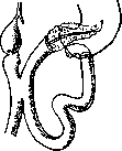
Figure 17. Resection of the vertical part of the duodenum. Pancreato-duodenostomy, duodeno-jejunostomy, cholecysto-jejunostomy and gastro-enterostomy. (According to Kausch.)
The approaches vary: for suprapapillary carcinomas, circular resection according to the second method of Billroth is applicable; for infrapapillary localization, circular resection with a circular suture in segments (Syme) or with both ends sutured tightly and an additional duodenojejunostomy. For tumors of the vertical part of the intestine, the condition is complicated by the involvement of the bile and pancreatic ducts; circular resection of this segment (Korte-Wilms) with circular suturing of the segments is possible; the bile and pancreatic ducts, previously isolated, are sutured into the restored intestine. If bile was previously diverted from the gallbladder into the intestine, the ductus choledochus can simply be ligated. Since circular suturing of the segments is a difficult task to accomplish, it is better to suture both ends tightly, apply a gastroenteroanastomosis, and suture the ducts into the peripheral segment of the Duodenum. But even this modification is too complex. Kausch and Kehr, and earlier Cott, proposed a new principle, the essence of which is that together with the Duodenum, the head of the pancreas is resected, and its cut end is sutured entirely into the lumen of the Duodenum. The operation is divided into two stages, the main task of the first stage being the diversion of bile from the gallbladder through an anastomosis with the intestine or stomach. In its completed form, the operation is shown in Fig. 16 and 17. Kausch's method was successfully carried out in one case by the author himself. Ampullary carcinomas, due to early manifestation of symptoms and early diagnosis, make it possible to operate on them at a stage when the tumor has limited dimensions and does not extend beyond the Duodenum; therefore, they can be removed by much simpler methods. For this, there are two paths: transduodenal and retroduodenal. I. In the transduodenal method, the intestine is opened with a longitudinal or transverse incision in front, the edges of the incision are pulled apart, and the tumor becomes visible and accessible. By inserting a finger behind the previously mobilized Duodenum and protruding its posterior wall into the incision of the anterior wall, the tumor can be almost completely pushed out of the intestinal cavity. After this, the tumor is outlined with an oval incision in the direction of the duct, through the mucous membrane or through the thickness of the intestinal layers, and is excised together with the ampulla and the end of the ducts; then the ends of the ducts, together or separately if they are cut above their junction, are sutured into the edge of the intestinal incision, and the excess is sutured with additional sutures. Finally, the incision of the anterior wall of the Duodenum is sutured. II. In the retroduodenal method, the Duodenum is mobilized according to Kocher and turned inward and downward, thereby gaining access to the ducts, isolating and excising their end together with a portion of the posterior wall of the intestine, and sometimes with a portion of the adhered head of the pancreas, after which the ends of the ducts are sutured into the incision or transplanted to another location of the Duodenum, where there is a serous covering (Mayo). To date, up to 60 cases of radical surgery for ampullary carcinomas have been described. Injuries, fistulas, and other diseases of the Duodenum. Injuries to the Duodenum from projectiles or stab wounds usually constitute only a part of multiple injuries and are extremely rare in isolated form, whereas subcutaneous injuries from blunt trauma are not uncommon; despite its deep position, the injury is favored by the fact that the Duodenum lies adjacent to the spine. Up to 100 cases of isolated subcutaneous injury to the Duodenum have been described, mainly in men, due to occupational conditions, as the usual cause of injury is a kick from a hoof, compression by buffers, carriage wheels, etc. The injury is usually located in the vertical or lower horizontal part of the intestine, is of a crushing or rupturing nature—partial or complete transverse (x/4 of cases), mostly intraperitoneal, but sometimes of retroduodenal type. The initial symptoms consist of shock phenomena, after which some relief sometimes occurs, patients can reach home on their own, but then rapidly follows the development of a picture of perforative peritonitis or bleeding into the abdominal cavity. In retroperitoneal rupture, a retroperitoneal accumulation can occur, palpable from the outside as a fluctuating tumor. Rudofsky collected 35 similar cases from the literature. The injury to the Duodenum cannot be established with complete certainty, but it is important to establish the presence of a severe injury requiring urgent surgical intervention, while the true nature of the injury is easily determined during laparotomy. For free access to the Duodenum, the median incision should be extended transversely to the right. A suture is applied to the ruptured intestine, and in case of narrowing, a gastroenterostomy is added; in transverse rupture, both ends are sutured tightly, and the operation is concluded with a gastroenterostomy; if there is a rupture in the lower part, a duodenojejunostomy can be performed—one or in combination with a gastroenterostomy. External fistulas of the Duodenum result either from injury to the intestine during operations on the intestine itself, on the bile ducts, or from the opening of perforating duodenal ulcers to the outside, or after traumatic paraduodenal abscesses. According to their course, two forms of fistulas can be distinguished: some have a rapid onset, give abundant discharge of food and digestive juices, and quickly lead to death from exhaustion; in the other form, the initially abundant discharges gradually decrease, and the fistula shows a tendency to heal and sometimes actually closes spontaneously. Diagnosis does not present great difficulty due to the characteristic discharges. Fistulas of the first group, as well as non-healing chronic fistulas, require surgical intervention, which should consist of an attempt to approach the fistula itself with the aim of excising its edges and applying sutures, which of course is a complex and difficult task, but nevertheless leads to complete recovery; in other cases, the immediate goal should be the application of a gastroenteroanastomosis and exclusion of the pylorus (Berg); as a last resort, jejunostomy (Mayo) should be applied. If fistulas form after gastric resection according to the second method of Billroth, i.e., from the stump of the Duodenum and therefore in the presence of a gastroenteroanastomosis, one should approach the stump of the duodenum, mobilize it, and apply sutures to the fistula, reinforcing them with omentum. Foreign bodies in the Duodenum (excluding gallstones). In the Duodenum, the lodging of swallowed objects is observed if they have a sharp end or considerable length (e.g., nails, pencils, spoons). Lodging usually occurs at the lower angle, and the foreign body stops in the vertical part. In half of the described cases, complete perforation was observed exactly at the lower angle; only in exceptional cases did foreign bodies remain in the Duodenum for whole years, but even after this, they caused either perforation or retroduodenal phlegmon. If a foreign body is visible on an X-ray, a characteristic feature of its location in the Duodenum is its position on the right outer side of the II or III lumbar vertebra and its vertical direction. For the operation to remove a foreign body lodged in the upper horizontal part, duodenotomy or gastrotomy can be applied; if located in the lower part, mobilization of the duodenum may be necessary to free its lower angle. Needles can be extracted by pushing them out from the inside. Operations have a high mortality rate, which is explained by the fact that they are almost always performed in complicated cases. According to Melchior, out of 17 cases, recovery was noted only in 11. Phlegmonous duodenitis is completely analogous to the same process in the stomach, but occurs much less frequently. Melchior was able to collect 11 cases from the literature. The cause was either the lodging of foreign bodies (fish bones) or the presence of a diverticulum of the Duodenum. The purulent infiltration, usually diffuse, less often limited, was most often found in the area of the papilla Vateri and in some cases extended to the retroduodenal cellular tissue or to the head of the pancreas. Bacteriologically, streptococci were usually found. The clinical signs of this disease are not very characteristic and are hardly accessible to precise diagnosis. The disease was more often diagnosed as peritonitis due to perforation of an ulcer; in other cases, acute pancreatitis or, in subacute course, obstruction of the common bile duct could be suspected. Indications for surgery are dictated by the above presumptive forms, but the prognosis is uncertain. The operation should consist of opening, after tamponade of the abdominal cavity, the retroduodenal phlegmon and creating a fistula with the jejunum for feeding the patient. Among the ulcerative processes developing in the Duodenum, besides round ulcers, other types of ulcers are also encountered, but some of them do not present great surgical interest due to either extreme rarity or inaccessibility for surgical intervention; another part of the cases reaches surgical clinics only at the stage when the condition has progressed to narrowing of the intestine or perforation, the true nature of which sometimes remains unclarified even during laparotomy.
Among such ulcers are ulcers of septic or embolic origin, for example, after burns or after operations, typhoid ulcers. Surgical intervention here can only be considered in cases of perforation or profuse bleeding, but with very doubtful hope of success. - Actinomycosis and tuberculosis can give rise to a palpable tumor from the outside (Wheeler, Cohen), usually mistaken for cancer or an inflammatory process. In other cases, tuberculous ulcers either gave no definite symptoms or ran their course like round ulcers with symptoms of stenosis; sometimes such ulcers perforated and were operated on due to peritonitis. The diagnosis of all these forms of the disease, as well as syphilitic ulcers, can only be made in a presumptive form, but more often is completely impossible. --Diverticula of the duodenum are apparently always of congenital origin, but clinically manifest themselves usually after the age of 40 and then only in cases when they have reached a considerable size and have caused narrowing of the intestinal lumen or given other complications. In most cases, diverticula are located in the vertical or lower horizontal part and have a size from a cherry to a chicken egg. Herbst collected 101 cases, of which 70 were located near the papilla Vateri [see figure 18 and separate plate (Vol. IX, article Yellow)], closely to the head of the pancreas, sometimes sinking into it. The clinical manifestation of diverticula is not sufficiently definite - a number of dyspeptic symptoms, vomiting, symptoms resembling an ulcer or narrowing of the intestine; when the bile ducts are compressed - remittent jaundice. Sometimes on the basis of a diverticulum, phlegmon or perforation of the intestine develops. A characteristic sign for a diverticulum, unlike ulcers, is the decrease in pain when pressure is applied or when the abdominal muscles contract. The radiological sign of a diverticulum is a sharp shadow next to the duodenum. Surgical treatment in the presence of stenosis is gastroenterostomy and, when feasible, resection. % g*

f&i
Figure 18. Duodenal diverticulum (According to Letulle.)

Fig. 19 Duodenum in the form of an hourglass. (From MoupШаа'a.)
Disorders of duodenal passage. Impaired duodenal passage, sometimes reaching the degree of complete obstruction, is caused, in addition to those already mentioned above, by a whole series of other reasons. One of the most common is narrowing of the intestine on the basis of an ulcer. According to patho-anatomical data, it occurs in 5% (Gruber)-10% (Collin), according to clinical material much more often: from 13% (Moynihan) to 25% (Melchior). The discrepancy in figures finds its explanation in that clinical manifestations can be observed even with such degrees of narrowing that are not taken into account patho-anatomically. The substrate of narrowing is not only the scarring of the ulcer, but also adhesive periduodenitis, and in some cases the ulcer itself is still unhealed. Corresponding to the favorite localization of ulcers, narrowing is usually located above the papilla Vateri; the clinical manifestation of such stenoses is identical with pyloric stenosis, but radioscopically one can sometimes confirm the presence of dilation of the initial part of the duodenum. With multiple ulcers, the stricture can be double, and the duodenum takes the form of an hourglass (see figure 19). With infrapapillary narrowing, in addition to the usual symptoms of stenosis, constant abundant reflux into the stomach of bile and pancreatic juice is noted; it is especially characteristic if the stomach is empty, after having been washed out the day before, contains bile in large quantities. If the narrowing of the intestine involves the excretory ducts of the liver, the picture is complicated by progressive jaundice with simultaneous dilation of the gallbladder and sometimes by the addition of cholangitis phenomena. Dilation of the proximal part of the duodenum sometimes reaches the degree of true megaduodenum. In differential diagnosis from other similar processes, radioscopic examination remains of decisive importance. As the basic method of surgical treatment of strictures, gastroenterostomy is applied, which can or should be supplemented by duodenojejunostomy (in infrapapillary strictures). Plastic surgery - duodenoplasty according to the pyloroplasty type - can only be applied under particularly favorable local conditions. Similarly, in the case of a duodenum in the form of an hourglass, resection (Mayo) with suturing 'end to end' or according to the second method of Billroth is rarely possible. In the case of scar narrowing involving the papilla Vateri, the outflow of bile must be ensured by creating an anastomosis between the gallbladder and the stomach or the jejunum, or the common bile duct must be sutured into the duodenum at a new place (choledochoneostomia). As for periduodenitis, in addition to ulcers they can also be caused by other reasons1; cholecystitis (according to Ochsner 3/„ of all cases), colitis of the hepatic part of the large intestine, sometimes appendicitis, operations on the bile ducts (performed with tampons), injuries, acute or chronic; among the latter - professional trauma in work associated with pressure on the epigastric region. As a result of the above reasons, either separate strands or diffuse adhesions compressing the duodenum are formed. Such adhesions can sometimes give a series of painful local sensations, occasionally exacerbating, often indefinite, but in the presence of the above etiology already acquire diagnostic significance. Sometimes all the symptoms of intestinal stricture develop (chronic duodenal ileus), confirmed also radioscopically. Simple division of adhesions in such cases is hardly likely to give a permanent cure, therefore it is recommended to supplement the operation with duodenolysis with free omentoplasty (Oppel). The cause of stenoses is sometimes compression of the intestine by pathologically enlarged neighboring organs (lymph glands, etc.) or tumors (pancreas, liver, kidney). This also includes cases of compression of the duodenum or jejunum by the mesentery of the intestine. Surgical treatment here, depending on the peculiarities of the case, is causal or palliative. -As for congenital narrowings or atresias of the duodenum (causes - see Intestine), their manifestation depends on the degree of intestinal patency and on the location of the stricture. Incomplete strictures may manifest themselves already in adulthood, sometimes only during an accidental examination of the patient. In complete atresia, from the very first days, the picture of duodenal ileus develops. A characteristic sign of them is that the abdomen remains not swollen, and white meconium is found in the rectum if the atresia is located infrapapillaryly. In incomplete atresias, the child can live for several months. Surgical intervention in the form of gastroenterostomy or duodenojejunostomy should always be considered indicated, but the prognosis in cases of complete atresias is very poor due to the exhaustion of the child, since the operation is undertaken late. Boiling described a case of duodenojejunostomy in a 2-week-old child, which ended in recovery. Among other congenital changes, which manifest themselves mainly also as impaired patency of the duodenum, duodenoptosis and duodenum mobile deserve mention. On the basis of abnormal mobility of the duodenum, cases (35 cases) of its presence in hernias (diaphragmatic) and even inguinal ones according to the so-called 'sliding' hernia type have been observed. In other cases, abnormal mobility of the intestine depends on the presence of a well-expressed mesentery throughout the entire upper horizontal part; sometimes the mesentery continues further onto the upper 1/3-% and even onto the entire length of the vertical part; thus mobility becomes complete; these latter forms have less clinical significance, but with partial mobility and with the simultaneous presence of gastric prolapse, a bend can form, mainly in the vertical position of the body (see figure 20). Th

Figure 20. Partial duodenoptosis (with simultaneous prolapse of the stomach, schematically according to the X-ray picture): 1-navel; 2-flex. sup.; 3 - bulbus; 4-duodenum; 5 - stomach; 6-flex. duod.-jejun. (From Melchior's.)
These forms can give a number of characteristic symptoms: pains in the upper half of the abdomen radiating to the back, often beginning in young years and having the character of colic; a feeling of heaviness in the abdomen; all these phenomena sometimes disappear after vomiting, but what is especially characteristic is - when lying down. In addition, due to twisting of the bile duct, jaundice in a mild degree or intermittent jaundice is observed. Banzet points out that even with complete displacement of the Duodenum, difficulty in emptying it can also occur due to the fact that the duodenojejunal angle is strongly stretched upward by the tense Treitz ligament, while the transverse colon compresses the second part of the Duodenum. In this form, the operative intervention should consist in cutting the Treitz ligament, but if this is not sufficient, a duodenojejunostomy should be added. With partial mobility with the formation of a bend in the vertical loop of the intestine or at the border of the vertical and upper horizontal part, one can limit oneself to duodenopexy (Perthes) or perform direct suturing to the parietal peritoneum. As a form not fully clarified, cases of idiopathic expansion of the Duodenum are described, where any mechanical factor is excluded, including compression by the mesenteric artery. The symptoms of such megaduodenum consist of attacks of vomiting of food or bile, attacks of pain in the right hypochondrium, protrusion of the hypochondriac region, sometimes visible peristalsis; usually constipation and general exhaustion are added. The intermittence of the picture is characteristic. The operative intervention in such cases should consist in the application of a duodenojejunostomy below the mesocolon.
V. Dobrotvorsky.
Radiodiagnosis of diseases of the Duodenum. Significant successes in the radiological recognition of diseases of the Duodenum became possible only thanks to the improved technique of recent times. In addition to good tubes and powerful apparatus for this purpose, instantaneous exposures with an exposure time of no more than 0.1-0.2 seconds, exposures 'in series' (a series of consecutive radiographs on the same film) and a device for instantaneous transition from fluoroscopy to radiograph ('selector') are also necessary. The intestine can be filled with a liquid, uniform and stable suspension of contrast substance by the usual method through the mouth or through a duodenal tube (David). In addition, Přibram proposes to inflate the bulb of the Duodenum with a small amount of air, thanks to which the clear visibility of the folds of the mucous membrane ('relief') is achieved. For diagnosis, it is equally necessary to use both fluoroscopy and radiograph. During fluoroscopy, the correct expansion of the bulb and the other parts of the Duodenum when filled with contrast mixture is observed. The contours of the bulb are examined not only en face but also in profile, for which the patient is turned into an oblique position. The careful use of palpation allows for better visualization of irregularities in the contour and relief of the mucous membrane of the Duodenum, as well as its passive mobility. The normal bulb has the shape of a 'mitre', a sugar loaf, a vertical and horizontal oval, more rarely a sphere (see figure 21). Its contours should be everywhere even and clear, without defects or protrusions. When refilled, the normal bulb retains its shape. When the walls contract, the entire bulb empties at once or

Figure 21. Normal bulb of the Duodenum: 1 - pylorus; 2 - recessus internus; 3 and 6 - contour of the bulb, corresponding to the lesser and greater curvature; 4 - apex of the bulb; 5' - recessus externus. l sh ~1 -^
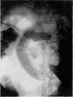
Figure 1. Megabulbus. Carcinoma of the greater curvature of the stomach. The bulb of the 12-finger intestine reaches the size of an apple. Figure 2. Periduodenitis in the region of the middle part of the duodenum. Patient with symptoms of stomach ulcer. Enhanced peristalsis of the greater curvature of the stomach. Constant constriction in the descending segment of the duodenum. Above - the lumen of the duodenum is widened, contours are smoothed. Figure 3. Periduodenitis bulbi duodeni. Patient with unclear symptoms of duodenal ulcer. On each of the 4 films of the series, the bulb has a different configuration. Its contours are uneven, as if tousled. Figure 4. Periduodenitis on the basis of cholecystitis. Patient with complaints of attacks of pain in the right hypochondrium. Insufficient expansion of the bulb of the 12-finger intestine with uneven edges. 4 stones in the gallbladder. Figure 5. Ulcus bulbi duodeni from the side of the lesser curvature. Patient with a distinct duodenal symptom complex. Asymmetrically located pylorus with a narrow canal. At the base of the bulb on the inner side there is a defect in the contour with a constant spot of Ba, visible on all 4 films of the series. Figure 6. Diverticula of the 12-finger intestine in the region of bulbi and in the region of papillae (verified during operation).
K" st. Duodenum.
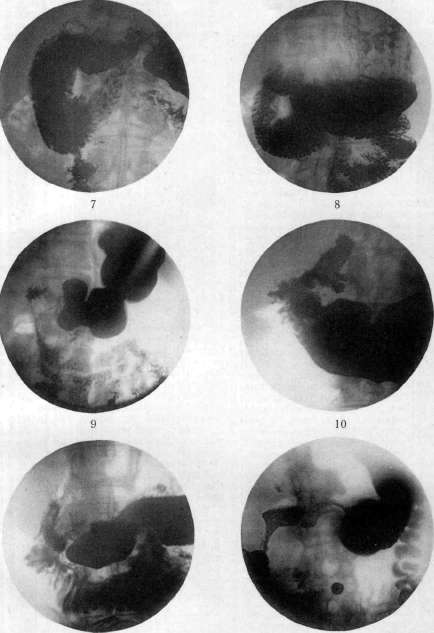
Figure 7. Megaduodenum. Stomach without special changes. 12-finger intestine is very wide. However, no delay of Ba mixture occurs in it. Figure 8. Periduodenitis in the region of flex. duod.-jejunal. Suspicion of stenosis of the stomach outlet. Stomach slightly widened. Pylorus is passable. 12-finger intestine is filled with a large amount of Ba mass. The lower part of the duodenum is especially wide. During fluoroscopy, antiperistaltic movements of the contours are noticeable. The first loops of the jejunum are outlined only after palpation of the 12-finger intestine. Figure 9. Ulcus bulbi duodeni. During operation, a large scar was found on the walls of the bulb. Enhanced peristalsis of both contours of the stomach (duodenal type). The bulb has the shape of a star spot. Pars descendens is narrowed. Figure 10. Ulcus bulbi duodeni. Typical duodenal anamnesis. Stomach is radiologically normal. Asymmetric bulb with retraction of the outer wall. On the inner wall three protrusions in the shape of a cloverleaf. The same changes after 2 and after 4 hours. Figure 11. Defect on the inner wall of bulbi duodeni due to compression by a tumor of the adjacent retroperitoneal gland. Figure 12. Compression and displacement to the periphery of the entire 12-finger intestine by a large tumor of the pancreas. K st. Duodenum. which achieves the clear visibility of the folds of the mucous membrane ('relief'). For diagnosis, it is equally necessary to use both fluoroscopy and radiograph. During fluoroscopy,
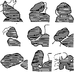
Figure 22. Various forms of the bulb of the Duodenum (From Schinz).
the correct expansion of the bulb and the other parts of the Duodenum when filled with contrast mixture is observed. The contours of the bulb are examined not only en face but also in profile, for which the patient is turned into an oblique position. The careful use of palpation allows for better visualization of irregularities in the contour and relief of the mucous membrane of the Duodenum, as well as its passive mobility. The normal bulb has the shape of a 'mitre', a sugar loaf, a vertical and horizontal oval, more rarely a sphere (see figure 21). Its contours should be everywhere even and clear, without defects or protrusions. When refilled, the normal bulb retains its shape. When the walls contract, the entire bulb empties at once or
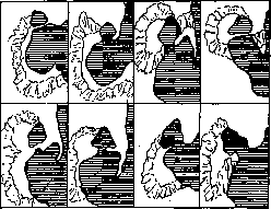
Figure 23. Various forms of the passage of a normal Duodenum (From Schinz).
in parts. Thus, when the upper half contracts, the lower half takes the shape of a 'hammer'. Various forms of the normal bulb of the Duodenum are shown in figure 22, and the passage of contrast food through the entire 12-finger intestine is shown in figure 23.

Among congenital changes of the Duodenum, anomalies of position, length and width of the lumen can be detected. With mesenterium ileo-colicum commune, the entire Duodenum lies in the right half of the abdomen (see figure 24). With congenital elongation of the mesentery, the Duodenum forms extra loops and is easily displaced (duodenum mobile). Expansion of the bulb or the entire Duodenum gives the forms megabulbus and megaduodenum (see separate table, figures 1 and 7). Congenital strictures in this section of the intestine are rare. Secondary displacements are observed with pyloroptosis, tumors of neighboring organs and with adhesions on the basis of inflammatory changes. In periduodenitis of a congenital nature ('essential' according to the terminology of French authors), the X-ray picture varies depending on the location and extent of the films and adhesions. Periduodenitis in the region of the bulb is characterized by its improper expansion, variability and peculiarity of contours (see separate table, figures 2 and 3). Adhesions in the middle part of the 12-finger intestine cause expansion of the proximal sections with narrowing of the distal ones, sometimes enhanced peri- and antiperistaltic movements of the stomach walls (see separate table, figure 8). The Duodenum reaches a special width with adhesions in the region of the 3rd part. At this time, a delay of the contrast mass in the widened intestine is observed. It is not always possible to distinguish by the X-ray picture periduodenitis of the 'essential' type from inflammatory adhesions. Sometimes filling of the gallbladder with tetraiodophenolphthalein can help here, thanks to which the mutual position of these organs can be determined exactly (see separate table, figure 4). With stenosis in the lower part of the Duodenum due to its compression by the root of the mesentery or by arteries passing through it (art. mesenterica superior and art. colica media), the proximal part of the Duodenum is widened, filled with contrast mixture, peristalsis is enhanced, while the distal part is empty and narrowed. The dividing line runs right along the midline, along the spine. When the patient is in the knee-elbow position or when the small intestines are lifted en masse upward with the hand, the compression of the 12-finger intestine is eliminated, and the contrast mass quickly passes into the small intestines.
In ulcer of the Duodenum, 'direct' signs are distinguished, i.e., immediate changes in the configuration of the bulb caused by anatomical or spasmodic deformations of its wall, and 'indirect', or 'secondary' signs, expressed in reflex changes in the configuration or functional activity of the stomach. The most convincing are the 'direct' signs of ulcer (see separate table, figure 5). These include: 1. Niche. It is caused by the penetration of barium into the crater of the ulcer and therefore is expressed by a small defect in the contour of the bulb, which is filled with contrast substance and is visible during the evacuation phase. 2. Deformation of the bulb. 3. Retraction of the wall. 4. Constant defect of the contour. 5. Delay in evacuation. 6. Changes in the stomach: a) hypertonicity, b) spastic narrowing of the antrum, c) spastic peristalsis, d) delay in evacuation. Duodenum liberum. The entire Duodenum is located to the right of the spine.

with a protrusion on the contour of the duodenal bulb (see separate table, Fig. 10) or has the appearance of a small rounded speck in the plane of the bulb under moderate ("dosages" according to Berg) compression of it (ulcer on the anterior or posterior wall). Not only the depth of the defect plays a role in the formation of the niche, but also those mucosal folds around the ulcer crater which, according to Forssell, actively arise as a result of irritation of the muscularis propria. The simultaneous appearance of several niches on the anterior and posterior or lateral walls is possible. In perforating callous ulcers, the niche has the appearance of a small speck connected by a narrow passage with the bulb.
J, ,, minor curvature; indentations of the contour (defects So-g - indentation on 1e). According to some authors, of the greater curvature; they are caused by cicatricial ne; 3-"pocket" Pylorus wide open. asymmetrical shrinking or partial - ™ ^ 5 H « - p y rk \ " spasm of the muscularis mucosae (similar to the indentation on the 3. normal con- greater curvature of the stomachfiguration bulbi. with an ulcer on the lesser), (According to Akerlund.) according to others - by the protrusion inward of the lumen of the bulb of mucosal folds. 3. Flattening or smoothing of one of the contours of the bulb, more often the medial one, while the pyloric canal is located asymmetrically. The cause of this phenomenon is segmental contraction or cicatricial shrinking. 4. "Pockets", or diverticulum-like protrusions between the retracted or contracted sections of the wall. According to Berg, periodic increase plays a certain role in their occurrence.
pressure in the cavity of the bulb. 5. Radial arrangement of mucosal folds with incipient or already pronounced scarring (see separate table, Fig. 9). The indicated changes occur in various combinations in the ulcer, as a result of which characteristic forms of the bulb arise, e.g., in the form of an "hourglass", "trefoil", etc. (see figure 25 and separate table, figure 10).-Indirect signs of a duodenal ulcer consist in increased tone, hypersecretion, and increased motor function of the stomach in the initial phase of the study with subsequent deceleration. However, it has been proven that these symptoms can also appear with diseases of other, often very distant organs. Therefore, at present time, they are given significance only as additional evidence. The symptom of coincidence of the painful point upon pressure with the duodenal bulb must also be used with a certain caution. Particularly accessible to X-ray examination are ulcers proceeding by the type of a chronic recurring process, with scars and shrinking, and ulcers penetrating deep enough into the mucosal and submucosal tissues. Mucosal erosions or flat fresh ulcers give less convincing symptoms. Duodenal diverticula are found in any of its sections, but most often in the region of the Vater's papilla and near the flexures (see figure 26). Their size is from a pea to a hazelnut. The shape is various, predominantly spherical or oval. Clairmont and Schinz characterize the duodenal diverticulum as follows: 1) a clearly contoured shadow of a rounded shape, 2) part of the contrast mixture passes next to or above this formation, 3) upon emptying of the duodenum, part of the contrast mass remains in the diverticulum and is retained from several hours to several days (see sep. table, fig. 6).--Primary tumors of the duodenum are very rare. Carcinomas in the region of the papilla have been described, causing phenomena of stenosis with dilation of the bulb and pyloric insufficiency. Carman cites a case where an X-ray revealed a shadow defect in the contour of the bulb; at operation, it turned out to be a polypoid angioma. Tumors originating from neighboring organs cause various deformations of the bulb depending on the degree of lesion and compression of the wall (see sep. table, fig. 11 and 12).-Foreign bodies sometimes get stuck in the duodenum, and only metallic objects are accessible to X-ray diagnosis. The casuistry in this regard is very rich. A wide variety of objects have been detected by X-ray in the duodenum and extracted operatively, ranging from small nails to a thermometer (Schinz's case).
Figure 26. Scheme of the position of duodenal diverticula: 1 - diverticulum at the opening of the accessory pancreatic duct (S); 2 - diverticulum-like expansion in the region of the Vater's papilla; 3, 4, 5 - diverticula in the region of the inferior duodenal flexure; 6 - diverticulum of the duodenojejunal flexure; W - pancreatic duct; Z - cystic duct; H - hepatic duct; Ch - common bile duct; F - gall bladder. (From Schinz.)
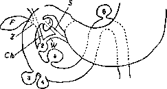
A. Zeitlin.
MOTOR CENTERS
Duodenum
Duodenum
446 Notes on the duodenum and the pylorus, Journ. of anatomy and physiology, v. XXXI, 1896-97; Jonnesco, Sur l'anatomie topographique du duodenum, Progres medical, t. IX, 1889; Luschka H., Die Lage der Bauchorgane des Menschen, Carlsruhe, 1873; Schiefferdecker P., Beitrage zur Topographie des Darmes, Arch. f. Anat. u. Physiol., Anat. Abt., 1886, p. 335; Toldt C., Bau und Wachstumsveranderungen der Gekrose des menschlichen Darmkanals, Wien, 1879; Tschaussow M., Bemerkungen uber die Lagerung der Bauchspeicheldriise, Anat. Anzeiger, B. XI, 1895; Villemin F., Recherches d'anatomie comparee sur le duodenum de l'homme et des mammiferes, Arch, de morph. gen. et exper., 1922, № 3. Pathology. General works.-Gal'perin Ya., Dobrokachestvennye zabolevaniya zheludka, Tver', 1910; Gautier R., Les maladies du duodenum, P., 1910; Guleke N., Nieden H. u. Schmidt H., Die Chirurgie des Magens u. des Zwolffingerdarmes (Die Chirurgie, hrsg. v. M. Kirschner u. O. Nordmann, B. V, Berlin-Wien, 1927); Handbuch der spez. path. Anatomie und Histologie, hrsg. v. F. Henke und O. Lubarsch, B. IV, T. 1-Magen u. Darm, B., 1926 (lit.); Hausmann Th., Die syphilitischen Tumoren des Magens u. sonstige syphilitische Tumoren der Oberbauchgegend, Erg. d. inner. Med., B. VII, 1911; Ohne, Die luetischen Erkrankungen der Bauchorgane, Halle, 1913; Meichior E., Die Chirurgie des Duodenums (Neue deutsche Chirurgie, hrsg. v. H. Kiittner, B. XXV, Stuttgart, 1917). Ulcer.-Anrirez E. i Durand G., Yazva zheludka i 12-perstnoy kisheki, M.-L., 1926; Bikhmatse E., O krugloy yazve zheludka i 12-perstnoy kisheki v Rossii, Sovremennaya meditsina, 1921, № 1; Voznesensky V., Kruglaya yazva 12-perstnoy kisheki, diss., M., 1913; Gal'perin Ya., 548 operatsiy na zheludke, Novyy khirurgicheskiy arkhiv, t. III, 1923; Mogil'nitsky V., K voprosu o patogeneze i etiologiyevrogennykh form krugloy yazvy, Trudy 1 Syezda patologov, Moskva, 1926; Petrovsky G., Proboydnye yazvy zheludka i 12-perstnoy kisheki, Vestnik khirurgii i pogranichnykh oblastey, t. XIV, kn. 42, 1928; Petrov N., K lecheniyu proboydnykh yazv zheludka i 12-perstnoy kisheki, Novyy khirurgicheskiy arkhiv, t. XII, kn. 1, 1927; Savkov N., Gastro-enterostomiya kak operatsiya vybora pri yazve zheludka i 12-perstnoy kisheki, Ibid., t. II, kn. 4, 1922; Tager N., Yazva 12-perstnoy kisheki, L., 1925; Fink'shteyn B., K voprosu ob operativnom lechenii neproboydayushchikh yazv 12-perstnoy kisheki, Novyy khirurgicheskiy arkhiv, t. II, kn. 3, 1922; Khesin R., Yazva 12-perstnoy kisheki, diss., P., 1914; Shelkov B. i Ilyin S., Yazva zheludka i 12-perstnoy kisheki i otdalennyye rezul'taty gastro-enterostomii pri ney, Novyy khirurgicheskiy arkhiv, t. XVII, kn. 3, 1927; Enel' G., Yazva zheludka, M.-L., 1929; Yarotsky A., Diteticheskoye lecheniye krugloy yazvy zheludka i 12-perstnoy kisheki, L., 1928; Aschoff L., Uber die peptischen Schadigungen des Magendarmkanals, Med. Klin., 1928, №50; Balfour D., The results of operation for duodenal ulcer in physicians, Annals of surgery, v. LXXXVI, 1927; Duval P., Roux J. et Besliere H., Etudes medico-radio-chirurgicales sur le duodenum, P., 1924; Finsterer H., Zur Therapie des Kardiospasmus und der Kardiostenose, Wien. klin. Wochenschr., 1922, p. 471; Gruber G., Die pathologische Anatomie des Ulcus duodeni, Mitteil. aus den Grenzgebieten der Med. u. Chir., Supplementheft 4, 1923; Jewett H., Chronic duodenal ulcer, Journ. of the Amer. med. assoc., v. XCI, 1928; Mayo J., End-results in ulcer of the stomach and duodenum, Surgery, gynecology and obstetrics, v. XXXVI, 1923; Moynihan B., Duodenal ulcer, Philadelphia-London, 1910 (German edition-Dresden-Leipzig, 1913); Schrijver G., Ulcus duodeni, B., 1914; Singer G., Ulcus duodeni (Spez. Pathologie und Therapie, hrsg. v. F. Kraus u. Th. Brugsch, B. VI, T. 2, 1922); Starlinger F., Ergebnisse 25-jahriger operativer Therapie der Geschwurskrankheit des Magens und Zwolffingerdarms, Archiv f. klin. Chirurgie, B. CXLVII, 1927. Duodenitis.-Oppel V., Periduodenitis adhaesiva chronica, Vrach, gaz., 1914, № 13; Einborn M., The diagnosis of chronic duodenal catarrh, Archives of diagnosis, v. III, 1910; Ungermann E., Duodenitis phlegmonosa, Virchows Arch., B. CXCIII, 1908. Cancer.-Dobrotvorsky V., Carcinoma papillare duodeni, Novyy khirurgicheskiy arkhiv, t. I, 1921; Deaver J. and Ravdin I., Carcinoma of the duodenum, Amer. journ. of med. sciences, v. CLIX, 1920; Forgue E. et Chauvin E., Le cancer primitif et intrinseque du duodenum, Revue de chirurgie, t. XLIX, 1929. Sarcoma.-Terent'yev A., Sarcoma 12-perstnoy kisheki, Voyennyy meditsinskiy zhurnal, t. CCXVII, 1906; Salis H., Uber das Sarcom des Duodenums, Deutsche Zeitschrift f. Chirurgie, B. CLX, 1920. Displacement and dilatation.-Mints S., O duodenum mobile, Vestnik khirurgii, t. XIII, kn. 39, 1928; Barber W., Dilatation of the duodenum, Annals of surgery, v. LXII, 1915; Gregoire R., Megaduodenum, Bull, et mem. de la Societe de chirurgie de Paris, v. XLVII, 1921; Meichior E., Das Megaduodenum, Arch. f. klin. Chir., B. CXXVIII, 1924. Diverticula.-Clairmont P. u. Schinz H., Zur Diagnose und Chirurgie der Duodenaldivertikel, Deutsche Zeitschrift f. Chirurgie, B. CLIX, 1920; Orelet J., Etude sur les diverticules du duodenum, P., 1921; Heitemeyer H., Uber Duodenaldivertikel, Appelhiiisen, 1918.
Related articles
Mentioned in
Cite this page
“Duodenum.” Soviet Medical Encyclopedia. English translation of Bolshaya Meditsinskaya Entsiklopediya, 1st ed. (Moscow, 1928–1936), ed. N. A. Semashko. https://sovietmedicalencyclopedia.pages.dev/article/duodenum/