Duodenal Tube (duodenal sounding, and duodenal)
Historical document, translated for reference. It reflects medical knowledge of the 1920s–30s and is not medical advice.
Summary
This article from the 1928–1936 Great Medical Encyclopedia discusses the history, construction, and initial procedure of duodenal sounding and obtaining duodenal contents using a duodenal tube, highlighting the contributions of Einhorn and Lyon.
Encyclopedia article (1928–1936)
DUODENAL TUBE, duodenal sounding, and duodenal contents. Attempts to obtain duodenal contents for diagnostic examination began in the late 1880s, but it was only in the 1920s that sounding of the duodenum became the simple and easy diagnostic procedure that it is today, thanks primarily to the works of Einhorn and Lyon. The modern duodenal tube consists of a long (150 cm or more), narrow rubber tube (with an internal lumen of 1.5 mm in diameter) that is sufficiently thick-walled so that its lumen does not close due to collapse of the walls or light pressure. At the end of the tube, there is a metal olive with holes on the sides. At a distance of 40 cm from the olive on the tube, there is a first mark; at a distance of 70 cm from the olive, the second; and at a distance of 80 cm, the third (Figure 1). In the morning on an empty stomach, the patient is placed in a sitting position, and the olive is introduced into the entrance of the pharynx, making...
duodenal sounding using a duodenal tube. Figure 1.
one must bear in mind the encountered dangers, such as hemorrhages, swallowing of the tube, and other complications.) When the first mark aligns with the edge of the teeth, it can be considered that the olive is in the region of the cardia; when the second mark reaches the same level, the olive is in the region of the pylorus, and when the third mark reaches the edge of the teeth, it can be considered that the olive is at the level of the ampulla of Vater. When the olive has entered the stomach, the patient is laid on their right side so that the olive can travel by its own weight into the region of the pylorus. Half an hour to an hour after the introduction of the tube, the olive ordinarily penetrates into the duodenum, and then through the outer end of the tube there begins to bleed a more or less sticky, golden-yellow fluid, completely transparent and possessing a distinctly alkaline reaction. If the spontaneous discharge of the characteristic duodenal contents [see separate table (pp. 107-108), Fig. 5-II] does not occur for a long time, aspiration of it is resorted to with a 10-gram syringe. The correctness of the position of the olive can be easily verified by X-ray examination, which is especially important when the duodenal contents fail for a long time to be obtained either by gravity or by aspiration and doubts arise as to whether the tube is truly in the duodenum. If, despite a sufficient time of stay of the tube in the stomach, the olive still does not pass through the pylorus, one has to think either of a narrowing of the pylorus, spasmodic or organic, or of a very sharply expressed gastroptosis. To overcome pylorospasm, 0.01-0.02 of novocaine or 0.04-0.06 of papaverine sulfate or hydrochloride is sometimes successfully injected. After an amount of fluid sufficient for examination has been obtained from the tube, the so-called "bile A", consisting chiefly of bile with a greater or lesser admixture of pancreatic and intestinal juice, the so-called Meltzer-Lyon test is performed, i.e., 30 cm3 of a 25-40% magnesium sulfate solution is injected into the duodenum, after which, under normal conditions, a dark brown, more viscous "vesicular bile" ("bile B", according to Lyon's terminology) appears from the tube [see separate table (pp. 107-108), Fig. 5-II]. After the discharge of this dark bile has ended, a lighter and less viscous, golden-yellow fluid is again discharged. This is the so-called "bile C", or "hepatic bile", again with a greater or lesser admixture of pancreatic secretion and intestinal juice [see separate table (pp. 107-108), Fig. 5-III]. Sodium sulfate and glucose act a little weaker than magnesium sulfate, a 10% peptone solution acts almost just as strongly; the discharge of "vesicular" bile is obtained especially easily upon the introduction into the duodenum through the tube of warmed Provençal oil in an amount of 15-20 cm3; solutions of sodium phosphate, magnesium citrate, and sodium chloride act significantly weaker. Pituitrin and other pituitary preparations administered subcutaneously (2 cm3 of Hypophysin solution) cause a significant contraction of the gallbladder and the discharge of "vesicular" bile, or "bile B". According to Meltzer-Lyon, "bile B" consists predominantly of vesicular bile and is obtained due to the fact that magnesium sulfate from the duodenum causes a reflex contraction of the gallbladder with simultaneous relaxation of the sphincter of Oddi. With complete blockage of the bile ducts, only intestinal juice without bile is discharged [see separate table (pp. 107-108), Fig. 5-IV]. The Meltzer-Lyon test therefore makes it possible to subject the contents of the bile ducts, gallbladder, and the bile freshly flowing from the hepatic ducts to macro- and microscopic, bacteriological, and chemical examination separately (although with the admixtures indicated above). Although against the views of Meltzer-Lyon, Rost, and many other authors that the dark "bile B" is the result of reflex contraction of the gallbladder there are also objections (Einhorn and others), nevertheless the vast majority of clinicians stand by the point of view of Meltzer-Lyon, since experiment and clinical observations speak in favor of the fact that "bile B" truly consists in a significant part of vesicular bile. Still, the question of whether a more concentrated bile cannot be secreted by the liver itself under the influence of the introduction of magnesium sulfate into the duodenum cannot yet be considered definitively resolved in the negative sense. Kalk and Schöndube propose pituitary preparations (Pituitrin-Parke-Davis and Hypophysin-Höchst) to induce the vesicular reflex, which are injected under the skin in an amount of 1.0-2.0. The reflex occurs in 20-30 minutes. Causing contractions of the gallbladder, these preparations lead to a more perfect emptying of it, and "bile B" is obtained after the injection of pituitrin more abundantly and more concentrated. Kalk and Schöndube propose calling this reflex the "pituitrin test". This test has the advantage over the introduction of magnesium sulfate and the like for obtaining "vesicular" bile in that with it the duodenal contents are obtained immediately without the admixture of these substances. Examination of the duodenal contents obtained by means of the tube plays a paramount role in the recognition of diseases of the duodenum and the organs opening into it. Normal duodenal contents consist of a mixture of intestinal and pancreatic juices and bile with a greater or lesser admixture of gastric juice. The ratio of the constituent parts of the duodenal contents varies depending on the predominance of one or the other part. The duodenal contents are straw-yellow or golden in color, transparent, and viscous. Specific gravity 1.010-1.012. Its chemical composition is determined by the secretions entering into its composition: of the pancreas, intestines, and liver (bile acids, cholesterol, bilirubin, mucin). Coagulable protein is usually absent. Urobilin is present only in pathological cases. The amount of bilirubin determined in "bile A" ranges from 5 to 20 units, on average 10-12 units. The number of units of bilirubin in "bile B" depends on the concentration of bile in the gallbladder, normally 50-100 units. In pathological cases it reaches up to 300 units and higher. Larger numbers already indicate stagnation of bile in the gallbladder. The coloration of "bile B" is dark brown, sometimes almost black. Microscopic examination of the duodenal contents is carried out in smears from the centrifugate and must be performed immediately after extraction. If this is impossible for any reason, measures must be taken to prevent the destruction of formed elements by the enzymes of the pancreatic juice. For this, it is best to add to the duodenal contents a third of its volume of 10% formalin and heat to 80°. Under normal conditions, the duodenal contents are very poor in both cellular forms and crystals, but already, for example, in achylia gastrica, the matter changes, since in achylia, due to the absence of peptic digestion in the stomach and the accelerated transition of the gastric contents into the duodenum characteristic of achylia, epithelial cells of the gastric mucosa, squamous epithelial cells from the esophagus, and even from the oral cavity appear in the centrifugate; sometimes single leukocytes from the nasopharynx or from the inflammatorily altered walls of the stomach are encountered; for the most part, however, leukocytes coming from the stomach are recognized with difficulty, since they manage to undergo significant digestion even in the duodenal contents, and only nuclei are determined under the microscope. In addition, in gastric achylia, bacteria ordinarily absent in the duodenum appear in the duodenal contents, especially the colon bacillus. In duodenitis and duodenal ulcer, large masses of mucus, goblet cells, columnar epithelium, and leukocytes can be found under the microscope. In angiocholitis, the first portion ordinarily contains leukocytes, epithelial cells, and flakes of mucus permeated with a multitude of rod-shaped bacteria in large numbers. As the jaundice disappears, the leukocytes become fewer and fewer. In cholecystitis, "bile A" is ordinarily very poor in formed elements, whereas in "bile B" there are very many leukocytes, so that they frequently cover the entire field of vision. On an unstained preparation, it can be noticed that epithelial cells and bacteria in this case are frequently impregnated with bile pigment. In cholelithiasis, a multitude of crystals and fragments of cholesterol crystals can frequently be found in "bile B" and even in "bile A" (Fig. 2). In calcareous stones and in biliary sand, amorphous black "poppy" grains of bilirubin lime are sometimes encountered in smears from the centrifugate.
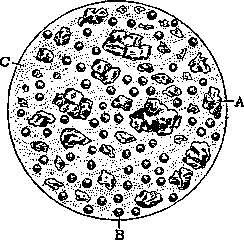
Figure 2. Smear from the centrifugate of "bile A" in cholecystitis calculosa: A - cholesterol crystals; B - pus corpuscles; C - mucus.
Clinical study of the bacteriology of the biliary tract became possible only since the introduction of duodenal intubation, performed with all possible bacteriological precautions in this case. Normally, the duodenum contains no or almost no bacteria. In gastric diseases associated with subacidity and anacidity, numerous colonies of Bact. coli and enterococcus (Streptococcus faecalis) can very often be grown from the duodenal contents. In hyperacid gastric diseases, however, the duodenal contents usually turn out to be sterile. In cholecystitis, enterococci (Streptococcus faecalis) are often found (60%), while in so-called catarrhal jaundice, nothing characteristic is usually found. In typhoid fever and especially in typhoid «bacillary carriers», typhoid bacilli are found more often than upon examination of feces. Parasites are sometimes found in the duodenal contents, of which Lamblia intestinalis is encountered most frequently. In the diagnosis of liver diseases, the examination of duodenal contents makes it possible to judge the amount of bile entering the intestine. Furthermore, the duodenal tube makes it possible to monitor with great precision the excretion of the components of bile, in particular bilirubin and urobilinogen in the bile, and moreover, both «vesicular» and «hepatic» bile. Fluctuations in the excretion of these substances sometimes shed light on the state of the concentration capacity of the gallbladder mucosa and contribute to the differential diagnosis between various types of cirrhosis. Thanks to the duodenal tube, it has also become possible to approach the question of protein excretion in bile (albuminocholia). Finally, the use of the duodenal tube has made it possible to place the so-called chromocholoscopy on a more rational basis, i.e., the study of functional disorders of the liver by means of studying the absorption and excretion of various dyes by the liver. For this purpose, as is known, various dyes have been used, such as methylene blue, tetrachlorophenolphthalein, and many others, but indigo carmine and azorubin have proven most convenient. These substances, when injected into a vein, are excreted almost exclusively by the bile and, moreover, as the duodenal tube shows, very quickly. Indigo carmine, for example, appears after just 15–30 minutes. It should be noted that sometimes (certain cases of jaundice, tumors), after the introduction of the duodenal tube into the stomach or into the duodenum, an admixture of blood visible to the naked eye is secreted along with the duodenal juice. In such hemorrhages, it is necessary to carefully remove the tube and stop further intubation. In the diagnosis of functional and anatomical diseases of the pancreas, the use of the duodenal tube promises to yield much that is new. In the therapy of diseases of the biliary tract, intestines, and indirectly of other organs, the duodenal tube renders ever greater services. By energetic evacuation of the gallbladder and the remaining biliary tracts using magnesium sulfate, peptone, and subcutaneous administration of pituitrin, one can combat pathological bile stasis. In this way, not only the biliary tracts are evacuated, but to a certain extent also the excretory ducts of the pancreas; delays of normal and pathological secretion are eliminated, which makes it possible to prevent or inhibit the development of chronic pancreatitis. Lyon called this method of treatment and prevention «nonsurgical drainage of the biliary tract». He used it in two forms: in the form of intermittent and continuous drainage. In the first case, drainage is used periodically for two to six hours, and in the second, the duodenal tube is left in place for 2–3 weeks or even more after insertion. Intermittent drainage is performed 1–2 times a week, and each time, having waited for the appearance of «bile A», there is injected through the tube at half-hour intervals first 75, then 50, and finally 30 cm3 of a 35% magnesium sulfate solution, trying each time, a few minutes after the injection, to withdraw back as large a quantity of the injected solution as possible. Usually, after each injection, a certain amount of vesicular «bile B» flows out, in total sometimes up to 150 cm3. At the end of the entire procedure, it is desirable to perform «duodenal lavage», i.e., to pour in through the tube in several portions from 1/4 to 1/2 liter of physiological NaCl solution, extracting each portion back if possible. Indications for intermittent drainage serve: 1) chronic or subacute cholecystitis without stones, 2) chronic cholecystitis with stones without colic with catarrhal inflammation of the biliary tract, 3) acute cholecystitis complicating typhoid fever or another infectious disease, 4) atonia of the gallbladder and biliary tracts, 5) incomplete obstruction of the cystic duct caused by catarrh of its mucous membrane. With continuous drainage, the tube is left for three weeks, and every day the gallbladder is emptied and the duodenum is washed out in the above-described manner. Continuous drainage, under the condition of the patient staying in a clinic or hospital under the supervision of experienced medical personnel, does not cause the patient any particular inconvenience. Indications for continuous drainage are all those cases where the need for daily drainage of the biliary tracts for a long time is foreseen, as for example in typhoid bacillary carriers. Nonsurgical drainage (intermittent) has currently won a firm place in the therapy of diseases of the biliary tract. In cholelithiasis, especially when the stones are located not in the gallbladder but in the common bile duct, in some cases after several sessions of drainage, even spontaneous passage of gallstones was observed. In biliary surgery, preliminary repeated nonsurgical drainage is often the best preparation for surgical intervention. Good therapeutic results are also given by the use of the duodenal tube for intestinal lavage. For this purpose, a physiological NaCl solution heated to 35° is used. Having inserted the duodenal tube and waited for the appearance of «bile A», this solution is poured in in an amount of about 5 liters from a large bottle raised on a stand to the desired height. The infusion is carried out slowly, approximately at a rate of one liter per quarter of an hour. The effect usually appears about two hours after the infusion or later, 5–6 hours later, in other cases even before the end of the infusion. Indications for intestinal lavage through the duodenal tube are, first of all, all kinds of intestinal autointoxications, both acute and chronic, as well as enteritis and enterocolitis, sometimes stubbornly not yielding to any other treatment. Excellent results are also obtained when expelling worms, and after the introduction of the duodenal tube and the appearance of «bile A», fern extract is injected into the duodenum first, and an hour later a physiological solution is poured in. The parasite paralyzed by the fern is removed by washing on average in 2–2.5 hours without any symptoms of intoxication, since transduodenal lavage helps to remove not only the worms, but also the remains of the toxic anthelmintic. Transduodenal lavage in various modifications also renders excellent services in the fight against pinworms (Oxyuris vermicularis).
Related articles
Cite this page
“Duodenal Tube (duodenal sounding, and duodenal).” Soviet Medical Encyclopedia. English translation of Bolshaya Meditsinskaya Entsiklopediya, 1st ed. (Moscow, 1928–1936), ed. N. A. Semashko. https://sovietmedicalencyclopedia.pages.dev/article/duodenal-tube/