Diverticulum
Historical document, translated for reference. It reflects medical knowledge of the 1920s–30s and is not medical advice.
Summary
Diverticulum refers to blind-ending hollow appendages or sac-like protrusions of tubular and hollow organs. This article describes the types, locations, and complications of diverticula, particularly in the digestive tract and urinary bladder.
Encyclopedia article (1928–1936)
Diverticulum (from Latin diverticulum - a road aside), a medical term for blind-ending hollow appendages or sac-like protrusions of tubular and hollow organs (the philologically more correct term would be appendix). Diverticula of the digestive tract are most common; among them, the greatest clinical significance belongs to diverticula of the esophagus, intestine, in particular: diverticula of the duodenum (see separate table attached to the article Stomach, fig. 2), Meckel's diverticulum of the small intestine, and multiple diverticula of the large intestine, designated by some authors (mainly French) as diverticula of the colon. Diverticula of the urinary bladder are also of great importance. In other hollow and tubular organs (heart, trachea, stomach, ureters, fallopian tubes, etc.) diverticula occur very rarely and represent only a casuistic interest. In the formation of a diverticulum, all layers of the wall can participate, in which case one speaks of a true diverticulum. If there is a hernia-like protrusion of one mucous membrane through a defect or gap in the muscular wall, it is called a false diverticulum. By origin, diverticula can be congenital or form during life as a result of internal and external mechanical influences. Among the latter, pulsion and traction diverticula are distinguished, i.e., protrusions that form due to a force acting from inside the cavity or from outside. The basis for the formation of pulsion diverticula is either mechanical damage or a congenital defect of the muscular wall. Traction diverticula form because there is an adhesion of the wall to surrounding tissues in which some scarring process occurs, gradually pulling the wall. Pulsion diverticula can reach large sizes and usually have a sac-like shape, traction diverticula are never large and have a funnel-like shape. In diverticula, due to difficult emptying, various substances can stagnate (e.g., food masses - in esophageal diverticula, fecal masses - in intestinal diverticula, urine - in diverticula of the urinary bladder, etc.), which in turn can cause local inflammatory processes - ulcers, bedsores and perforation of the diverticulum with all the resulting consequences. Along with local inflammatory processes in diverticula, general inflammations are also observed - so-called diverticulitis similar to appendicitis, which can also end in perforation. Sometimes diverticula can be the starting point for the development of malignant tumors (cancer).
M. Alekseyev. DIGITALIS, Digitalis purpurea, foxglove, a plant of the Scrophulariaceae family, used since ancient times as a folk remedy in some localities; introduced into scientific medicine by the English physician Withering, who pointed out all the essential aspects of its action. D. grows wild in the forests of mountainous regions of Western Europe; it is cultivated for its beautiful flowers to decorate gardens and for medical purposes. A biennial plant, whose stem is 30-50 cm high, develops fully in the second year and bears at its apex during flowering a long spike of large drooping red-violet flowers of a tubular-inflated shape. The leaves are alternate, covered with hairs, elongated-egg-shaped, at the base of the stem large (up to 25 cm), with a long petiole, the upper ones small (up to 5 cm), almost without a petiole. The leaves are collected from wild-growing plants in the second year of growth during their flowering, and from non-flowering yet (annual) ones a month later compared to flowering ones. In addition to age and flowering period, the place of growth and weather affect the content of active ingredients in the leaves. Along with D. purpurea, since 1915-16, when the import of D. from Germany to Russia ceased due to military operations, the leaves of Digitalis ambigua, s. D. grandiflora, also began to be widely used in our country. D. ambigua, large-flowered foxglove (Fig. VII), grows in a significant part of the USSR, but is rare in the central zone; it grows especially abundantly in the Urals, in high mountains and their slopes and is known there under local names - yellow bells, meadow buttercup, stone buttercup, but most often under the name - zmiyeva grass. In D. ambigua the stem is annual, herbaceous, erect, 50-120 cm high, simple, with a terminal multi-flowered spike; the flowers are large, bisexual, hypogynous, drooping, sitting on short glandular-hairy pedicels; the flower consists of a persistent green calyx and a falling corolla of light yellow color, with
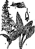
Digitalis purpurea: 1-apex with flowers and leaves; 2-basal leaves; 3-longitudinal section of the flower; 4-longitudinal section of the ovary.
with purplish veins on the inner surface of the petals; the flower has four stamens and a naked, thread-like style ending in a bilobed stigma. The leaves are alternate, light green on top, grayish-green below, lanceolate, unevenly serrated, slightly pubescent on both sides; the lower ones are petiolate; the upper ones are sessile, almost stem-clasping; the length of the leaves is up to 30 cm, and their width up to 6 cm. The collected leaves of D. purpurea or ambigua are dried and dehydrated until their water content does not exceed 1.5%; in this form they can be preserved for medical purposes for up to 2 years, and when dried by the ordinary method for up to 1 year. The leaves ground into powder should be stored in glass vessels filled to the top without access to light and moisture. Due to the large difference in the content of active principles in leaves of different origins, it is necessary to check their strength of action. In the absence of a chemical criterion, various methods of biological research are used for this purpose. According to Ph VII, the smallest dose of an alcoholic extract from the leaves, prepared according to the precise instructions of Ph VII, is determined, which within 1 hour in a male frog (Rana temporaria) weighing about 30 g causes systolic arrest of the heart. Such a smallest dose is called a 'unit of action' (U. A.); the pharmacological value of the preparation is measured by the amount of U. A. in a unit of its volume or weight. Leaves containing 66-50 U. A. in 1 g of leaves are considered suitable. The question of the active principles of D. remained quite unclear for a long time, since the substances allegedly found by some authors to be independent subsequently turned out in some cases to be a mixture of several substances, and in other cases to be identical with previously found substances. According to Straub, the active principles of the leaves of D. include the following glycosides: 1. GITALIN (Kraft), which is an independent fraction of the extraction from the leaves, but according to the latest data (Kiliani) represents a mixture of several substances; it is difficult to dissolve in cold water (1:600), easy to dissolve in chloroform; it is decomposed by boiling water and alcohol. 2. DIGITALEIN (Kiliani, Windaus) - an amorphous substance, easily soluble in water and alcohol, insoluble in chloroform; its aqueous solution does not change upon boiling, but decomposes upon prolonged standing. 3. DIGITOXIN - a crystalline stable substance, C36H56O14, insoluble in water, easily soluble in chloroform and alcohol. French digitalin (Nativelle) is very close to digitoxin. - In addition to the listed glycosides, the leaves of D. contain saponins - digitasaponin and gitin (also in the form of glycosides), possessing the general pharmacological properties of saponins, but not belonging to the number of specifically active principles of D. The seeds of D. contain active glycosides: 1) Digitalinum verum, an amorphous substance, difficult to dissolve in water, insoluble in ether and chloroform and well soluble in alcohol, and 2) digitalein, apparently a substance identical with the digitalein found in the leaves, and the saponin digitonin, devoid of the specific properties of cardiac agents. The strength of action of the specific substances of D. can be characterized by the following figures, indicating the amount of substance per 1 g of weight of the frog, which causes systolic arrest of the heart upon subcutaneous administration: gitalin - 0.00585 mg, digitalein - 0.005, digitoxin - 0.00365; the content of individual glycosides in the leaves amounts to (Straub): gitalin - 0.375%, digitalein - 0.37%, digitoxin - 0.24%; on average their total content is close to 1%. Locally, preparations of D. cause irritation, followed by anesthesia, for which reason foxglove should be classified among the anaesthetica dolorosa. The irritation is particularly strong when foxglove is introduced into the eye; in this case severe pain, hyperemia and acute inflammation are observed, followed by anesthesia. The same effect is observed on the mucous membrane of the tongue and upper respiratory tract. When introduced under the skin, inflammation occurs, often with a result in suppuration. These phenomena of irritation are most strongly caused among other active principles by digitoxin. - When administered per os, the active principles of D. are not absorbed in the stomach, but only after they pass into the small intestines, where however they are partly decomposed, which is more pronounced when absorption is reduced, e.g. caused by stagnant phenomena in the area innervated by the n. splanchnicus. Although when introduced into the blood the active principles of D. disappear very quickly (after 4 minutes 1/5 of the administered amount remains), however the maximum effect when a toxic dose of digitoxin is administered occurs late (after 2 days). When other components of D. are administered, the effect of action occurs faster. Having reached the maximum effect, the action of foxglove on the heart gradually weakens, however it can still be detected for a very long time - apparently depending on the slow destruction of the poison fixed in the heart. And here a significant difference is observed for the individual active principles of foxglove. The duration of action of foxglove on the heart explains its cumulative action, consisting in that successive small doses sum up and can give phenomena of poisoning. In this case it depends on the accumulation of poison in the heart. If D. is prescribed in such doses and with such intervals that the administered doses have time to be destroyed, then long-term prescription of foxglove without manifestations of cumulation is possible. - No habituation to foxglove is observed. With resorptive action, the active principles of foxglove possess a sharply expressed selective ability in relation to the organs of circulation - the heart and vessels - and certain special departments of the nervous system, exerting a strongest effect on them, while other organs, including the central nervous system in those departments that have no relation to circulation, are little sensitive to the substances in question. Action on the circulation. The points of application of the poison when acting on the circulation are in the heart the muscles and the excito-motor nodes, outside the heart - the center of the n. vagi, the vascular walls, probably the vascular center. - Action on the heart. A characteristic feature of the action on the heart of foxglove, as well as of other cardiac agents of this pharmacological group, is the dual character of this action. Foxglove on the one hand excites the motor elements of the heart, which determines its systolic action, and on the other hand excites and inhibits the apparatus of the heart, mainly the system of the n. vagi, on which depends its diastolic action. The latter appears in man and higher animals exclusively as a passive phenomenon and consists in weakening or cessation of the systolic action. In the frog Schmiedeberg recognizes an active diastole, depending on the contraction of the peripheral layers of the cardiac muscles. Depending on the dose, phase of action and preliminary state of the heart, different ratios are observed between the exciting (systolic) and inhibiting (diastolic) action of foxglove, consequently the total its effect on the heart can be very different. If the dose is large and causes a toxic effect, then usually three phases are distinguished. With therapeutic doses only one phase is observed, corresponding to the initial phase of toxic doses. This first phase is characterized by strengthening of contractions during systole with greater relaxation of the heart during diastole and slowed pulse. On the electrocardiogram the action of foxglove on the heart is expressed by an increase in the T wave (V. Zelenin). The strengthening of heart contractions depends on the direct action of foxglove on the cardiac muscle. At this time both more perfect contractions of the heart and greater their force are observed, which can be determined by the indications of a manometer connected with a balloon placed inside the ventricle. Strengthened contraction is observed on both ventricles and can be detected both on the whole animal and on the isolated heart. The systolic action of foxglove is particularly sharply expressed when it is applied to a tired (resp. poisoned, e.g. by chloral-hydrate) heart with insufficient systolic contractions, which foxglove brings more or less to normal. Furthermore, the strengthened stretching of the atria, as well as the strengthened stretching of the ventricles themselves during diastole, also causes their more energetic contraction in the following systole. The diastolic action of foxglove, expressed in strengthening of diastolic relaxation, is manifested much weaker than its systolic action and in higher animals and man depends mainly on the action of D. on the center of the n. vagi, and therefore is little noticeable on the isolated heart. It is more strongly expressed on the heart with complete systole and insufficient diastole (Cuslmy). Both the contraction of the heart during systole and its relaxation with transition into diastole, under the influence of foxglove, proceed faster, consequently the duration of diastole somewhat increases; however the transmission of excitation from the atrium, where it arises, to the ventricles occurs more slowly, so that the interval of time between the contraction of the atria and ventricles increases. This depends on the action of the excited n. vagus on the His bundle, the conductivity of which is hindered.
However, the direct action of digitalis on the same muscle bundle at the beginning of the therapeutic phase increases its conductivity, which can be detected in experimental conditions on an isolated rabbit heart. The slowing of the pulse with digitalis in humans and warm-blooded animals depends on the action of the vagus nerve, the center of which is excited by digitalis. The central action of digitalis is evidenced by the experiment with section of the vagus nerve, after which slowing of the pulse hardly occurs. At small doses, the slowing of the pulse is expressed less strongly than the strengthening of contractions, so that a therapeutic effect of digitalis can be observed without slowing of the pulse. On a diseased heart, in addition to the central action of the vagus nerve, a peripheral effect of digitalis may also occur, consisting, according to some authors (Rothberger and Winterberg), in the heart becoming especially sensitive to the action of the inhibitory apparatus. Some peripheral action of the vagus nerve with digitalis even on a healthy heart is proven by the fact that after this poison, irritation of the sectioned vagus nerve gives a greater effect than before digitalis. The increase in amplitude gives an increase in the amount of blood ejected with each cardiac contraction (increase in Schlagvolumen). The slowing of the rhythm, by reducing the number of contractions, decreases the amount of blood ejected per unit of time (Minutenvolumen). In the therapeutic phase of action, the first moment prevails over the second, and Minutenvolumen is increased. With poisoning doses, a phase may subsequently occur during which the amount of blood ejected per minute decreases. The increase in Minutenvolumen in the therapeutic phase is the main goal of prescribing cardiac drugs. On a healthy, well-functioning heart, it may be weakly expressed: the heart works optimally even before digitalis and pumps all the blood from the venous system into the arterial system. In a diseased heart, for which the task of proper circulation before taking digitalis seems difficult to accomplish, after digitalis it becomes feasible. Such an action of digitalis is best proven by means of Starling's method, which consists in replacing the systemic circulation with a system of tubes in which resistance to blood flow can be established at the desired height. With such an experimental setup, a heart that cannot cope with the given resistance and is excessively stretched by it, after digitalis returns to a normal volume and successfully pumps blood through the increased resistance. The further, second phase with toxic doses is characterized by a frequent pulse, and irritation of the vagus nerve no longer causes stopping or slowing of the heart. This, however, does not depend on paralysis of the vagus nerve, but on excitation of the excito-motor apparatus of the ventricles, which at this time do not obey the vagus nerve, while the atria are still regulated by it. The fact that the observed increase in pulse frequency occurs even after preliminary poisoning of the endings of the vagus nerve with atropin serves as proof that the observed increase in pulse frequency does not depend on paralysis of the vagus nerve. In this phase, systoles become even more energetic, and diastolic relaxation less expressed. In the further, third phase, as a result of excitation of the excito-motor nodes, extrasystoles occur, and the increasing difficulty in conductivity of the His bundle leads to a violation of the synchronism of contractions of the atria and ventricles. A rhythm of 2:1, 3:2 and 4:3 is established. Then the conductivity of the His bundle completely ceases, complete heart block occurs. At this time, the ventricles, due to strong excitation of the third-order nodes located in the wall of the ventricles, can contract very frequently and finally their contractions take the character of flutter. The heart stops in the state of strongest systole. The action of digitalis on the vessels in the therapeutic phase is expressed as follows: constriction of vessels of the viscera, innervated by the splanchnicus nerve, and dilation of vessels of the kidneys, muscles, skin-muscular layer, brain and coronary vessels. This action is not sharply expressed and experimental data are not always the same. With toxic doses, the vasoconstrictor effect is much more definite. Digitoxin in these doses directly causes constriction of all vessels. Digilan, like strophanthin, gives constriction only of vessels of the viscera. The vasoconstrictor effect with digitalis depends, firstly, on the direct action on the vessel walls. Such is the action of all cardiac drugs on the vessels of the viscera, and of the digitoxin group on all other vessels. Of the same origin, from the local action of digitalis, is the dilation of renal vessels and probably coronary vessels by most cardiac drugs. The dilation of skin and muscle vessels, as well as cerebral vessels, partly occurs passively as a result of displacement of blood from the visceral vessels, and partly due to reflex dilation in response to constriction of visceral vessels. The experiment on a dog, in which a limb with artificial circulation and with preservation only of the nervous connection with the trunk, when cardiac drugs were introduced, showed the same dilation of vessels as in a normal limb, speaks in favor of the latter. Blood pressure under the influence of digitalis in a healthy person with therapeutic doses changes little, despite the increase in the amount of blood ejected by the heart in 1 minute. According to Cushny, this can be explained by the fact that better blood supply to the vasomotor center lowers its excitability, which determines the decrease in obstacles from the vascular system for circulation. In experiments on animals, with toxic doses, in the 1st phase pressure usually increases, but sometimes it may not be increased. With excessive slowing of the pulse, pressure may even fall below normal, so that with an increase in pulse frequency in the second phase it rises above normal and reaches a maximum. When the 3rd phase sets in with its characteristic irregularities in cardiac activity, pressure finally falls. In patients, the phenomena are significantly more complicated, since insufficient circulation causes abnormality in the composition of blood (increase in hydrogen ion content), and this in turn affects the excitability of the vasomotor center, the center of the vagus nerve, etc. In order to correctly evaluate the significance of digitalis for circulation, it must be borne in mind that under normal conditions the general speed of circulation is established not by the heart, but by the total activity of all organs. When transitioning from rest to work, the tissue chemistry changes, which causes dilation of the vessels of this area and increased blood flow to the working organ. As a result, the amount of venous blood flowing to the heart correspondingly increases. The latter then pumps this blood into the arterial system. If the heart is weak or the work demanded of it is excessive, the heart copes with the task with difficulty. Externally this is manifested by stretching of the heart, and with stretching, as indicated above, the work of the heart increases. With an even greater discrepancy between the working capacity of the heart and the task demanded of it, the heart does not pump all the blood flowing to it. Stagnation of blood occurs in the venous system and small circle (with weakness of the left heart) with subsequent formation of edema. With digitalis, the working capacity of the heart increases, and the task that was beyond the power of a weak heart becomes feasible for it. Both Schlagvolumen and Minutenvolumen increase, and as a result redistribution of blood occurs: 1) blood is pumped from the venous system into the arterial system, 2) from the small circle (with stagnation in it) into the large circle, 3) from the vessels of the abdominal cavity innervated by the splanchnicus nerve, which constrict with digitalis, into the peripheral vessels. At the same time, abnormally elevated diastolic pressure falls, both in the right heart, which improves coronary circulation and consequently nutrition of the heart, and in the arterial system, which facilitates the work of the heart. With restoration of normal circulation, the conditions for the formation of edema disappear (insufficient nutrition of capillary walls with an increase in their permeability and asphyxia of tissues with an increase in their acidity, which, according to Fischer, promotes tissue swelling). The edema fluid, entering the blood, causes hydremia, which is one of the causes of the increased diuresis after taking digitalis in edematous patients. On the other hand, normal and even increased compared to normal (due to dilation of renal vessels) renal circulation restores the disturbed nutrition of the kidney, eliminating here also asphyxia. The latter is important both for the normal work of the organ and because, by reducing the swelling of tissues caused by their acidity, it eliminates excessive constriction of renal vessels. With restoration of normal circulation, the blood pressure, which before taking digitalis was at an abnormal height, usually returns more or less to normal. In this connection, two categories of cases are observed.
The lowered pressure, which is connected with heart weakness, rises in parallel with improvement in cardiac function, and conversely, the elevated pressure, caused by asphyxia of the vasomotor center which had received an insufficient amount of blood before treatment, decreases with the elimination of this asphyxia. Digitalis is prescribed to strengthen cardiac activity in various decompensated heart defects, with the effect not always being the same. The worst effect is given by aortic insufficiency, perhaps because the lengthening of diastole caused by digitalis allows more blood to flow back into the heart from the arterial system during its diastolic relaxation. Further, the not always favorable effect in mitral stenosis is noted, in which digitalis can cause undesirable overfilling of the lesser circulation. It should be kept in mind that digitalis does not give new strength to the heart, but only calls upon its reserve forces for work. Therefore, in their absence, e.g., in myocarditis, success cannot be expected. The further meaning of prescribing digitalis in insufficiency of atrioventricular valves lies in the fact that under the influence of digitalis the heart decreases in volume and accordingly the mismatch of the openings also decreases. In addition to increasing the strength of heart contractions, digitalis also has a regulating effect on them. This is particularly sharply manifested in arhythmia perpetua, when atrial fibrillation causes secondarily extremely irregular ventricular contractions. Digitalis in these cases decreases the conductivity of the His bundle, as a result of which only the strongest impulses arising in the sinuses reach the ventricles, while the others are as if filtered out. As a result of such blocking, the ventricular contractions acquire a regular character, while atrial fibrillation may even intensify. Digitalis acts similarly in 'flutter' of the atria, when the contractions of the latter are very frequent but regular. In this case, the ventricles cannot keep up with the atria, although they also contract very frequently, which causes their exhaustion. Digitalis here also, by hindering transmission along the His bundle, slows the rhythm of the ventricles. Finally, in complete heart block, which arose independently of the action of digitalis, when due to complete non-conductivity of the His bundle impulses from the atria do not reach the ventricles, and the impulses arising in the latter are too weak and infrequent, digitalis can strengthen these impulses arising in the third-order nodes (in the walls of the ventricles).-In addition to the circulatory organs, digitalis affects temperature, lowering it (which probably depends on the excitation of a special cold center), and on the vomiting center, which is proven by attacks of vomiting with intravenous administration of digitalis.-The symptoms of poisoning from digitalis are expressed in vomiting: 1) early, occurring soon after administration and caused by reflex from the gastric mucosa, in which besides the specifically cardiac principles of digitalis, the saponins contained in it also play a role, and 2) late-central, which is an expression of digitalis intoxication. Further, an extreme slowing of the pulse is noted, which can suddenly transition to a very rapid one, which, as was indicated above, is characteristic of the toxic phase of digitalis action. The amplitude of the pulse can decrease in poisoning, probably from constriction of peripheral vessels, and a decrease in diuresis can also occur from constriction of the kidney vessels, which also constrict with large doses of digitalis. Preparations. Folia Digitalis- leaves of D.-are most often used in the form of powder or aqueous infusion; many authors consider infusion the best method of extracting the active principles from the leaves. Only freshly prepared infusion should be used, since it loses up to 1/2 of its strength within 24 hours at room temperature; neutralization of its organic acids with sodium bicarbonate or addition of a small amount of alcohol can make it more stable. Dosage: leaves-up to 0.2 per dose; 1.0 per day; Inf usum-0.5-1.0 : 200.0 by the tablespoonful 3-4 times a day or 6-1 times a day every 2 hours.-Tinctura Digitalis contains not all the gitalin found in the leaves, but much digitoxin; is given up to 1.0 per dose, 3.0 per day.-Digipuratum-dry extract of D., freed from ballast substances but containing all therapeutically active principles; insoluble in water, easily soluble in diluted alkalis; 1 tablet Digipuratum corresponds to 0.1 leaves.-Verodigen consists mainly of amorphous gitalin, a white, amorphous, water-soluble powder, available in tablets containing 0.0008 each (=0.1 leaves).-Digalen-Digitoxinum solubile-according to Straus, consists of gitalin and digita-lein, available in flasks containing 15 cm³ of aqueous solution with 28% glycerin (1 cm³ of solution corresponds to 0.0003 digitoxin = 0.15 leaves), as well as in ampoules of 1.1 cm³ for intramuscular and intravenous administration.-Diginorm-extract from D. freed from ballast substances.-Digifolin-contains mainly digitoxin and digita-lein, in ampoules and tablets, 1 cm³ of solution or 1 tablet = 0.1 leaves.-Digitalinum-extract freed from saponins, 1 cm³ of solution or 1 tablet corresponds to 0.1 leaves.-Digitoxinum crystallisatum-a compound that breaks down into digitoxigenin and digitoxose; a white crystalline powder, insoluble in water; 0.00025 per dose.-Russian preparations: Gitalen-corresponds to Digalen and Verodigen, contains as active principles gitalin and digita-lein; 20-30 drops per dose.-Diginorm R. contains digitoxin and digita-lein, 1 cm³ corresponds to 65 E.D., also available in ampoules of 1 cm³; 15-20 drops per os or 1-2 cm³ intravenously per dose.
Related articles
Mentioned in
Cite this page
“Diverticulum.” Soviet Medical Encyclopedia. English translation of Bolshaya Meditsinskaya Entsiklopediya, 1st ed. (Moscow, 1928–1936), ed. N. A. Semashko. https://sovietmedicalencyclopedia.pages.dev/article/diverticulum/