Phimosis
Historical document, translated for reference. It reflects medical knowledge of the 1920s–30s and is not medical advice.
Summary
Phimosis is a pathological condition of the prepuce where it cannot be retracted over the glans penis. This article discusses its causes, complications, and various surgical treatments from a 1930s Soviet medical perspective.
Encyclopedia article (1928–1936)
PHIMOSIS (phimosis, preputial stenosis), a pathological condition of the prepuce in which it cannot be retracted over the glans penis. This occurs due to congenital or acquired narrowing of the preputial orifice, the small size of the prepuce, or adhesions between the glans penis and the inner lamina of the prepuce. In children, phimosis is physiological, as they are born with a narrow opening of the preputial sac and also have epithelial adhesions between the glans and the inner lamina (Bokai). With time (as the child grows), the ring of the prepuce stretches, and the adhesions are mechanically destroyed, and by the time of puberty the glans penis is usually freely exposed. However, in cases where adhesions are not destroyed, smegma can accumulate between them, and when it decomposes, it causes inflammatory processes, which leads to the development of very strong adhesions between the glans and the inner lamina. Sometimes the preputial orifice does not coincide with the external urethral opening, and the child urinates into the preputial sac, which also leads to the development of inflammatory processes with all their consequences. The shape of the preputial sac also contributes to the formation of phimosis. Atrophic forms are distinguished, where the prepuce is so thin and narrow that it tightly fits the glans penis, and when attempting to open it or during erections, it constricts the glans and causes pain. In cases of hypertrophic forms, the prepuce is extremely elongated, with its end hanging in the form of a nipple or trunk. Urine is retained in the folds of the prepuce, causing inflammation with ulceration and scarring, which leads to narrowing. Diseases caused by soft chancre, located on the inner lamina or at the edges of the preputial orifice, lead to scarring and scar contraction of the prepuce. Frequently recurring balanitis and balanopostitis serve as predisposing factors for the development of phimosis. Phimosis causes difficulty in urination, especially if the preputial orifice is extremely narrow. Sometimes it reaches the size of a pinhead. Phimosis is credited with the development of dilatations and diverticula of the urethra, urinary bladder, ureters, and even the development of hydronephrosis, on the basis of difficulty in urination. In inflammatory conditions, phimosis can cause reflex urinary retention. Some (Englisch) associate difficulty in urination with phimosis with the formation of inguinal and other hernias, hydrocele of the testicle, prolapse of the rectum, etc. Retention of urine in the preputial sac (with a very narrow orifice) can cause its distension to the size of a chicken egg or larger (fig. 1).
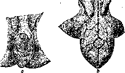
Fig. 1. Sac-like prepuce in phimosis in collapsed (a) and filled (b) condition.
Kirmisson associates phimosis with enuresis nocturna and frequent erections. The development of itching of the glans is observed, due to which children rub the glans, press against it with the penis, try to lie on their stomachs, all of which also causes erections and subsequently the habit of masturbation. Phimosis can lead to frequent inflammations of the inner lamina of the prepuce (see Balanitis and Balanopostitis), to the formation of preputial stones, etc. During sexual intercourse, under the influence of increased sensitivity of the glans covered by phimosis, ejaculatio seminis praecox is possible. During sexual intercourse, the narrowed prepuce, when retracted behind the glans, can constrict it, cause edema, and create a condition of paraphimosis, the most common and unpleasant complication of phimosis. Some (Kaufmann, Czerny, Federl, Butyagin and others) attribute to phimosis a predisposing role in the development of cancer of the penis. The treatment of phimosis pursues several goals. In children, sometimes it is necessary to take some preventive measures that do not allow the development of phimosis in the future, namely - the separation of epithelial adhesions between the inner lamina of the prepuce and the glans penis using a probe. After this, and generally in children to avoid possible inflammation, it is necessary to wash the preputial sac with a solution of alum (2%), boric acid (2%) or potassium permanganate (1:5,000-6,000). Irrigations are done every 5-6 days from a small rubber bulb with a soft tip and can be entrusted to the child's mother. These irrigations are also usefully recommended to adults who have a long prepuce and abundant accumulations of smegma. Indications for operation of phimosis, according to Zarkevich, should be considered: pain during erection and sexual intercourse, recurrent balanitis and balanopostitis, recurrent constriction of the glans, unbearable itching of the glans, and finally difficulty in urination with an extremely narrow preputial orifice and spraying of the urine stream. Sometimes (rarely) an operation for phimosis has to be performed out of necessity: 1) when it is necessary to treat gonorrhea, if the external urethral opening is located deep in the preputial sac, and 2) when it is affected by soft chancre. In surgical interventions for phimosis, the surgeon aims to achieve not only a functional but also a cosmetic effect after the operation (fig. 2). König advises performing a bloodless reposition of the prepuce, which is possible only with a sufficiently wide and elastic ring. The manipulation should be repeated until free exposure of the glans penis is achieved. He also recommends the simplest operation of incising the prepuce along the dorsal surface. Both laminae are incised simultaneously, and then sutured to each other. The disadvantage of this operation is partly cosmetic: prominent flaps of the prepuce are formed, which are not subsequently smoothed out. If the wound heals with scarring, this operation does not give a functional success either: a scar ring is formed that does not allow complete exposure of the glans penis. Therefore, Roser proposed making a small plastic addition: the incision is made along the dorsal surface, but the incision of the inner lamina stops, not reaching 1-11/2 cm to the sulcus retroglandularis. From the end of this incision, two lateral incisions of the inner lamina are made, and thus a triangular flap is created. The apex of the flap is sutured to the angle of the incision of the outer lamina, and ordinary sutures are made on the rest of the extent. With this plastic surgery, Roser achieves elongation of the edge of the inner lamina and smoothing of the angles. The above-mentioned incision operations along the posterior surface (discisio) are performed in small and thin preputial sacs. In hypertrophic forms, with a massive and long prepuce, it is better to resort to the operation of circular circumcision of the prepuce (circumcisio). The simplest method of operations of this kind is as follows. With two Kocher clamps, the prepuce is pulled back as much as possible and both laminae of the prepuce are excised with a scalpel or scissors, and then after the bleeding stops, they are sutured to each other. When making the incision, it is necessary to spare the frenulum and glans penis, therefore some authors first grasp the prepuce in front of the glans with some clamp and only then make the incision along it. For the best cosmetic and functional effect, a number of plastic operations for phimosis have been proposed. These include: 1) Schloffer's method. The incision is made along the posterior surface of the prepuce, with the outer lamina being cut obliquely in one direction, and the inner lamina obliquely as well, but in the other direction. When the prepuce is retracted, a rhomboid wound is formed, which is sutured transversely (fig. 3). This method elongates the edge of the preputial orifice. 2) Hagedorn's method. An oval incision of the outer lamina is made, which runs parallel to the retroglandular groove (fig. 4). The skin flap is pulled toward the root of the penis, and then a similar oval incision of the inner lamina is made, but on its posterior surface a triangular flap is excised, the apex of which is sutured to the center of the incision of the outer lamina. The rest of the wound is sutured in the usual way. The excision of a flap from the inner lamina elongates its edge. This is done on the assumption that the skin is more elastic than the mucous membrane, and the excess mucous membrane prevents subsequent narrowing. 3) Drüner's method. The outer lamina of the prepuce is incised with a straight incision along the posterior and inferior surface of the prepuce. Two skin flaps are obtained, located on the lateral surfaces. The skin is dissected from the inner lamina, and the preputial orifice is bypassed by the incision. The inner lamina is incised along the lateral surfaces, and here two flaps of the inner lamina are formed - upper and lower. The suturing is done in the following order: the apex of the upper flap of the inner lamina is sutured to the angle of the skin incision; the apex of the lower flap of the inner lamina is sutured to the angle of the lower skin incision, and the apices of the lateral skin flaps to the angles of the corresponding incisions (right and left) of the inner lamina. The remaining parts of the incisions are sutured to each other. A very wide ring of the preputial sac is obtained (fig. 5). 4) Doiteau's method aims not to completely expose the glans penis. The incision of the outer lamina is made slightly behind the constricting ring. The skin is pulled as far as possible toward the root of the penis. The inner lamina is excised parallel to the sulcus
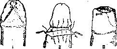
Fig. 3. Operation for phimosis according to Schloffer's method.
The inner lamina is excised parallel to the sulcus


Fig. 4. Operation for phimosis according to Hagedorn's method.
The inner lamina is excised parallel to the sulcus
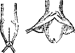
Figure 5. Operation according to Drüner's method. retroglandularis at a distance of no more than 0.5 cm from it. The flaps are sutured to each other, and when fusion occurs, the skin will fold inward and replace the inner flap, covering the glans penis with its excess. Phimosis operations are performed under local anesthesia in adults and under general anesthesia in children. Careful hemostasis is necessary, as extensive hematomas may form here, hindering smooth healing. Catching the cellular tissue in the suture is not desirable, as it prevents close contact of the flaps. For the same reason, it is better to ligate individual vessels of the cellular tissue rather than relying on catching them in the suture. To protect the dressing from being soaked with urine, it is most convenient not to bandage the penis, but to apply a strip of gauze over the tied sutures and secure its ends with the sutures. With the Duato method, a dressing can be dispensed with, and the suture should be dusted with talc or smeared with vaseline.
Related articles
Mentioned in
Cite this page
“Phimosis.” Soviet Medical Encyclopedia. English translation of Bolshaya Meditsinskaya Entsiklopediya, 1st ed. (Moscow, 1928–1936), ed. N. A. Semashko. https://sovietmedicalencyclopedia.pages.dev/article/phimosis/