Soft Chancre
Historical document, translated for reference. It reflects medical knowledge of the 1920s–30s and is not medical advice.
Summary
Soft chancre is a venereal disease caused by the streptobacillus Ducrey-Unna, primarily transmitted through sexual contact. The article details its historical recognition, morphological characteristics, laboratory identification methods, and cultivation challenges.
Encyclopedia article (1928–1936)
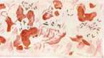
SOFT CHANCRE, venereal ulcer (ulcus molle, ulcus venereum), a venereal disease caused by the streptobacillus Ducrey-Unna, transmitted mainly through sexual contact, easily transferable from place to place and from person to person; soft chancre has been known since ancient times. Hippocrates already mentions buboes; Galen speaks of them in great detail; he distinguishes between hard and soft ulcers and requires different therapy for them; in the Middle Ages, many physicians considered soft chancre a disease sui generis, but the severe epidemic of syphilis that spread through Europe in the 15th-16th centuries caused great confusion among physicians; the subtle differences between hard and soft chancre blurred; with incredible speed (Vella; 1508), the idea of the identity of hard and soft chancre took root; having the most authoritative followers (Hunter, Ricord), this theory reigned supreme until the middle of the 19th century, when thanks to the works of Bassereau and others, the difference between these diseases was finally established. The causative agent of soft chancre (Streptobacillus Ducrey, Bac. Ducrey-Unna-Krefting) was first described by Ducrey in pus (1889) and soon after by Unna in the tissue of soft chancre. In pure culture, the causative agent of soft chancre was obtained by Russian researchers Istomanov and Askipyants (1897) and independently of them almost simultaneously by Lenglet in France. The morphology of this microorganism, despite significant polymorphism, is very characteristic and depends on the environment (pus, tissue, artificial medium); it is well revealed by staining. The streptobacillus of Ducrey takes up (but not easily) basic aniline dyes (methylene blue, fuchsine, etc.); it is decolorized by Gram's method. In the classical form, the streptobacillus of Ducrey is found in pieces of tissue, easily obtained with a platinum spatula from under the undermined edge of the ulcer (see separate table, fig. 1), in a less characteristic form, the microorganism is observed in pus (inside and outside cells) (see separate table, fig. 2). The indicated starting material is carefully smeared on a slide with a loop (even better - with the edge of a slide), dried in the air or in an incubator, and fixed for 1-2 minutes with formalin vapors or by careful triple flaming. For practical purposes, in order to detect morphological properties, the preparation is stained with diluted Ziehl's carbolfuchsin (1 part dye to 4 parts distilled water) with heating (until the first vapors appear) and then for 2-3 minutes on a cold plate; for differential distinction from other similar microorganisms, Gram staining is performed (additional staining with heating according to Finkelstein). Finkelstein generally recommends staining the Ducrey bacillus with heating, as this method ensures the detection of the characteristic bipolarity of the Ducrey bacillus and the poorly staining degenerative forms1(swelling, disintegration). More complex methods have also been proposed (methylgreen-pyronin according to Pappenheim-Unna: bacteria are reddish, pus cells are greenish-blue). In preparations with the indicated stains, various forms of the Ducrey bacillus can be seen, representing different stages of its development and degeneration: 1) Bacilli of various lengths (1-2 μ), located singly, in pairs, in short or long chains, inside (granulocytes, monocytes) or outside cells; egg-shaped and boat-shaped forms with clear bipolarity are also found. 2) Classical long (ten-fifteen or more segments) streptobacilli, arranged in parallel rows (found mainly in tissue). The Ducrey bacillus has the same appearance in pure culture (see separate table, fig. 3), which is traced in so-called smear preparations (Klatschpraeparat). In liquid media, the Ducrey bacilli often stretch into long, unsegmented threads. The technique for cultivating the Ducrey bacillus does not seem to be fully studied; obtaining a pure culture of it succeeds depending on methods in very different ways: most researchers (Moore, Reenstierna) note 5-8% success; individual authors (Saelhof, Deibert) give higher figures (65-85%). In media for growing the Ducrey bacillus, the presence of hemophilic factor is mandatory. The most commonly used media are: 1) Blood medium according to Besançon-Reinschern, for which two parts of 3% agar plus one part of defibrinated rabbit blood are heated for five minutes in a water bath, poured into tubes in 5-10 cm3 and slanted; rubber caps must be placed on the tubes to prevent drying out ft» *^ V{
^~ ».


/

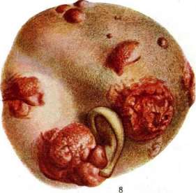
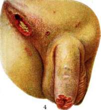
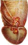
.:.- L Maecenas on "veia iety soft chancre: griptopashmi Decree, located in water alkaline. Fig. E. Wowi from groya at mnkod ttiuJiKpe: Llydchnid form st]1£.ch1t p111nlln Lykrekhy Fig. 4. Chancre of soft chancre and chancre-like ^b^b^i. IJnc. 5. Nar mщ-kago щilkri ni krdGшsL flesh, Put 6. Ya: iMTWpro muiiKpn in the area of the frenulum. Fig. ". iViMi Enu(MiiK(.'jiiLLSi pplt<^нтн.M1.цг|]| niM|iui;yi[ij[nyM reaction when injected with Ъаящиш and i streyshim Dtк'ren. Phne, i, ^vcii^H fnu^-lJc!*r E'is. ii. G Enk11lli?v-111 -i i [iji.[|H'.t kidney: pyra (l)--yellow color, medullary substance (ft) - Oledneyerogo > in the parenchyma. vessels (jure. lh 'I and 3 - by Talsh-czewskf, fig. I, r,h (i and 7 - by SLnmpku), K st Mvcosi? fittigni>, Soft chancre, RoditOHfWUX. nia. 2) Environment with defibrinated sheep blood. 3) Environment of protein: rabbit blood and 3 parts agar. 4) Clotted blood at an angle: ram (Durand), guinea pig (Himmel). 5) Nikolay's medium: agar, rabbit blood and one percent starch. Most authors note that the growth of Ducrey's bacillus occurs best on media containing blood of young animals. Frey points to the growth-enhancing effect of adding glycerin (one percent), gelatin (one-two per thousand). The active acidity of the medium (pH) ranges from 7.2 to 7.6. Extreme humidity is extremely important; temperature around 37°; develops better in the presence of O2, but relative anaerobiosis is acceptable (Frey, Lipinski); does not form a true toxin; endotoxin is proven; in some cultures a weak hemolysin has been found (Frey, Brams). Colonies of Ducrey's bacillus (on agar medium) grow within 24-48 hours; they are small, round (diameter 1-2 mm), with even edges, whitish-gray (and sometimes brownish). The consistency of the bacterial coating is mucoid-viscous due to the presence of a mucoid substance (substance glacieuse Lenglet) in it; on liquid media, flaky formations appear. Ducrey's bacillus does not show active motility. The viability of cultures varies greatly: the first generations often die quickly, but after five-six passages on artificial media, Ducrey's bacillus becomes accustomed to the medium and can then be maintained with regular (every 1-2 days) transfers for months, and according to some authors (Frey, Reinscherna)-even years. The stability of cultures to disinfectants (lysol, sublimate, carbolic acid) in ordinary solutions is insignificant; death occurs after several minutes; temperature stability is also not great: sterilization occurs at 50° after 5 minutes, at 42° after 1-2 hours. The method of cultivating Ducrey's bacillus to this time cannot be considered practically important. Of the many methods proposed for isolating the microbe in pure form, the two-moment method is probably the most practical: primary seeding on semi-liquid blood medium (Durand, Himmel, Davis), bactericidal for accompanying competitor-microbes, and then transferring to solid agar medium. By this method, some authors (Brams, Selgof) have found Ducrey's bacilli in healthy people, i.e., the question of bacillus carriage has been raised; the latter, as is known, was first discovered in apparently healthy women by Brook. In connection with bacillus carriage in healthy people, the differential diagnosis of morphologically similar microorganisms to Ducrey's bacillus becomes particularly relevant; of the latter, the following are known: 1) Streptobacillus urethrae Pfeifferi, a Gram-positive microbe, which differs from the Gram-negative streptobacillus of Ducrey; this saprophyte of the urethra is often found in the navicular fossa of the urethra in men. 2) Lehn and Neumann's microbe, a Gram-positive streptobacillus, found in the vagina. 3) Miyahara's streptobacillus, Gram-negative, never forms long chains. 4) Frey's microbe, isolated from a case of gangrenous ulcer; it is much more delicate than Ducrey's bacillus, monomorphic, and does not show parallel arrangement. The last two microorganisms, being Gram-negative, most resemble Ducrey's bacillus and therefore certainly represent practical interest; in general, they are still little studied. Animal inoculation. According to data from both old authors (before the discovery of Ducrey's bacillus) (Auzias-Turenne, Thibierge, Welz) and more recent researchers who used pure cultures of Ducrey's bacillus for inoculations, monkeys (macaques) contract a mild form of M. sh., which runs quickly (1-2 weeks) and histologically (superficial process, Reenstierna) differs from human soft ulcer. In macaques, the entire skin is susceptible, but inoculation on the upper eyelids is easier and more successful; mucous membranes usually do not become infected. Of other animals, Fontana obtained rudimentary forms of soft ulcers in rabbits (inoculation with corneal pus), Reinscherna (inoculation of pure culture on the scrotal skin), Terebinsky, etc. A number of authors reported positive inoculations in cats. Vaccine from cultures of Ducrey's bacillus is prepared either in the usual way (Ito), from a 24-hour agar culture in the form of an emulsion in physiological NaCl solution, heated to 60° for two hours, or according to Reinscherna; the latter instead of heating treats the culture emulsion with 0.5% carbolic acid (phenol) for two weeks. Such a vaccine can be used both for vaccinodiagnosis (intracutaneous reaction) and for vaccinotherapy (see below).
10. Finkelstein. Clinic of M. sh. For the virus to penetrate, damage to the skin is always necessary. Infection in the vast majority of cases occurs from a carrier of the same ulcer. The presence of the streptobacillus Ducrey-Unna bacillus in the urethra, vulva, and cervical canal of clinically healthy women apparently gives the right to speak of bacillus carriers in soft chancre, although this question requires further research (histological confirmation, cultures, transfers) and clinical observations (confrontation-simultaneous examination of a patient with M. sh. and the source of his infection). The incubation period is short: on the 3rd-4th day after infection, the ulcer is already fully formed. First (after 12-18 hours) a red spot appears at the site of virus penetration; by the beginning of the second day-a red pointed nodule in the center of the constantly expanding spot; by the end of the second day, a vesicle appears on the top of the nodule, which turns into a pustule within a few hours; the red spot becomes edematous; the pustule quickly ruptures, exposing a small crater-like ulcer. This stage is easiest to observe in autoinoculations and especially in CHANCRE
20 but in transplantations. Upon microscopic examination of soft chancre in the initial stages, phenomena of purulent infiltration are discovered both in the thickness of the epidermis and subepidermally; two abscesses arise, as it were, connected with each other by strips of the same infiltrate (chancre en bouton de chemise Nicoll'); the surrounding tissue is hyperemic, with diffuse infiltrates, mainly from plasma cells; many streptobacilli are present, lying in chains. The abscesses soon merge into one, and then ulcers develop. During the following 2-3 weeks the ulcer continues to increase both in depth and width, after which it cleans up and heals with a scar within several days. At the height of its development it possesses the following characteristics: shape-at first usually round; then it can take various outlines; size-from a pinhead to the palm of the hand and larger (in the serpiginous forms of M. gl.); edges-vertical, undermined, eaten away, jagged; bottom usually of soft consistency, rather deep, uneven, covered with a yellowish-salvage coating and sharply painful on touch; around the ulcer an inflammatory halo is often visible, and in rare cases a second, eccentrically located yellowish rim (Petges). Soft chancre is rarely solitary, usually several ulcers are encountered, sometimes very many (autoinoculation, "daughter ulcers"), in different stages of development. During the ulcer period, from the central purulent-necrotic focus with streptobacilli and other bacteria, strands of infiltrates extend in all directions, also with necrobiotic changes and streptobacilli; such dispersal of infiltrates leads to the fact that the visible boundary of soft chancre is not its actual boundary. In the depth of the skin a dense ridge of plasma cells is discovered. Healing proceeds in the usual order with the formation of a small scar. The clinical picture of M. sh. is influenced by a whole range of factors: size of ulcers, their depth, quantity, localization, combination with other infections (syphilis), developing complications, treatment. With localization on the frenulum (frequent rupture of it) in the coronal sulcus, along the edge of praeputii [see separate table (pp. 15-16), figures 5 and B] and along the ridge of the labia majora, the ulcers often acquire some, sometimes considerable density; with localization around the anus (most frequently encountered in women) radially arranged very painful ulcers on walking and especially on defecation, difficult to treat, are formed. The mucous membrane of the rectum (with the exception of isolated cases) is usually not affected. With localization of soft chancre along the edge of praeputii numerous cracks often arise. In women M. sh. localizes on the labia minora and majora and in the navicular fossa; lesions on portio uteri, in the cervical canal have been described; localization on the mucous membrane of the vagina is very rarely encountered. Extragenital chancres can localize everywhere. They arise either primarily (professional infection on the fingers of hands of doctors and midwives, perverted sexual intercourse) or secondarily (significantly more frequently) by inoculation of the virus from the primary focus. Clinically they do not differ from genital chancres. They are rarely encountered: Sinani and Shapiro in 2,858 cases of M. sh. found extragenital localization in 4 cases. In women extragenital M. sh. is encountered more frequently than in men (according to statistics 14:1). The following varieties of M. sh. can be noted: 1) Ulcus molle elevatum-with strong development of granulations of the bottom; more often than other forms of M. sh. it is solitary. 2) Ulcus molle folliculare, ulcera mollia miliaria Vorner-with penetration of the virus into a sebaceous gland; most often in the coronal sulcus, along the edge of praeputii and on the labia minora; narrow, deep ulcers with a dense bottom. 3) Ulcus molle infundibuliforme-from the base of the ulcer into the subcutaneous or submucous tissue a dense cone extends; with superficial palpation it can be easily confused with hard chancre; most often localizes in the coronal sulcus. 4) Ulcus molle impetiginosum-a rare form when a pustule dries in the form of crusts. 5) Ulcus molle diphthericum (not to be confused with diphtheric superinfection)-a pseudodiphtheritic coating on the bottom of the ulcer. 6) Ulcus molle serpiginosum-a sharp change in the course of M. sh.; lasts for months and years; extremely resistant to treatment; the ulcer scars from one edge and grows from the other, spreading to the scrotum, abdomen, thigh and sharply changing the clinical picture of M. sh.: the starting point in the overwhelming majority of cases is a chancroid bubo; ulcus molle serpiginosum is a rare form, usually encountered in patients receiving careless treatment; indication of a longer incubation period (8 days) still needs verification; upon transplantations an ordinary chancre (Reichel) or, more rarely, a serpiginous one (Luithlen) can be obtained; the cause of such a peculiar course of M. sh. is unknown. 7) Ulcus molle gangraenosum*-not frequent, but a very severe complication of M. sh., changing beyond recognition both the clinical and microscopic picture of it; usually begins suddenly, develops rapidly; in several days a grayish-black deep eschar forms, leading to loss of entire parts of tissue (prepuce, glans); sometimes the purulent process goes beyond the fascia, exposing the cavernous bodies; hemorrhages are rare; in the vicinity-strongly expressed inflammatory phenomena (redness, swelling); the general condition of the organism is sharply disturbed (high temperature, chills, general weakness, sometimes sepsis); more often encountered in untidy, untreated persons, in persons with a long and narrow prepuce; quickly complicated by phimosis with abundant fetid discharge; the cause is probably in a fusospirochetal symbiosis (in the sense of angina Plau-Vincent). It is possible that here factors of hyperergy are of importance. * Phagedenism describes both the deep ulcerative process (rule. m. gangraenosum) and the superficial, serpiginous one. Therefore Frei, when he proposes to completely destroy the term phagedenic chancre, as introducing obscurity. from the side of the organism itself. 8) Ulcus mixtum, mixed chancre; sometimes elements of both soft and hard chancre predominate, sometimes it has the character of an indifferent ulcer. 9) Combination of M. sh. with diphtheria (confirmed by bacterioscopic examination and cultures); a rare form with predominance of either the diphtheritic coating or the character of M. sh., usually uncharacteristic, poorly granulating, difficult to treat ulcers. Complications of M. sh. 1) Phimosis, which in turn extremely unfavorably affects the course of M. sh. (duration of the process, development of ulcus molle gangraenosum). 2) Perforation of the prepuce either with ordinary M. sh. developing in depth or, much more frequently, with ulcus molle gangraenosum. 3) Hemorrhages. 4) Lymphangitis either diffuse (in the vicinity and at the base of the ulcer, along the edge of phimosis) or in the form of dorsal and (rarely) lateral strands (bundles), on which small tumors (bubonulus) can develop, which then resolve or open up and give deep ulcers (chancre Nisbet); in women neither lymphangitis nor bubonulus occur. 5) Bubones-inflammation of regional lymph glands in response to penetration into them from the primary focus of Ducrey-Unna streptobacilli; with localization of the ulcer on the sexual organs the inguinal, especially those closest to the symphysis, glands become ill, usually on one side, rarely on both (see separate table (pp. 15-16), fig. 4]; bubones have also been observed with extragenital localization of M. sh. and upon transplantations; the frequency of bubone formation varies from 8-10% (Wolf and Mulzer) to 28% (Sinani and Shapiro) and 35% (Miekley); more often the left inguinal glands become ill (Miekley); in women they arise significantly less frequently than in men; increased walking, absence of treatment, localization of ulcers on the frenulum, in the coronal sulcus, on the clitoris, phimosis, strong cauterizing agents can contribute to the formation of bubones. With soft chancre bubones most often arise on the 3-4th week of the existence of the chancre, sometimes in the first days of the disease and in rare cases already after healing of the ulcers; bubones d'emblée without M. sh., phlegmonous bubone arising many months after healing of Ml., and recurrent bubone are described. If a bubone arises with a mixed infection with syphilis, it is not acutely inflammatory, at first hard, then slowly suppurates, gives little pus, lasts long (mixed bubone Diday-bubon mixte Diday); sometimes typical scleradenitis occurs only after the disappearance of inflammatory phenomena; in the process with soft chancre the tissue surrounding the gland is quickly involved (periadenitis); rarely is the matter limited to one gland (monoadenitis); usually a group of glands is involved (polyadenitis et polyperiadenitis). Outcomes of bubones with M. sh.: a) the bubone, fluctuating or non-fluctuating, resolves without opening; b) the fluctuating bubone opens and heals with a scar; c) the edges of the opened bubone become chancroidous due to infection with Ducrey-Unna streptobacilli; d) proceeding as a cold abscess, the bubone gives long-lasting non-healing fistulas (strumous bubone-unsuccessful term, since it is usually used in tuberculous infection of glands); the average duration of the bubone is 2-3 weeks with large fluctuations in one direction or another.
Correct behavior of the patient and appropriate therapy, especially vaccine treatment, have a favorable effect on the course, duration, and outcome of the bubo; some authors (Lennhoff, Frey) note a definite dependence between the bubo and soft chancre in the sense that ulcers heal faster when a bubo develops. 6) Finally, soft chancre can be complicated by eczema, erysipelas, acute condylomas, etc. Diagnosis and differential diagnosis. In typical cases, diagnosis is not difficult. In doubtful cases, vaccination is an excellent, but not always harmless (due to the possibility of serpiginous chancre, buboes, resistance to treatment) diagnostic method, to which recourse should be had only when clinical picture and microscopy do not resolve the question. After disinfecting the presumed site of inoculation with alcohol, ether, and then physiological solution, several scratches are made, wherefrom the same lancet transfers pus from the edge parts of the ulcer (chancre dermo-papillaire №co11e'я). The site of inoculation is covered with a cover glass. Fischer recommends making deep incisions and waiting for bleeding to stop (chancre dermique №co11e'я). Heteroinoculations, i.e., vaccination from person to person, must be abandoned due to the possibility of syphilitic infection and the possibility of complications of the vaccinated ulcer. Of greatest practical importance and often most difficult is the differential diagnosis with syphilis, especially with hard and mixed chancre. Clinical picture and microscopy do not always give a decisive answer. Then one has to resort to confrontation, vaccinations, sometimes waiting. RW can (indeed, rarely) give temporary positive fluctuations in complicated bubo and sometimes even in uncomplicated S. ch. lasting 14-26 days; therefore, repeated blood examinations are necessary; but even repeatedly positive serological reactions do not always decide the nature of the ulcer (e.g., S. ch. in a latent syphilitic with positive serological reaction); even in unmistakable S. ch., repeated serological examinations for at least 6 months are necessary (this has now become an axiom), as there may be a delaying effect (lengthening of the period of second incubation) of S. ch. on syphilitic infection. A single soft chancre can simulate a superficial gumma, however with the latter there are fewer reactive phenomena, less pain, and it has a denser consistency. Tuberculo-ulcerative syphilides, especially if they serpiginize, can also simulate soft chancre, especially when located on the female genital organs; to resolve the question in such cases, repeated RW, and sometimes trial specific treatment, are necessary. Ulcus vulvae acutum (Lipschutz) differs from S. ch. by the larger size of the ulcers and more vault-like edges; upon examination, Bac. crassus is usually found. In differential diagnosis of genital diphtheria (especially in women), it is resolved by bacterioscopic examination and cultures. Herpes progenitalis, balanitis et vulvitis are easily distinguished from S. ch. by their characteristic appearance and rapid course. Ulcus simplex Buschke or pseudo-ulcus veneris Jadassohn, with which S. ch. sometimes also has to be differentiated, represent superficial erosions without any characteristic appearance and with rapid course. Differential diagnosis with diabetic ulcers on the genital organs is facilitated by examination of urine and blood. When phimosis has developed rapidly and in gangrenous process, the original nature (ulcus molle, durum, mixtum, balano-postitis) b. ch. cannot be established immediately. Buboes are easily distinguished from inguinal hernia, cryptorchidism, aneurysm of art. cruralis; banal buboes in non-venereal lesions run less acutely, resolve more easily, and in the pus pyogenic cocci are found, cuti-reaction with streptobacillary vaccine is negative. In typical cases, soft chancre bubo is easily distinguished from painless syphilitic polycleradenitis; in doubtful cases (mixed infection with syphilis, superinfection of syphilitic scleradenitis with Ducrey-Unna's streptobacillus or other microorganisms), differential diagnosis is sometimes impossible (repeated serological reactions). In the 4th venereal disease of Nicolas-Favra (see Lymphogranulomatosis, Lymphogranulomatosis ing. subacuta) (there is packet-like swelling of inguinal and then iliac glands) cuti-reaction is negative, Frey's reaction is positive. If rare extragenital soft chancre buboes take a chronic course (fistulas), they may resemble tuberculosis; in such cases, the general condition, condition of other organs should be taken into account, and tuberculin test should be performed. The cuti-reaction of Ito-Reinshern with streptobacillary vaccine is extremely interesting theoretically and important practically. In patients with S. ch. and especially with bubo, after intradermal injection of 0.1 vaccine, an urticarial-erythematous spot with induration and papular elevation in the center [see separate table (pp. 15-16), fig. 7] appears, reaching maximum development in 24-48 hours and disappearing in 8-10 days; sometimes slight elevation of t°, lymphangitis and even lymphadenitis is noted; in healthy individuals who have not had S. ch. and in persons with other diseases of the genital organs, a small redness appears that disappears in 2-3 days. This reaction is almost absolutely specific; it gives about 1% non-specific results. It appears no earlier than 6-8 days after the appearance of S. ch., gradually increases, is most pronounced in patients with bubo and lasts for many decades (up to 50 years t; Reinshern's case). It makes it possible in doubtful cases to diagnose soft chancre, without however excluding mixed infection with syphilis, to differentiate vulgar and other buboes from soft chancre buboes, to make a retrospective diagnosis of S. ch. many years after infection. It reveals the hypersensitivity that appears in S. ch. and lasts for a long time, not only of the skin but of the whole organism (a more vigorous, local and general reaction after intramuscular and especially intravenous injection of vaccine than in healthy and non-S. ch. individuals); it radically changes the concept of S. ch. as a local infection. Cultivation of Ducrey-Unna's streptobacillus and preparation of vaccine encounter very great difficulties. However, Krivchevsky and Sinyelnikov (1926) proposed for diagnostic purposes pus-vaccine, i.e., pus of soft chancre buboes diluted 10 times with physiological solution and inactivated. Cuti-reaction with this pus-vaccine proved to be specific. The prognosis is usually favorable in typical uncomplicated forms of S. ch. In ulcus molle serpiginosum due to the duration of the course, resistance to therapy, possibility of intercurrent diseases, and amyloid degeneration of organs, and in ulcus molle gangraenosum the prognosis is poor. Phimosis and buboes prolong the course of i\<. sh.; buboes leave scars and can become the starting point of a serpiginous process and therefore worsen the prognosis. The patient should be warned about possible complications and the possibility of mixed infection with syphilis. Therapy. The better the drainage of pus, the less chance of lymphatic apparatus involvement; therefore, it is important to change dressings frequently (5-6 times a day), preceding them with potassium permanganate baths (10-15 min.). In fresh, uncomplicated ulcers without marked inflammatory phenomena in the surroundings, one can try one of the numerous cauterizing agents (cauterize the edges and the sacs under them, but not every day - danger of buboes): liquefied carbolic acid, 50% solution of zinc chloride, copper sulfate. After cauterization and in between them - dressings. The most energetic, perhaps even specific action is exerted by iodoform; however, its smell sharply limits the sphere of its application; the action of its derivatives and analogues is incomparably weaker: Dermatol, Xeroform, Airol, Vioform, Europhen, etc. A good dusting powder is Sodium sozojodolicum. All these agents are used in the form of dusting powders, paste with physiological solution, alcohol, ether (well retained), emulsion (10% emulsion of iodoform in glycerin), urethral sticks and rectal suppositories (2.0 iodoform each), pastes and ointments (2-5-10%). In the State Venereal Institute in Moscow, the following paste is used with success: Dermatoli, Zinci oxyd. aa 2.0, Natr. sozoiodol. 1.0, Ac. carbol. 0.1, Lanolini 10.0. Suteev recommends 50% pyrogallic ointment (control urine!), Almkvist-2-10% ointment from Cuprum amidato-aceticum, Gudelis from the clinic of I Med. institute-fatty acid copper; one can try smearing with salvarsan solution, etc. When sloughing ceases, agents promoting granulation are prescribed: 3% boric, 1% lunar ointment, 5% ointment from Peruvian balsam and especially x/4-1/2% lunar lotions.-In involvement of the lymphatic apparatus and in marked inflammatory phenomena, cauterizing agents are not suitable; lotions are necessary (potassium permanganate 1:1000, 1:3000, Arg. nitr. V*-V2%, 3% boric solution, etc.).
Sometimes a good effect is given by a 10% solution of methylene blue (Brocq) or a solution of 7 parts of carbolfuchsin and 3 parts of a 10% solution of the same blue (Queyrat). Some authors (Schprecher, Kolomoytsev and others) are generally against cauterizations, powders and ointments and treat only with compresses. More energetic methods of treatment are rather harmful than beneficial: snow CO2 (del Portillo), galvano- and thermocautery, scraping with a sharp spoon, etc.; they retain some significance in ulc. m. serpiginosum. In stubborn cases, ultraviolet rays are recommended (small quartz lamp, 10 cm distance, 3-:5 min., 2-3 times a week - Stümke); x-ray (x/3 H..E.D. 7a-1-mm aluminum filter, 2 - 3 times every 5 - 6 days); spraying of chloroform ether for 2 min. 5 times a day in buboes (Dub),- in serpiginous ulcers (Wright); hot air (60-70°) or iodine vapor 1-2 times a day for 15 min. (Muller, Welander, Belot), 2-3 sessions, especially in ulc. m. serpigin. Azua reports a good effect from intramuscular, and Goodman - from intravenous injections of a 1% solution of Tart. stibiati; locally 1/2% solution of it was successfully used by Rupel. General methods of treatment (see below), used mainly in buboes, are also applied in uncomplicated soft chancre. In inflammatory phimosis - hot baths, irrigation several times a day under strong pressure of the preputial sac (KMnO4 - 1 : 5 000, Ag. nitr. -1 : 1 000, 0.3% solution of hydrogen peroxide, etc.) with subsequent injection of iodoform emulsion or drainage of the preputial: sac with thin wet strips of gauze. Small incisions to the edge of the prepuce, to the dorsal incision (to the coronary sulcus), as well as to circumcision are resorted to only in exceptional cases (danger of chancreous degeneration of the wound edges). Buboes. With them, conservative treatment is also desirable. Treatment with heat (hot compresses, warming compresses, diathermy) in combination with pressure (sandbag) can have an effect in the initial stages. By adding general treatment to this treatment, it is possible in many cases to achieve regression even of fluctuating buboes. Autogemotherapy acts well (from 3 to 10 cm3 every 2-3 days); even better - lactotherapy (2-3-4 injections of 5.0 each); Cubero, Sinani and Shapiro and others report a good effect from tuberculin; intramuscular injections of 7% iodoform emulsion on buboes are ineffective (Bugarsky). Injections of Ol. Terebinthinae are rather suitable for opened, chancreous buboes; Fay and Gaal injected under the skin pus obtained by puncture from one's own buboon (0.25-1.0 every 4-5 days). In clearly fluctuating buboes, aspiration of pus as early as possible is recommended, followed by introduction into the cavity of a 5-10% iodoform or iodoform-xylol (10:1:90) emulsion, iodoform ointment, 1-2% ointment from Cuprum amidato-aceticum (Almqvist), etc.; in France, a thread-like drainage (a thread passed through the buboon with the help of a needle) with a compress on top is recommended; for incisions, the smallest possible incisions (V,-1 cm) are desirable, followed by introduction of ointment (see above) or drainage with thin wet strips of gauze. Radical operations (excision) retain some significance only in stubborn strumous buboes. X-ray (also in chancreous and fistulous buboes) and ultraviolet rays have a good resorptive effect. But especially rapid and certain effect, making surgical treatment almost unnecessary, is produced by intramuscular and even better intravenous administration of non-specific (typhoid) and especially specific vaccine (0.25-0.5-1.0 no more than 2.0 pro dosi every 1-2 days); even in clearly fluctuating buboes, 3-4 injections are sufficient. Reinschner prepared by intravenous administration to a sheep over many weeks killed and living streptobacilli of Ducrey-Unna an anti-streptobacillary serum, 1-2 injections of which (10.0 each) are sufficient for resorption even of fluctuating buboes; injections are made into the muscles. The unquestionable success of vaccine therapy, which reduces the duration of a bubo to 10-15 days (instead of 5-7 weeks with surgical treatment), urgently requires its wider application. Prevention in M. sh., besides socio-prophylactic measures, consists in hot soap washes of the genital organs after any suspicious intercourse and in the use of other personal preventive measures, in as early treatment as possible and in the quickest possible identification of all carriers of M. sh. As for the preventive treatment (course of salvarsan injections) proposed by Müller, and then Neisser for every case of M. sh., such treatment retains its significance only in cases of the undoubted possibility of simultaneous infection with syphilis (confrontation). To prevent buboes, Agronik recommends in every case of soft chancre from the very beginning of the ulcer's appearance to use intramuscular injections of milk.
V. Feldman. Epidemiology of M. The main epidemiological feature of M. sh. is the predominant disease of the urban population; in rural areas, M. sh. is as a rule registered significantly less frequently. In the period 1902-1913, in Russia a minimum of 37.1, a maximum of 55.8 cases of soft chancre per 10,000 urban population were registered; in rural areas respectively - 1.93-- 3.22. In the RSFSR, soft chancre was registered (table 1): Table 1. Years
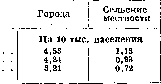
Thus, in the three-year period 1926-28, compared with pre-war years, a decrease in the registration of M. sh. is noted, especially sharply expressed among the urban population; in the pathology of the rural population, M. sh. in view of its extremely rare registration (0.72°/ooo) has no significance. An equally sharply expressed decrease in the registration of M. sh. in the Red Army compared with the tsarist army (in the period from 1888 to 1926 - a decrease of 88%); at present M. sh. in the Red Army is registered as an extremely rare disease. The predominance of morbidity from soft chancre among the urban population is also noted in Western Europe. Haustein gives the following data on the morbidity of soft chancre in Denmark (table 2): Table 2. Years 1911 1922 Copenhagen Provincial cities Rural areas Per 10,000 population 15.0
1.5
0.2 According to the data of a questionnaire conducted from 1/X 1920 to 30/IX 1921, in Switzerland was registered in cities with population over 50,000 inhab.-1.2 patients with M. sh. per every 10,000; with population less than 10,000 inhab. and in industrial points-0.2; in villages-0.07. Changes in the curve of registration of M. sh. among the urban population occurred also in years of relatively high its registration. Poray-Koshits, observing for 16 years (from 1870 to 1885) the movement of M. sh. in Kharkov, noted the "extinction" of M. sh.: in the seven-year period 1870-76 M. chancre constituted 13.6% of all registered venereal diseases, in the seven-year period 1879-85-only 2.4%. At the same time, as is evident from table 3 (according to Poray-Koshits), soft chancre constituted according to Western European and Russian statistics on average more than 1/3 of all registered venereal diseases (table 3). Table 3. Frequency of registration of M. sh. in the serological era (in %). Authors Gonorrhea According to Mischaud-before 1865 g. . . | 39.7 According to ZeissPio-1872 G.....| 37.1 According to Tarkovsky (Peter| burg)-1866-69 g......! 49.0 Report of Myasnitskaya b-tsy | (Moscow)-1880 g According to Poray-Kosh-1 1870/76 g itsu (Kharkov) . (1879/85 g According to Grinfeld (Odessa city b-tsy)-1903 g....... . . 9.1 46.4 42.2 14.2
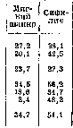
In later years, the proportion of Soft Chancre among venereal diseases has decreased: Blaschko, based on an analysis of data from 37 German cities where special statistical surveys were conducted, established that in 1913 Soft Chancre constituted 10% of all venereal diseases, syphilis-20%, gonorrhea-70%. The war of 1914-1918 and the immediate subsequent years are characterized by increased registration, along with other venereal diseases, of Soft Chancre as well. Borchardt notes the increase in Soft Chancre in Berlin from 1918 to 1920, Jambon and Tzanck observed the same in France. Gaushtein provides data on the incidence of Soft Chancre in Copenhagen from 1907 to 1924 (see figure); the greatest increase after 1916 falls in the period 1919-20, after which the Soft Chancre curve drops sharply. The temporary increases in Soft Chancre in the post-war years did not have a long-term character, and as a rule, Soft Chancre is rarely registered among venereal diseases. According to data from venereal dispensaries of the RSFSR in 1924-25, the proportion of Soft Chancre was 1.8% (Galperin and Isagv); in venereological institutions of the Ukrainian SSR for 1924-2.3%(Fedorovsky). These indicators are somewhat higher for large metropolitan centers and ports: in Moscow in 1928, 25,106 cases of venereal diseases were registered, of which Soft Chancre-5.5% (gonorrhea 52.3%, syphilis 42.2%); in Leningrad in 1928-13,969 cases of venereal diseases, of which Soft Chancre-3.1% (gonorrhea 70%, syphilis 26.9%); in the institute named after Chief in Odessa from 1917 to 1926, a total of 51,279 cases of venereal diseases were registered, of which Soft Chancre-7.3% (gonorrhea 33.8%, syphilis 58.9%). Despite the relatively low prevalence of Soft Chancre compared to gonorrhea and syphilis, epidemic outbreaks have serious significance in connection with the fact that in such cases, an increase in syphilis morbidity may also occur. The latter is explained by the fact that the main focus of Soft Chancre-prostitution (see below table 5)- is also one of the main sources of sexual infection with syphilis. The frequency of mixed chancre, which occurs with such simultaneous infections, varies according to various authors within the range of 13-1-% of all cases of Soft Chancre; however, there are indications that Soft Chancre in 58% may be a mixed chancre. An isolated epidemic outbreak of Soft Chancre was noted in 1929: according to Podvysockaya, through the inpatient department of the hospital named after Tarnovsky in Leningrad, in absolute numbers for 19 8 year 323 patients with Soft Chancre passed (6.5% in relation to all hospitalized skin and venereal patients), in 192» year-994 patients with Soft Chancre (33% in relation to all hospitalized or 60% in relation only to Ben.6-Hbix). Rottmann observed an epidemic outbreak of Soft Chancre in the port area of Louisburg-Ruhrort (Lower Rhine region in Germany), where from March to September 1928, a total of 7 cases of Soft Chancre passed through the venereal dispenser (a total of 1 Soft Chancre infection among 180 new venereal diseases on average per month), and from November 192S to September 1929, 343 patients with Soft Chancre were admitted (a total of 31 new infections on average per month). Such a sharp increase in the number of patients with soft chancre, Rottmann partially explains by the growth of prostituting women in port cities; these women infected sick sailors not only in the German port of Duisburg but also in foreign countries, in particular in Holland, which is also reflected in the increase in morbidity from soft chancre during the navigation period. Among various professional groups of the population, Soft Chancre is spread unevenly: the most vulnerable in terms of infection are workers of railway and water transport. According to data from Khodakovsky, among the entire population of Rostov n/D, in 1926, 10.0 cases per 10,000 inhabitants were registered, and among the transport population-23.0. When registering Soft Chancre in 1927, equal to 4.04 per 10,000 urban population, the corresponding number of transport population, according to data from 31 medical sections of the North-Caucasian Railway-6.4. An epidemiological feature of Soft Chancre is the significantly higher morbidity in men than in women, noted both in the pre-serological era and in subsequent years (table 4). According to Blaschko, in Hamburg, with an average annual morbidity from venereal diseases equal for men 45.4°/oo and for women 10.8°/oo, the morbidity from Soft Chancre was in men 7.2°/oo, in women 0.6°/oo. The rare attendance of women for soft chancre has led to attempts to explain this phenomenon by special bacillary carriage (see above). The smaller number of women with Soft Chancre can also be explained in part by the limited foci of Soft Chancre, the carriers of which, unlike gonorrhea in the vast majority, are prostitutes (table 5). Table 5. Sources of Soft Chancre infections (in%). Prostitutes Acquaintances Wives Khodakovsky (transport population of Rostov railway)-56.3 31.3 6.2 Frenkel (urban population of Kharkov)-1924-26 yrs. 78.9 15.6 2.9 Borshchevsky (workers of water transport)-1926 77.3 22.7 The percentage of infections from prostitutes given in table 5 should be increased to some extent due to infections from so-called 'casual acquaintances'. Thus, according to pre-war statistics of Peterson (quoted by Frenkel), when prostitution in Russia was regulated, a survey of 579 patients with Soft Chancre revealed 517 infections from prostitutes (90%). In Vienna in the period from 1906 to 1910, among regulated prostitutes, 1,888 cases of venereal diseases were found, of which 458 (24.2%) were Soft Chancre. The higher morbidity of the population in port cities is connected with the greater presence of prostitution in these places (so-called port prostitution): according to Borshchevsky's data from the Odessa outpatient clinic of water transport for 1924-26, 91 cases of soft chancre were registered, of which 31 (38.9%) occurred in Soviet and foreign ports, and all infections in foreign ports took place in brothels. Soft Chancre, both among men and women, most often occurs in the age group 20-30 years. The distribution by sex and age (in absolute numbers of patients with soft chancre registered in Moscow) is given in table 6 (according to data from the Moscow Statistical Bureau). In table 7 (see art. 31) Blaschko gives the distribution of morbidity from soft chancre by sex and ages in 1913 in Hamburg (per 1,000 population). Table 6. 1924 1927. Age m. f. total m. f. total m. f. total Children under 15 yrs. 8 1 9 15-19 .... 11 5 16 48 20-29 .... 138 38 176 30-39 .... 40-49 .... 50-59 .... 60 and older . - - Age unknown ~ Total adults 1,049 1,156 1,207 8 1 9 154 1,086 155 1,241 Excluding age groups below 15 and above 50 years, Blaschko establishes for the adult population aged 15-50 years, for which venereal diseases have the greatest significance, an annual morbidity from Soft Chancre equal to men-12.8°/oo, women-1.0°/oo. Among 400 patients with Soft Chancre registered in the State Venereological Institute in Moscow in 1924-26, in the age group 15-20 years-9.2%, 20- Table 7. Age 0-10 yrs. 10-15* »
. 40-50 » 50-60 » above 60 years, of Men Women 0.1 6.1 21.8 25.8 18.7 6.7 2.6 0.9 0.2 : 0.1 ! 1.2 2.3 2.2 1.2 0.4 0.1 - under 30 years-65.5%, 30-40 years-19.2%, 40-50 years-3.9% and over 50 years-2.2%. In childhood (under 15 years) S. c., as a venereal disease transmitted almost exclusively through sexual contact, occurs extremely rarely. Roeb collected in the literature up to 1920 descriptions of only 6 cases of soft chancre in children. Soft chancre is observed among married persons relatively less frequently than other venereal diseases; according to the data of Galperin and Isaev, who analyzed the marital status of 6,753 syphilis patients (1924-25), among the latter 57% were married; among 4,823 gonorrhea patients-51%; according to Frenkel's data, out of 308 S. c. patients, 34.1% were married. Analyzing the marital status of 1,400 soft chancre patients separately among men and women, Hatene-ver and Gutina found that among the former there were: married - 24.8%, unmarried -76.2%; among the latter there were: married-67.4%, unmarried-32.6%. In addition to the general factors influencing the spread of venereal diseases, such as war, associated with it the movement of masses of people, the intensification of prostitution in port and railway centers, - the spread of S. c. is also facilitated to a certain extent by the low sanitary level of the population. Frenkel among 308 S. c. patients registered in the Kharkov Venereal Institute for 1923-26 found that 18.8% lived in a separate room, 51.6% in a shared apartment and 29.6%-in flophouses; of all patients 71.7% did not use a separate bed, towel, dishes. In accordance with this are the author's indications that 36.9% of all male S. c. patients were manual workers, living predominantly in flophouses. The largest percentage of S. c. patients are persons with lower education. According to the data of Hatene-ver and Gutina for 1924-26, illiterate and semi-literate were 11%, with lower education-72.7%, with secondary education-15% and with higher education-1.3%. - The fight against S. c. in the USSR is one of the tasks of the venereological organization (see Venereal diseases). The fight against S. c. in the USSR is facilitated by the progressive improvement of the socio-economic living conditions of the working people and the elimination in the process of socialist reconstruction of all factors causing the periodic increase in S. c. in capitalist countries. The elimination of prostitution in the USSR and the enormous growth of the cultural-sanitary level of the masses of working people already at the present time have led to a sharp decline in the incidence of soft chancre, which is a rare disease in the USSR.
Related articles
Mentioned in
Cite this page
“Soft Chancre.” Soviet Medical Encyclopedia. English translation of Bolshaya Meditsinskaya Entsiklopediya, 1st ed. (Moscow, 1928–1936), ed. N. A. Semashko. https://sovietmedicalencyclopedia.pages.dev/article/soft-chancre/