Gonococcus
Historical document, translated for reference. It reflects medical knowledge of the 1920s–30s and is not medical advice.
Summary
The gonococcus Neisser is a bacterium discovered in 1879 that causes gonorrhea. This article describes its morphology, staining properties, and various atypical forms, including the so-called diplococcus Asch.
Encyclopedia article (1928–1936)
Gonococcus Neisser (Gonococcus s. Diplococcus Neisseri) and gonococcus-like microbes. G. was discovered by A. Neisser in 1879 and isolated in pure culture by Bumm in 1881; in its typical form (fresh urethral secretion, 24-hour culture) G. in stained preparations is characterized as a diplococcus consisting of a pair of half-cocci (biscuit-shaped, bean-shaped); between the half-cocci a clear gap is visible, weakly or not at all stained. As in pathological material, and especially on artificial media, due to the variability of environmental conditions (acidity, bactericidal properties, temperature, fluctuations in oxygen saturation), G. easily changes its morphology, giving rise to so-called atypical forms [diplococcus of the Ash type]. This variability is expressed in variations in size (microgonococci, macrogonococci), with possible combinations of small and large individuals in the same pair; instead of bean-shaped forms, spherical forms may appear as a result of microbe swelling. However, all these rather sharp morphological changes do not go beyond the limits of coccal forms. Depending on changes in the chemistry of the protoplasm of G., demonstrated in cultures by Schumacher, the staining reaction may also be altered (weak staining of degenerative small forms, more intense staining of larger, more stable forms). There is no specific stain for G. In the living state it is stained with neutral red, in smears it is well stained by all basic aniline dyes; best of all by methylene blue (see separate table, fig. 2). For differential diagnosis from morphologically similar microbes, Gram's decolorization method is used (see Gram's method). According to this method, G. is decolorized under the influence of alcohol and stained with a counterstain (see separate table, fig. 1); its ability to be stained by Gram in a state of degeneration has not been proven. On the basis of essential data from Moscow researchers (Ivanov, Finkel'shtein, Fronshtein and others), and partly from foreign authors (Adler, Asch, Ganet and others.), the following position regarding the so-called diplococcus Ash can be formulated: 1) in 95% (approximately) this is an atypical G., in 5% - a gonococcus-like microbe; 2) in such an atypical form, G. is most often observed in chronic gonorrhea (in women, little girls) as a stable form (according to some authors) as an adaptation to some long-term applied .4S a

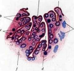
Figure 1. Gonococcus in a smear from the urethral discharge: numerous gonococci are visible inside the leukocyte, individual gonococci are also found outside the cell (stained by Gram's method). Fig. 2. Gonococci Pfeiffer are located extracellularly (stained by methylene blue). Figure 3. Tooth pulp: three macrophages filled with fatty grains (in the center): around them are marked cells (a) and polymorphonuclear leukocytes (b) (from a preparation of the pulp). Figure 4. Small intestine of a human: a - villi; b and c - follicles; d - glands; e - glands of Lieberkühn; f - lung-like crypts; g - connective tissue (weak magnification). According to Granzhem. The gonococcus can lose its virulence: 1) through repeated cultivation; 2) under the influence of a medicinal preparation or other environmental factors; 3) cases of carrier state of this type have been described; 4) the diplococcus of Neisser was found in subepithelial infiltrates in chronic gonorrheal vaginitis (Ivanov), as well as in the organs (liver) of white mice that died from toxic cultures of G.; 5) in experiments on mice, atypical G. is less toxic than typical. G. is cultivated only under certain conditions of the nutrient medium: 1) a certain degree of acidity, 2) the presence of native protein found in ascitic fluid, human and animal serum (pigeon, rabbit, etc.); 3) recently the special importance of vitamins has been proven (Levinthal). The most suitable media are ascitic agar (1 part ascites and 3 parts agar), blood agar, Levinthal's medium. On ascitic agar at 37° after 24 hours, small transparent colonies are visible, in which a semi-transparent center subsequently appears. In subsequent transfers, the colonies become more coarse, semi-transparent, merging into a fairly thick coating (grayish). The gonococcus is very sensitive to drying; it quickly dies without moisture. Ordinary disinfectants even in very weak solutions (Kalium hypermanganicum, preparations of Ag and Hg) quickly kill it. In the body, in the presence of proteins enveloping G., as well as in the presence of tissue antibodies and antivirus, the conditions for sterilizing G. are much more complex and in general little studied. Some cultures of G. in artificial conditions easily autolyze (even in physiological solution of sodium chloride). In relation to temperature, G. has a special property: in artificial conditions, the optimum for its development is around 37-38°; at this temperature, cultures can sometimes remain for months even without transfers; at temperatures below 33° G. quickly degenerates and dies. G. in most cases is an aerobe, i.e. needs the constant presence of O2, but sometimes it reproduces better or even exclusively (in the first generation) under conditions of relative anaerobiosis. For this purpose, a very simple method of growing G. on a medium (agar-broth) covered with a layer of sterile oil (vaseline, liquid paraffin) is used. This fact fully harmonizes with the observations of Buschke and Langer that such conditions of relative anaerobiosis exist in the deep infiltrates of the urethra. G. on artificial media does not produce a true toxin. The poisonous products found in cultures (filtrates of liquid cultures, bacterial extracts) belong to the type of endotoxins. The endotoxicity of gonocultures can be very different. According to experiments by Jotten, it is more or less constant and characteristic of certain strains: some cultures cause death of white mice when injected into the abdomen 2-3 loops of daily agar culture; others cause the same effect when injected with 8-10 loops; third may not kill mice at all. Virulent strains most often cause a more severe course of gonorrhea in humans (complication with epididymitis, adnexitis); low-virulent ones do not give complications. Experience has shown that gonococcal vaccines made from toxic cultures with a small admixture of low-toxic ones give the best practical results. In old (8-10-14 day) broth cultures both in vitro and in vivo on mice, the presence of the so-called antivirus of Bezredka can be proven (see Antivirus). In a test tube experiment, this gonococcal antivirus delays the development of G. In an experiment on mice, antivirus mixed with a lethal dose of gonococcal culture often delays the process of poisoning, and the animal survives (Finkelstein, Timokhina). In experiments on women suffering from gonorrhea, no therapeutic effect from gonococcal antivirus was established. G. in the human body promotes the formation of certain antibodies (mainly substances that bind complement), to a lesser extent - bactericidal substances, as well as opsonins. In animals (rabbits, horses) by artificial immunization with increasing doses of live cultures of G. (intravenously), strongly agglutinating sera can be obtained, which also have other serological properties (opsonizing, complement-binding, etc.). These sera are used for retrospective serodiagnosis: such a serum agglutinates G. and does not agglutinate gonococcus-like. Of other biological properties of G., its relation to other microbes with which it meets in the body and in artificial media in a test tube is interesting. In this respect, there is a big difference between toxic G., which is capable of suppressing competitors (staphylo-, diplo-, streptococci, rods of Doderlein), and atoxic or low-toxic G., which itself is displaced by saprophytes of this area. G. causes an abundant leukocytic reaction in tissues; some authors (Pozner) consider the eosinophilic reaction of tissue to be characteristic of G. Gonococcus-like microbes. Gram-negative diplococci resembling gonococci, observed in the healthy genitourinary system, have also been found in pathological discharge from men, women, and little girls. Their role as causative agents of local inflammatory processes of the genitourinary system is very probable, but not finally established. Gonococcus-like microbes cannot be distinguished from G. morphologically in the original material; most often the assumption of gonococcus-like microbes arises in the absence of proportionality between the presence of a large number of Gram-negative diplococci located on the flat epithelium and the relatively small neutrophilic leukocytic reaction. In general, this group of microbes has not been sufficiently studied and represents two main types. 1. The type Micrococcus fallax (Wormser), approaching the species Micrococcus catarrhalis. This type on simple agar after 24-48 hours gives round, coarse, medium-sized semi-transparent colonies, which subsequently easily merge and form a grayish coating; in broth - general turbidity; blood media are not hemolyzed. 2. The type Diplococcus crassus (Königsfeld, Salzmann) approaches the type of meningococcus (type Eger). On simple agar it gives small, opaque, whitish-gray colonies, growing slowly (3-4 days), not merging; on media with blood and ascites the growth is abundant; it does not hemolyze blood media. Both types are typical aerobes; they grow, although slowly, at room temperature. General signs distinguishing them from G.: 1) they grow slowly (2-3 days), on simple media, often at room temperature; 2) they decompose not only maltose (like G.), but also other sugars - sucrose, lactose, levulose; 3) they are not agglutinated by gonococcal agglutinating serum and do not give a complement-binding reaction with it; 4) in the human body in pathological discharge, eosinophilia is not observed. The two main types of gonococcus-like mentioned differ from each other not only in some morphological differences but also serologically: in cross experiments with heterologous sera, agglutination and complement-binding are not observed at all or are observed only to a negligible degree.
Related articles
Mentioned in
Cite this page
“Gonococcus.” Soviet Medical Encyclopedia. English translation of Bolshaya Meditsinskaya Entsiklopediya, 1st ed. (Moscow, 1928–1936), ed. N. A. Semashko. https://sovietmedicalencyclopedia.pages.dev/article/gonococcus/