Parametritis
Historical document, translated for reference. It reflects medical knowledge of the 1920s–30s and is not medical advice.
Summary
Parametritis is inflammation of the parametrium, the connective tissue surrounding the uterus. It is typically caused by infection, often following childbirth or abortion, and can present in different clinical stages with characteristic symptoms.
Encyclopedia article (1928–1936)
PARAMETRITIS (parametritis), inflammation of the parametrium. Due to different understandings of the term parametrium (see), the term "parametritis" also has different meanings. Since anatomically it is more correct to call the "parametrium" that part of the pelvic connective tissue that includes the entire complex of tissues constituting retinaculum uteri, including the adventitial sheath of the uterus, accordingly, P., unlike pelviocellulitis (inflammation of the entire pelvic connective tissue), is more correctly called inflammation of the connective tissue within retinaculi uteri and the connective tissue sheath of the uterus. P. is usually caused by infection. The infectious agents are the same as for phlegmons developing in other areas: more often (in approximately half of cases) pyogenic streptococci and staphylococci, much less often - E. coli, pneumococci, and bacteria from the anaerobic group (Bac. perfringens, Streptococcus anaerobius, etc.). E. coli and anaerobes in most cases cause mixed infection. The gonococcus independently causes P. only in exceptional cases (Wertheim), and even in these cases one can think of mixed infection (more often with the participation of streptococci and anaerobes). The bacteria reach the parametrial connective tissue by various routes, but most often through traumatic injuries to the walls of the genital tract during childbirth or miscarriage. In 2/3 of cases, P. is etiologically related to obstetric (resp. abortive) trauma. P. outside the postpartum or post-abortal period occurs much less often, and sometimes inflammation of the parametrial connective tissue joins diseases of the appendages of the uterus and the surrounding peritoneum; in other cases, P. are observed after surgical interventions (sounding, dilation of the cervix, curettage, incisions on the cervix, insertion of laminaria, etc.) performed without proper asepsis, or after accidental traumatic injuries (injuries to the vaginal vaults, bedsores); sometimes the starting points for P. are diseases of the intestine (appendicitis, sigmoiditis, paraproctitis), bladder (cystitis), pelvic bones, etc.; finally, P. are observed in the treatment of cervical cancer with radiant energy, in operative wounds after supravaginal amputation of the uterus, during removal of ovarian tumors, especially interligamentous ones, etc. The bacteria penetrate the parametrium and spread here mainly along lymphatic pathways. For their penetration, wide gates are usually required, which indicates their low virulence and the good reactivity of the body. Sometimes, however, the microbes causing P. penetrate the connective tissue by the hematogenous route - during angina, typhoid fever, severe influenza, and other diseases.
Pathological anatomy and clinical manifestations in P. Basically, path.-anat. changes in P. are observed in two main forms (Martin). In some cases, in the connective tissue, along with diffuse inflammatory phenomena, the lymphatic vessels are most affected, which over a large extent, starting from the uterus, are dilated and filled with thrombi and purulent masses, as a result of which they acquire a club-shaped appearance; the process is usually localized in the upper parts of the broad ligament along the course of plexus lymphaticus spermaticus, but it is also found in the base of the broad ligaments (plexus lymphaticus uterinus, s. paracervicalis), as well as in the mesentery of the ovary; the connective tissue through which the affected lymphatic vessels pass is also involved in the process. In other cases, with P., the lymphatic vessels are little affected or not affected at all, and the pathological process proceeds mainly along the course of the veins, appearing as hard cords or nodes, in the lumen of which bloody-purulent or purely purulent fluid can be found; the connective tissue, as a result of the accompanying periphlebitis, is usually impregnated with edematous fluid. Path.-anat. changes and clinical symptoms in P. proceed in several consecutive stages. 1. The initial stage is characterized by hyperemia, dilation of the blood vessels, resp. lymphatic vessels, serous impregnation of tissues, and the appearance and increase (depending on the severity of the infection) of edema. Clinically, in this stage there are feverish increases in temperature (up to 39° and higher), and the fever is either constant or increases in temperature alternate with decreases (Fig. 1); the pulse in this stage is somewhat accelerated, but always in full accordance with the temperature; patients experience slight independent pain and clearly expressed tenderness on examination; if the edema is sufficiently pronounced, the affected connective tissue on palpation appears doughy, pasty, resistant; even in this stage, it is sometimes possible to note the beginning characteristic displacement of the uterus (to the opposite side and upward).-2. The stage of infiltration, resp. exudative. The changes characteristic of the initial stage of P. can under favorable conditions disappear without a trace, under other conditions the path.-anat. changes do not limit themselves to edema, but progress further, and instead of soft edema, a dense exudate appears (due to the emigration of leukocytes, mainly neutrophils, from the vessels and the production of small-cell elements by the inflamed tissue itself). This exudate is leukocytic (neutrophilic), the blood remains sterile, patients experience slight independent pain, tenderness on palpation. On examination in the area of the affected connective tissue, one can find in place of soft edema a dense exudate, which at first is localized around the blood vessels, resp. lymphatic vessels, and then it spreads further and wider (Fig. 2). The localization, routes of spread, and direction of the inflammatory exudate in one or another segment of the parametrium will depend, on the one hand, on anatomical conditions and relationships, and on the other hand, on the portals of entry through which the infectious agents penetrated. Around the inflammatory focus, a granulation wall forms, which serves as a barrier to the further spread of infection. The clinical phenomena in this stage remain the same as in the initial one, in particular the temperature and pulse retain their previous character, in the blood there is a displacement of the uterus to the side and upward. The mobility of the vaginal mucosa in the area of contact with the inflamed connective tissue is limited, its folds are smoothed out; with significant exudate, the corresponding vault is also smoothed out, its concavity disappears. In some cases, there are disorders of the functions of neighboring organs (rectum, bladder).
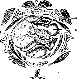
Fig. 2. Parametritis dextra. Horizontal section: 1-infiltrate in spatium parauterinum; 2-bladder; 3-anterior Douglas space; 4-uterus; 5-posterior Douglas space; 6-rectum.
3. Stage of exudate consolidation. The exudate formed in the second stage of P. is rich in fibrin, which soon precipitates, and the exudate acquires considerable density (density of wood, stone). In place of the granulation ridge, a powerful capsule appears, which firmly limits the inflammatory process. Clinically, the picture of the disease in this stage compared to the previous one represents only one difference, namely, upon palpation, the extraordinary density of the exudate, which has a consistency resembling myoma.-4. The final stage of the disease can proceed in two ways: in some cases, the exudate gradually resorbs and is completely eliminated, leaving behind adhesions, fusions, sometimes hardening (residual infiltration) in the area of the affected parts; in other cases, the parametritic exudate, melting down, undergoes suppuration, and at first the lymphatic vessels are filled with pus, and then separate abscesses, merging with each other, form purulent cavities of varying sizes, which in the end form one common cavity, delimited from the surrounding parts by a dense pyogenic membrane and filled with pus. Sometimes the parametric abscess passes into diffuse, phlegmonous inflammation of the pelvic cellular tissue (pelveocellulitis purulenta, s. phlegmona pelvis), and sometimes in patients the rupture of the parametric abscess occurs into the bladder, rectum, vagina, peritoneal cavity, the area of the buttocks, Scarpa's triangle, through the anterior abdominal wall, etc. In such cases, much time often passes before the rupture is finally eliminated. Clinically, the picture of the final stage of P. varies depending on the patho-anatomical substrate. If the exudate shows a tendency to resorption, all acute phenomena gradually subside: the temperature drops, the pulse returns to normal, the dense exudate slowly but steadily melts, its volume decreases, and in place of the exudate, scar connective tissue appears, which, shrinking, leads to displacements of the uterus—usually toward the inflammatory process. If the process ends in suppuration, characteristic changes are observed in the temperature curve: the temperature acquires a remittent type (suppurative fever), the pulse rate fluctuates in accordance with the size of the temperature, and neutrophilic leukocytosis is still observed in the blood. If the pus finds its way through the external coverings, swelling, limited redness, protrusion, and fluctuation appear in them. When rupturing into the rectum, among the prodromal phenomena there are tenesmus, discharge of mucus, and bloody discharge from the rectum. Perforation into the bladder is accompanied by symptoms of cystitis. Usually, the process after the rupture of the parametric abscess takes a favorable course; cases of perforation into the bladder proceed worse (threat of pyelonephritis). In some cases, suppuration after rupture remains prolonged, fistulas and sinuses form, the general condition of the patient worsens, and if she is not promptly given surgical help, the case can lead to amyloid degeneration of parenchymal organs with all the resulting consequences. As a rule, P. proceeds without chills. Only the first rise in temperature may be accompanied by moderate chills. The appearance of chills in the further course of the process indicates a complication that has joined (suppuration, thrombophlebitis, septicemia). Pain—and even quite significant ones—are observed in P. if the peritoneum is involved in the process; sometimes they also depend on the joined psoas muscle inflammation (in this case the pains radiate to the thighs), but in most cases, moderate pain in P. is caused by the stretching of the inflamed tissue by exudate and the compression of nerve trunks and plexuses. One cannot deny that in addition to compression, toxic influences may also play a role here, followed by the development of true neuritis. Clinical forms of P. Different authors distinguish various forms of this disease, depending either on the nature of its course, acute or chronic, or on the localization of the pathological phenomena in one or another part of the pelvic cellular tissue. Thus, the following forms of P. can be distinguished: 1) acute septic P. (septic phlegmon), which serves as a manifestation of general septic infection (septicemia, septicopyemia), 2) anterior P., parametritis anterior, which also has an acute course but with a greater tendency to localization, 3) lateral P., parametritis lateralis, of the same nature, 4) posterior P., parametritis poster., in most cases from the very beginning having a chronic course. 1. In the septic form, on the 3rd or 4th day after childbirth or miscarriage, rarely after moments of non-puerperal character, a picture of a severe disease rapidly develops—high temperature (up to 40°), frequent pulse, often an ominous crossing of the temperature curve with the pulse curve, sunken eyes, a pointed nose, cold extremities, dry, coated tongue, unquenchable thirst, insomnia, clear signs of peritonitis, prostration, etc. The causative agents of this form of P. are most often streptococcus circulating in the blood, or microbes of mixed infection. Usually, the portal of entry for the infection is the site of placental attachment, from where the infection, spreading immediately through the lymphatic and blood vessels, passes to the myometrium and cellular tissue, and also affects the peritoneum, at the same time flooding the blood and lymphatic systems of the entire body with microbes. Upon palpation, the parametric exudate here usually appears small. Clinically, the picture of septic P. is masked by the formidable signs of general sepsis. The prognosis of this form of P. is very poor, therapy—as in general sepsis.-2. Anterior P. (parametritis anterior)—inflammation of the cellular tissue within the limits of the anterior part of retinaculi uteri—is relatively rare. The portal of entry for the infection here is injuries to the anterior part of the cervical canal and the bladder mucosa, accidental injuries in the area of the anterior fornix, some operative interventions (e.g., sometimes vaginal cesarean section, etc.). The inflammatory exudate usually involves the anterior circumference of the cervix, more rarely—the anterior vaginal wall in the area of trigoni vesicae (Pavlik's triangle area). The exudate in anterior parametritis can pass into the paravesical space (cavum Retzii) and from there into the cellular tissue of the anterior abdominal wall (plastron abdominal). When an abscess forms, the pus can find an exit (rarely) into the bladder, vagina, peritoneal cavity (pericystitis), and as an exception into the symphysis. 3. Lateral P. (parametritis lateralis)—inflammation of the cellular tissue of the broad ligaments, including here pars media retinaculi uteri—the most frequently encountered clinical form of P. Infection enters the cellular tissue after childbirth or abortion primarily as a result of progressive local puerperal disease (puerperal ulcer, endometritis); in addition, the portal of entry for infection in parametritis lateralis serves as obstetric and gynecological injuries and damage in the area of the lateral walls and fornices of the vagina, as well as within the limits of the cervical canal (deeply penetrating tears of the cervix after forceps, after extraction of the fetus by the breech in elderly primiparas, etc.). If the infection enters from the anterior-lateral wall of the cervix and the corresponding fornix, the inflammatory exudate goes in a broad strip to the side, upward, and forward, in the direction of the round ligament of the uterus, then spreading in the form of a broad infiltrate along the lateral wall of the pelvis and above Poupart's ligament (the so-called 'abscess of Dupuytren'). With primary localization of the infection in the posterior-lateral parts of the cervix or in the lateral fornices of the vagina, the exudate usually spreads along the posterior part of the parametrial cellular tissue, reaching the walls of the small, and then the large pelvis; from here the exudate, going behind the cecum and S-Romani, can rise upward to the kidney, forming here a pararenal or even subdiaphragmatic abscess. When the m. psoas is involved in the process, psoas muscle inflammation can develop with the formation of contracture of the lower limb in the groin fold. Clinically, lateral parametritis is divided into upper and lower. The paths of pus spread in lateral P. and the places of its exit to the outside are indicated above. 4. Posterior P. (parametritis posterior). Meanwhile, in the first three forms of P. the disease has at all times or in the initial stage an acute course and is accompanied by the formation of a more or less significant exudate, in posterior P. this is very rare—usually in cases of accidental obstetric and gynecological injuries and damage. In most cases, this form of P. from the very beginning has a chronic course and is not accompanied by the formation of a significant exudate. This is because the infection here most often penetrates the cellular tissue from neighboring organs without prior damage to the latter. Such organs include primarily the peritoneum. Most often, the chronic form of posterior P. is encountered in combination with chronic intraperitoneal inflammation of the Douglas pouch. As a result of such inflammation, shortening of the sacro-uterine ligaments occurs, bringing the uterus into a retroverted position (the so-called parametritis posterior of Schulze). In the clinical picture here, the foremost are the painful pains in the sacrum, painful coitus, dysmenorrhea, and painful defecation.
Opitz sees the basis of these disorders as an excessively increased excitability of the smooth musculature of the uterosacral ligaments of constitutional origin, while Bischoff proposed a new name for this pathological condition—'spasmophilia genitalis.' E. Martin and other authors, however, tend to believe that here there is an actual inflammatory process. The diagnosis of posterior Schultze's parametritis is based on the results of palpation (shortened uterosacral ligaments, diffusely or nodularly thickened, painful when pulling the vaginal part of the uterus). Diagnosis. Parametritis in all its stages and forms is diagnosed relatively easily. It is most often necessary to differentiate it from pelveoperitonitis, especially that form of the latter in which the process is localized in the peritoneum covering the appendages of the uterus (periadnexitis exsudativa). For distinction, one must take into account the more sharply expressed picture of inflammation in pelveoperitonitis with signs of strong irritation of the peritoneum (pain, meteorism, nausea, vomiting, etc.), as well as pay attention to the relationship of the exudate to the walls of the pelvis (parametritic exudate fuses with the walls of the pelvis, inflammatory tumors of the appendages are isolated from them or come into contact with them with their small part) and to the mucous membrane of the vaginal vault (with periadnexitis exsudativa the mobility of the mucous membrane is not impaired, whereas with parametritis it appears impaired). The diagnostic sign of Henter can also be successfully used here: percussing the area of the anterior superior spines (spina ossis ilii ant. sup.), with parametritic exudates a dull percussion tone is obtained, or even completely dull (extraperitoneal process), which does not happen with inflammatory effusions around the appendages (the tone here remains tympanic in the area of spina ant. sup.). On palpation, the sharp outlines of the parametritic exudate are striking, and here there is always a coincidence of the percussion and palpatory boundaries; conversely, with pelveoperitonitic effusions, the palpatory boundary is always wider than the percussion one, since the inflammatory tumor on the periphery is covered by adherent loops of intestine. In the stage of induration, the parametritic exudate can be confused with myoma. For differential diagnosis, it is necessary to carefully collect the anamnesis and properly interpret it, as well as pay attention to the absence of connection of the tumor with the walls of the pelvis, its smooth rounded surface, the absence of a sharply expressed leukocytosis, acceleration of erythrocyte sedimentation, etc. The prognosis for parametritis, except for the septic form quoad vitam, is definitely good. Mortality is determined by tenths of a percent (0.2-0.4%). Somewhat clouding the prognosis are: 1) the presence of mixed or secondary infection, 2) the transition of parametritic exudate into suppuration (observed on average in 10-15%) and 3) ruptures of the parametritic abscess into the bladder, rectum, and peritoneum. On average, uncomplicated parametritis lasts no more than 6 weeks, but with suppuration, especially after rupture, the disease is prolonged twice as long, sometimes even more. In relation to residual phenomena, the prognosis for parametritis is also quite favorable. The only thing that remains after parametritis is sclerotic changes in the affected area (more often in the broad ligament) and usually a displacement of the uterus that remains for life in such cases (lateroversio, or lateropositio). The treatment of various clinical forms of parametritis is carried out according to one plan; the difference in therapeutic procedures depends exclusively on the stage of the disease. In the acute stage, during the period of formation and increase of exudate, therapy should be strictly conservative: rest, bed rest, ice on the abdomen, care for the intestines (enemas) and narcotics for pain (morphine, opium, pantopon, belladonna, papaverine). Narcotics should not be abused in this case. Ice should be kept until the temperature drops, and even after its drop, it should not be removed for another week. Some French gynecologists instead of ice apply heat (warm moist wraps, heating compresses on the abdomen, etc.): heat is supposedly better tolerated by patients, faster has an analgesic effect and with it the exudate resolves more reliably and quickly. Whether to use ice or heat in the acute stage of parametritis is a matter of each physician's experience. From the side of the general regimen, it is necessary to care for good nutrition, abundant drinking and maintaining good sleep in the patient. Any local intravaginal therapy in the acute stage of parametritis should be considered contraindicated. With prolonged fever, especially of hectic type, one must think about suppuration of the exudate. In the presence of such suppuration, it is necessary to ensure the emptying of the abscess, and it is not recommended to wait for a clearly expressed fluctuation, since the dense capsule surrounding the abscess often does not allow it. In doubtful cases, one has to resort to puncture in the area of greatest protrusion of the exudate. Parametritic abscesses are more often opened from the vaginal side, less often from the side of the Poupart's ligament and as an exception—in some other place. If no pus is found at the incision, there is nothing to fear. Sometimes such incisions themselves contribute to the faster dissolution of a long-persisting parametritic exudate. After opening the abscess, a gauze tampon or drainage tube is inserted into its cavity, which are changed daily. The abscess cavity should not be irrigated, at least for the next few days; later such irrigations are done with saline solution. For some time it was recommended to cut hard parametritic exudates even with a complete absence of a softening focus, however, against such an too early incision of the exudate, one should object (difficult stopping of bleeding in the sclerosed callus-like tissue, danger of embolism in the presence of not completely thrombosed veins, danger of injury to neighboring organs, etc.). One should proceed to the resorptive treatment of parametritic exudate no earlier than a week after discontinuing ice, and first ordinary anti-inflammatory heat treatment is applied: rest, sexual and physical (abstinence from work, movements), care for the intestines (enemas, mild laxatives), hot douches, tampons with ichthyol (10% solution in glycerin). To enhance the resorptive effect, heating compresses on the abdomen (for 6 hours, at night), hot water bottle or hot water bottles (2 times a day for 30-60 min.), general baths, sitting salt baths (38-40°, 10-15 min.), diathermy, ionotherapy can be prescribed. The best resorptive effect is given by mud therapy (mud cakes on the lower abdomen, vaginal mud tampons, especially in combination with diathermy). A good effect is also given by spa treatment (e.g., in Matsesta—sulfur baths, constant irrigations with Matsesta water, drop enemas with the same water). For long-unresolving exudates, treatment with weight (Belastungstherapie) is recommended: the patient is placed in a position with the pelvis highly elevated, at an angle of 30° to the horizon (to facilitate lymph outflow), a colpeurynter with heated (to 40°) mercury (1,000 cm³) is inserted into the vagina, a bag with sand weighing 1-3 kg is placed on the abdomen (for counterpressure). The session lasts from 30 to 60 min. Of other methods of treatment of exudative parametritis, one should mention autohemotherapy, treatment with turpentine according to Klingmüller, intravenous infusion of distilled water (300-450 cm³), etc. The transition from one method to another, especially the transition to more energetic therapeutic measures (diathermy, muds), is recommended to be controlled by measuring temperature and the reaction of erythrocyte sedimentation (SR), in particular, one should switch to diathermy in the case that erythrocyte sedimentation is not less than 1 hour (according to Linzenmeier). Preventive measures for parametritis in general coincide with those recommended and carried out during childbirth and in the postpartum period.
Related articles
Mentioned in
Cite this page
“Parametritis.” Soviet Medical Encyclopedia. English translation of Bolshaya Meditsinskaya Entsiklopediya, 1st ed. (Moscow, 1928–1936), ed. N. A. Semashko. https://sovietmedicalencyclopedia.pages.dev/article/parametritis/