Parametrium
Historical document, translated for reference. It reflects medical knowledge of the 1920s–30s and is not medical advice.
Summary
The parametrium is defined variously in medical literature, referring to different parts of the pelvic connective tissue surrounding the uterus. This article describes its anatomical structure, functions, and relationship to other pelvic spaces.
Encyclopedia article (1928–1936)
PARAMETRIUM (parametrium). The term "parametrium" is understood differently: some authors call the parametrium the connective tissue located on the surface of the uterus under its serous covering (the so-called adventitial vagina of the uterus); others denote by this term the connective tissue of the broad ligaments of the uterus - either entirely or only those connective tissue strands that are located at the base of these ligaments (pars cardinalis, s. basalis); Martin accepts as the parametrium an anatomical formation to which he gave the name retinaculum uteri; finally, some authors (French) under the parametrium in the broad sense mean all the pelvic connective tissue. - "Nowhere is there such an abundance of connective tissue as in the cavity of the small pelvis," notes Bichat. The pelvic connective tissue, of which the parametrium is a separate department, performs various functions here. First of all, the loose connective tissue fills the "empty spaces" (intervals, clefts, spaces) between various pelvic organs, ensuring them the mobility necessary for their physiological functions and the ability to expand and collapse. In addition, the loose parts of the connective tissue serve as a place for the deposition of fat masses, which is important for the tone and resistance of the tissues. The denser parts of the pelvic connective tissue (the so-called "zones of consolidation", fasciae) are formations that hold the uterus and other organs of the small pelvis in a mobile-suspended state (the fixing and suspending apparatus of the mentioned organs). Furthermore, covering the pelvic organs, including the uterus, like a mantle, the connective tissue is at the same time the anatomical substrate for the attachment of the peritoneum, a substrate ensuring the latter a certain mobility. In the pelvic connective tissue are then located blood vessels, an abundant network of lymph vessels and lymph glands, a dense network of nerves, ureters, etc. Finally, according to recent data (Roessle), connective tissue is considered an organized apparatus of metabolism; the so-called ret.-end. apparatus plays a particularly important role here. Corresponding to such a variety of functions, the pelvic connective tissue, resp. parametrium, appears by its nature and structure far from uniform. In it one can distinguish the following differentiated elements: 1) loose, wide-meshed tissue with abundant fat content; 2) connective tissue of the pelvic organs (adventitial vaginas of the uterus and other organs); 3) rather dense connective tissue sheaths for blood vessels, lymph vessels, and for the ureters; 4) fasciae of the pelvic muscles - connective tissue sheaths, which are essentially condensed connective tissue that covers one or another muscle; 5) the so-called "zones of consolidation" (W.A.Freund) - located deeper denser parts of the pelvic connective tissue, in intimate connection with the visceral leaf of the pelvic fascia and with the adventitial sheaths of the pelvic organs; in addition to connective tissue, these zones include elastic fibers with an admixture of smooth muscle fibers. Several such "zones of consolidation" are distinguished. First of all, through them is formed the retinaculum uteri with its three parts: the a-n-t-e-r-i-o-r part (pars anterior), representing dense connective tissue strands located in the vesicocervical ligaments (lig. vesico-cervicalia), the middle part (pars media), the most powerful, which is part of the so-called lig. cardinalia of the broad ligaments, and the p-o-s-t-e-r-i-o-r part (pars posterior), forming the connective tissue basis of the sacrouterine ligaments (lig. sacro-uterina).-Retinaculum uteri in
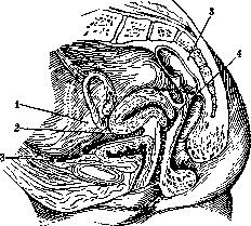
Figure 1. Topography of pelvic connective tissue: 1-spatium parauterinum; 2-spatium praecervicale; 3-spatium paravesicale; 4-spatium retrocervicale; 5-spatium pararectale.
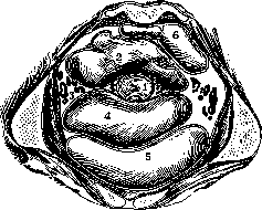
as a whole is a solid apparatus supporting the normal situs of the uterus. In addition to the "zones of consolidation" that make up the retinaculum, there are in the small pelvis other similar formations, which are usually interpreted as separate ligaments (lig. rotunda, vesico-umbilicalia lateralia, infundibulo-pelvica, s. suspensoria ovariorum, ovarii propria). In diseases of the parametrium, these consolidations together with the retinaculum play an essential role, being the boundaries of the so-called subserous connective tissue spaces, in which products of inflammatory processes (serous exudate, pus, etc.) accumulate and from which these products spread in one direction or another. Three connective tissue spaces are distinguished (spatia subserosa): 1) paravesical (spatium paravesicale), 2) parauterine (spatium parauterinum) and 3) pararectal (spatium pararectale) (figs. 1 and 2). They are all connected with each other, although not particularly closely; certain boundaries can be drawn between them.--The paravesical space is bounded in front by the anterior abdominal wall and the posterior wall of the symphysis, behind by the urinary bladder (fascia propria Velpeau), on the sides by fibrous plates located in the mesentery of the round ligaments of the uterus (mesodesma lig.rotundi), below by the pubovesical ligaments (lig. pubo-vesicalia) and finally above by the peritoneum, which passes from the anterior abdominal wall to the urinary bladder. The paravesical space anatomically coincides with Retzius space. This part of the pelvic connective tissue has no direct relation to the parametrium. In inflammatory processes in the area of Retzius space, the exudate as a rule spreads upward under the anterior abdominal wall, appearing in the form of a triangle, the apex of which is directed toward the navel (the so-called plastron abdominal of French authors). The spread of phlegmon of the paravesical connective tissue into the parauterine connective tissue is prevented by the mesenteries of the round ligaments. The parauterine space - right and left - is significantly larger in size than the paravesical. Its boundaries: in front - the urinary bladder and the fibrous framework of the round ligament (mesodesma), on the sides - the lateral walls of the pelvis, where the parauterine connective tissue at the terminal line (1. terminalis, s. innominata) merges with the subperitoneal connective tissue of the iliac wing, behind - the "zones of consolidation" that are part of the infundibulopelvic ligament and the proper ligament of the ovary, above - the apex of the broad ligament and below - the fascia endopelvina. The parauterine space, compared to others, is less delimited from neighboring parts. The exudates accumulating here have two paths for their spread: one along the round ligament, in the direction of the internal opening of the inguinal canal (above Poupart's ligament), the other - directly to the iliac fossa and further in the direction of the so-eum, resp. S-Romanum, to the area of the kidneys. In practical terms, the parauterine space is divided into two parts: the upper - the fimbriae of the fallopian tube (ala vespertilionis) and the lower - the root of the broad ligament (pars cardinalis). Within the parauterine space, two smaller parts of connective tissue are also distinguished - one in front of the uterus, between the bladder and the cervix (spatium praecervicale), the other behind, between the posterior surface of the uterine cervix and the rectum (spatium retrocervicale). C86 The boundaries of spatii praecervicalis: in front - the posterior wall of the bladder, behind - the cervix of the uterus, above - the peritoneum of the anterior Douglas space, below - the vesicocervical ligaments; on the sides, the connective tissue of the precervical space, embracing the cervix of the bladder, communicates with the paravesical connective tissue, and behind passes into the parauterine space. The boundaries of spatii retrocervicalis: in front - the posterior wall of the cervix of the uterus, behind - the rectum, above - the peritoneum of the posterior Douglas space, below - the mucous membrane of the posterior fornix; this part of the connective tissue communicates on one side with the pararectal connective tissue and on the other - with the parauterine.-The pararectal space contains connective tissue located on both sides of the rectum and on the anterior surface of the sacrum. Its boundaries: in front - the "zones of consolidation" that are part of the infundibulopelvic ligaments and the proper ligaments of the ovaries, behind - the sacrum, above - the promontory, below - the fascia of the levator ani. The pararectal connective tissue has no direct relation to the parametrium.
Related articles
Mentioned in
Cite this page
“Parametrium.” Soviet Medical Encyclopedia. English translation of Bolshaya Meditsinskaya Entsiklopediya, 1st ed. (Moscow, 1928–1936), ed. N. A. Semashko. https://sovietmedicalencyclopedia.pages.dev/article/parametrium/