Vagina
Historical document, translated for reference. It reflects medical knowledge of the 1920s–30s and is not medical advice.
Summary
The vagina is a muscular canal in the female pelvis connecting the uterus to the external body. This article describes its anatomy, embryological development, blood supply, innervation, and normal acidic secretions.
Encyclopedia article (1928–1936)
VAGINA (Latin-vagina, Greek-kolpos), represents a tube located in the middle of the small pelvis, which in the standing position of a woman is positioned obliquely to the vertical line, directing downward and forward upward and backward. This tube is almost straight, with a slight curvature, the convexity of which is directed backward, and the concavity forward (see figure 4). The lower boundary of the vagina is the hymen, separating it from the entrance to it (introitus vaginae). Above it is limited by the vaginal part of the uterus (portio vaginalis uteri), which protrudes into its lumen so that a ring-shaped depression forms around it-the vaginal fornix (fornix); usually four parts are distinguished: anterior, posterior, and two lateral, with the posterior fornix being significantly deeper than the anterior one. In front, the vagina borders: below with the urethra, from which it is separated by the urethro-vaginal septum (septum urethro-vaginale), and above-with the bladder, separated from the vagina by the vesico-vaginal septum (septum vesico-vaginale). Behind, adjacent to the vagina, lies the rectum, which below is separated from it by the perineal body, above-adjoins its posterior wall, which here forms part of the recto-vaginal septum (septum recto-vaginale), and even higher again recedes from it; between the vagina and the rectum here is located the posterior Douglas pouch of the peritoneum, the bottom of which is located, on average, at the boundary between the upper quarter of the posterior vaginal wall and the lower three quarters. On the sides, the vagina is separated from the bony walls of the small pelvis partly by muscles, partly by connective tissue, in which blood vessels and nerves are located.-Regarding the embryonic origin of the vagina, there has been disagreement until recently: most believed that the entire vagina together with the uterus in the female embryo develops from the Müllerian ducts, specifically from their lower, merging segments; however, some researchers believed that such origin has only the upper, larger part of the vagina, approximately its upper two-thirds, while the lower third develops at the expense of the urogenital sinus (sinus urogenitalis). The latest research by Chukalov resolves this question in favor of the first view, i.e., that the entire vagina develops from the Müllerian ducts, and even the covering of the posterior surface of the hymen comes from the epithelium of these.-The dimensions of the vagina in an adult woman are difficult to determine due to its great extensibility. Usually the length of its anterior wall is 8 cm, the posterior is 2 cm longer. Its width is even more variable; in general, the vaginal tube is narrowest at its lower end, upward it widens, and at the very upper end narrows again. The vagina is a hollow organ, almost directly communicating with the external environment; nevertheless, under normal conditions it does not contain air, with its anterior wall closely adjacent to the posterior one, so that on a cross-section the lumen of the vagina appears in the shape of the letter H (see figure 5). Since the upper part of the posterior wall of the vagina borders the peritoneal cavity, it is under the influence of intra-abdominal pressure, which in normal positions (standing, sitting, lying on the back)-is positive. Only under certain pathological conditions can outside air enter and exit the vagina (so-called garrulitas vulvae). If a woman is placed in the knee-elbow position, in which intra-abdominal pressure becomes negative, and the introitus vaginae is opened, air will enter the vagina and stretch its cavity; some old gynecologists used this circumstance during intravaginal operations, for example, when suturing vaginal fistulas. Structure of the vaginal walls. The vagina is lined inside with a mucous membrane covered by stratified squamous epithelium, the cells of which contain glycogen. The subepithelial layer consists of interwoven connective tissue fibers with a significant admixture of elastic tissue; on its surface it forms clearly expressed papillae. As a rule, there are no glands in the mucous membrane of the vagina, but lymph follicles are encountered. The surface of the vaginal mucous membrane is uneven: transverse folds or wrinkles (rugae) protrude on it, especially pronounced in its lower parts, on the anterior and posterior walls, where they form entire complexes-columnae rugarum ant. et post. (see figure 3). The anterior column under the urethra forms a keel-like projection-carina urethralis. Outside the mucous membrane, in close connection with it, lies the muscular coat (muscularis) of the vagina, in which longitudinal smooth muscle bundles predominate, upward passing into the musculature of the cervix. However, circular bundles are also found here, which gives authors grounds to distinguish in the muscularis vaginae two layers-an inner circular and an outer longitudinal. In the lower part of the vaginal tube, where it passes through the opening in the pelvic floor (hiatus genitalis), smooth muscles are joined by striated muscles from various layers of the pelvic floor. The outermost layer of the vaginal wall (adventitia vaginae) consists of connective tissue, which in places is dense, and in other places, especially in the upper part of the vaginal tube, behind and on the sides-loose (paravaginal cellular tissue). The total thickness of the vaginal wall varies in different parts, usually-3 mm. In the thickness of the lateral parts of the vaginal wall, especially in the area of the lateral fornices, remnants of the Wolffian ducts (Gartnerian canals) can sometimes be found in the form of small cavities of various shapes, lined with either single-layer or multi-layered epithelium. These cavities sometimes serve as starting points for the development of vaginal cysts.-The vagina is supplied with arterial blood: in its upper part-from the cervico-vaginal branch of the uterine artery (ramus cervico-vaginalis a. uterinae), in the middle-from branches of the inferior bladder artery (a. vesicalis inferior), while its lower third receives blood from the middle hemorrhoidal (a. haemorrhoidalis media) and internal pudendal (a. pudenda interna) arteries.-The vaginal wall is very rich in venous vessels, which form entire plexuses. Lymphatic pathways from the lower third of the vagina go together with those of the external genital parts, heading to the inguinal glands, while from the upper two thirds they go together with the lymphatic vessels of the cervix and lower part of the uterus, located in the lower parts of the broad ligaments of the uterus and flowing into the glands lying near the lateral wall of the pelvis (sub-umbilical, iliac, and lateral sacral).-The nerves innervating the vagina, like the nerves of the uterus and the nearest segments of the fallopian tubes, originate from the cervical nerve plexus of Frankenhauser, among which there are both sympathetic and spinal nerves. In the thickness of the vaginal wall, Sinitsyn discovered between the longitudinal and circular muscle layers a nerve plexus from myelinated and unmyelinated nerve fibers; unmyelinated fibers departing from this plexus, penetrating into the thickness of the muscle layers, in turn form dense plexuses on the surface of the muscle bundles, and from here branches originate, supplying the muscle cells with terminal motor apparatuses. In addition, in the musculature of the vagina, Gruz observed sensory nerve endings. In the mucous membrane of the vagina, Sinitsyn everywhere found terminal nerve bodies (Krause bulbs) of various types.-Although there are no glands in the mucous membrane of the vagina, its lumen always contains a certain amount of 'secretion' that makes the surface of the vagina moist. The flora of the vagina. The bacteria contained in the vaginal secretion are in close connection with the bacteria of the vulva; in married women, in addition, part of them are introduced during sexual intercourse. While the secretion of uterine glands has an alkaline reaction, the reaction of the vaginal secretion under normal conditions is sharply acidic. This depends on the presence of lactic acid in it, which is produced from the glycogen contained in the vaginal epithelium; however, there is no strict correspondence between its amount in the epithelial cells and the degree of lactic acid content in the vaginal secretion. For a time it was thought that lactic acid is produced here from glycogen due to the vital activity of the so-called vaginal rods (Bac. vaginalis) or Döderlein rods (Doderlein), non-pathogenic, non-spore-forming, facultatively anaerobic, gram-positive bacteria, which have the ability to produce lactic acid on media with grape sugar and which play a dominant role in the flora of a healthy vagina (according to Heurlin, they occur in 90% of all cases).
However, direct observations show that an acidic reaction in the vaginal secretion is also observed in newborn girls, while the contents of their V. (as established by precise observations) are completely sterile in the first hours after birth (microbes appear in it no earlier than 9-15 hours). Moreover, the presence of lactic acid can be detected in the secretion of the vagina even in those women whose V. does not contain Döderlein's bacilli. In any case, there is no doubt that both the acidic reaction of the vaginal secretion and the presence of Bacillus vaginalis in it are factors by which the female sexual canal is protected from the penetration of pathogenic microbes. Experience shows that if pathogenic microbes, for example, pus-forming streptococci, are introduced into the V. of a healthy woman, they disappear from there within 24-36 hours. The V., as is commonly said, is capable of self-cleansing, and in this self-cleansing the bacilli of Döderlein present in it and the lactic acid that is part of the vaginal secretion play an important role. The vaginal secretion has a specific smell, somewhat reminiscent of the smell of brine, which depends on the content of trimethylamine in it, which, according to Mikhin, on the one hand, is a product of the vital activity of microbes, and on the other hand, has bactericidal properties with respect to pathogenic microorganisms. All this refers to the normal vaginal secretion. However, even under these conditions, it varies both quantitatively and qualitatively within wide limits, depending on the age of the woman, her constitution, the state of the autonomic nervous system, the phenomena of the ovarian-menstrual cycle, pregnancy, etc. Of course, it changes even more under pathological conditions: its quantity increases to such an extent that it flows out of the genital cleft, stains underwear and often irritates the coverings of the parts surrounding the genital cleft (vaginal leucorrhea, fluor albus vaginalis); its color changes from milky-white to yellowish and greenish (purulent leucorrhea); the reaction becomes neutral and even alkaline; the number of leukocytes increases; the bacterial flora of the secretion changes, namely, the number of rod-shaped forms, and in particular, Döderlein's bacilli, sharply decreases due to an increase in the number of cocci, etc. From this point of view, it makes sense, together with Geurlin, to distinguish 4 degrees of purity of the vaginal secretion. The first degree is characterized by a sharply acidic reaction of it, a very small amount of it, the presence in it of only a small number of cells of the vaginal epithelium and leukocytes, the predominance among the microbes of vaginal bacilli, with only a small admixture of staphylococci, pseudodiphtheria bacilli and yeast fungi (see Figure 6). This degree of purity is found in
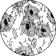
Figure 6. I degree of purity of the vaginal secretion (according to R. Schroeder).
healthy virgins and healthy pregnant women, more rarely—in non-pregnant healthy women, both those who have given birth and those who have not.- The second degree of purity differs from the first only in a somewhat greater content of leukocytes and epithelial cells, as well as the addition to the Döderlein bacilli of the curved bacilli of Mönchenberg (Comma variabile) and a small number of aerobic and anaerobic cocci (see Figure 7). We find this degree in most healthy women living a sexual 139
VAGINA
140 life.-With the third degree of purity, the Döderlein vaginal bacilli are pushed into the background, while the Mönchenberg bacilli are still present in large numbers,

Figure 7. II degree of purity of the vaginal secretion (according to R. Schroeder).
and mainly—anaerobic streptococci and other anaerobic microbes abound (see Figure 8).-Finally, if the Döderlein bacilli are completely absent in the vaginal secretion, and its flora consists mainly of anaerobic cocci, with a moderate admixture of anaerobic bacilli, then Geurlin speaks of the fourth degree of purity, at which the vaginal secretion already acquires the pathological properties noted above: its quantity increases, the color becomes yellowish, the reaction—neutral or alkaline, the content of epithelial cells, and especially leukocytes, increases. However,

Figure 8. III and IV degrees of purity of the vaginal secretion (according to R. Schroeder).
even this degree of purity of the vaginal secretion, not to mention the third, can be observed in women with a completely healthy vagina, but with impaired general nutrition and a weakened constitution; more often, however, we encounter this degree of purity of the secretion in gynecological patients. For a long time, the presence of streptococci in the vaginal secretion (apparently of perfectly healthy women) has been of great interest to obstetricians, which, at least morphologically, do not differ from the streptococci that so often act as the causative agents of puerperal sepsis. According to some authors, these streptococci, previously harmless inhabitants of the female sexual canal, during pregnancy can acquire pathogenic properties—independently pass into the uterus in the puerperal period and cause the so-called "self-infection" of parturients, the result of which can even be the most severe forms of puerperal sepsis ("puerperal fever"). Although this question is still not fully clarified, however, most modern authors
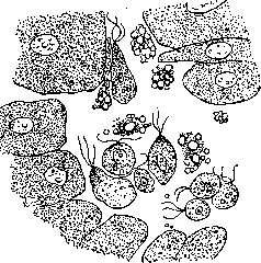
Figure 9. Trichomonads of the vagina in the vaginal secretion (according to a preparation by Dr. Belyaev).
inclined to deny 'self-infection' of puerperal women, at least in the form currently indicated. The question regarding the pathological significance of trichomonads (Trichomonas vaginalis), which are often found in the vaginal secretions of both healthy and women suffering from colpitis (see Figure 9), remains unclear to this day; some hold the view that trichomonads play a role in the etiology of catarrhs of the vagina, while others tend to see them as innocent inhabitants of the vagina affected by catarrh, being merely symbionts of those microbes that actually cause colpitis. Pathology of the V. Among anomalies of position of the V., the most important from a practical standpoint is that whose initial stages are known as prolapse, descensus, and further stages as prolapse, prolapsus. The pathological essence of this anomaly is that the walls of the V. are displaced downward from their normal position, and in prolapse are located outside the genital cleft; in this case, the normal pathological relationships of the various layers of the vaginal wall change (see Figure 2): the inner layer (mucous), instead of being turned inward, begins to look outward, while the outer layer (adventitia vaginae) becomes turned inward, i.e., prolapse of the V. (prolapsus vaginae) is simultaneously an inversion (inversio vaginae). In prolapse, the entire vaginal tube may be outside the genital cleft-complete prolapse of the V. (prolapsus vaginae completus), or only part of it-incomplete prolapse (prolapsus vaginae incompletus). Furthermore, with this anomaly, either the anterior wall of the sleeve may protrude from the genital cleft (descensus s. prolapsus vaginae anterior), or the posterior wall (descensus s. prolapsus posterior), or both together. Partial downward protrusions of the lower segment of the vagina, mainly its anterior wall, and of the upper segment, mainly the posterior wall, so-called enterocele vaginalis posterior, are also distinguished. In most cases of this anomaly, the uterus also participates in the displacement along with the V. Sometimes the V. begins to prolapse first, and its displacement then affects the uterus; the latter may react to the traction from the V. in various ways: if its ligamentous apparatus is relaxed, primary prolapse of the V. quickly leads to secondary prolapse of the uterus; if, on the contrary, the ligaments firmly hold the body of the uterus, it remains in place, and the descending V. protrudes the cervix-sometimes to such an extent that the apex of portio vaginalis begins to protrude from the genital cleft (elongatio colli s. pseudoprolapsus uteri); finally, if with primary prolapse of the V. the body of the uterus initially resists the traction from the latter, but then yields to it, both elongation of the uterine wall and true prolapse of the uterus develop. Cases of primary prolapse of the V. with secondary prolapse of the uterus are distinguished from cases where the uterus begins to descend first, and then pulls the V. downward-secondary prolapse of the V. after primary prolapse of the uterus. Finally, often the factors causing the anomaly under consideration act simultaneously on both segments of the genital canal-both on the V. and on the uterus. The immediate cause of the anomaly under consideration is an increase in intra-abdominal pressure, which is why prolapse of the V., like the uterus, is especially common among women forced to engage in heavy physical labor (peasant women, weight lifters, etc.); but what anatomical factors create favorable conditions for increased intra-abdominal pressure to cause prolapse, this question finds various answers in the literature. Among congenital anomalies, spina bifida with paralysis of the pelvic floor nerves, congenital hypoplasia of the latter, low position of levator ani, slight inclination of the pelvis, retroversion of the uterus, etc., are usually highlighted; among acquired ones-disruption of the integrity of the pelvic floor (perineal ruptures) with enlargement of the hiatus genitalis, acquired retroflexion of the uterus, stretching of the ligaments of the female genital apparatus under the influence of pregnancy, loss of their normal tone in old women, stretching of the vaginal walls during childbirth, senile atrophy of the muscles of the vaginal wall, disappearance of pelvic fat tissue on the basis of starvation, etc. Apparently, in most cases, prolapse is the result of the action not of any one of the listed factors, but of the combined action of a whole series of them.--Whatever the origin of prolapse of the V., in any case this anomaly represents a very serious disease, often completely depriving a woman of her ability to work. Naturally, to eliminate it, a whole range of different surgical methods have been proposed. Traumatic injuries to the V. occur during childbirth, and they are either in the form of ruptures or in the form of necrosis from pressure (the consequences of which are either vaginal fistulas, urinary and intestinal, or stenoses and atresias of the vaginal tube), or in the form of hematomas. Among ruptures of the V. during childbirth, two categories can be distinguished: ruptures of the upper and lower segments. As for the former, regarding their origin and significance, in general the same can be said as is usually said in obstetric textbooks about ruptures of the uterus, especially since injuries of this kind often represent a continuation of corresponding injuries to the uterine wall. To this same group of injuries of the V. belong those ruptures of its vaults, or more precisely, detachments of the sleeve from the uterus, to which the Russian obstetrician Gugenberger (1876) first drew attention, giving them the name kolpoporrhexis. Ruptures of the lower parts of the vaginal tube are usually combined with similar violations of the integrity of the perineum. Necrosis of the vaginal wall from pressure (Drucknekrose of German authors) is usually observed after childbirth with a narrow (especially flat) pelvis. In these cases, the fetal head often remains for a long time at the entrance to the pelvis, pressing the soft tissues of the genital canal and adjacent organs-the urinary bladder in front, the rectum in the rear-against the bony walls of the pelvis; the pressed parts first undergo ischemia, then necrosis, the dead areas of tissue slough off, and fistulas-urinary or intestinal-result. If the dead tissues do not slough off, they are replaced by scars, which, contracting, cause narrowing, and sometimes complete obliteration of the lumen of the V. Very often both fistulas and scar strictures of the V. are found simultaneously. Finally, hematomas of the V., like hematomas of the vulva, occur in cases where birth trauma leads to rupture of blood vessels located in the perivaginal fat tissue.--Besides childbirth, traumatic injuries to the vaginal tube can also occur depending on sexual intercourse, especially when there is a mismatch between the size of the vagina and the dimensions of the male penis (coitus with minors and old women), during too vigorous coitus (intoxication, etc.). In the Russian medical literature, cases of ruptures of the vagina and detachments of the vaults sub coitu (Bartel, Biryukov, Gimmel'farb, Volfovich, Lvov, etc.), formation of rectovaginal fistulas on the same basis l j; (Boryakovskii, Sinaiskii, Lezin, etc.; one such case was also observed by Gruzdov in the Kazan clinic), formation of vesicovaginal fistulas after sexual intercourse (Masalitin and Sheftel) have been described.--Furthermore, various kinds of traumatic injuries to the V. can be caused by operations-obstetric and gynecological, criminal manipulations in the female genital canal (for the purpose, for example, of interrupting pregnancy), accidents, such as falls of women with the vulvar area on stakes and other sharp objects. Cases of ruptures of the V. due to strong jolts, blows, etc., have also been described. Finally, foreign bodies introduced into the V. for various purposes, especially pessaries that have remained in the vagina for a long time, can play a role in the occurrence of traumatic injuries to the V. The starting point for neoplasms of the vagina is rarer than other organs of the female genital sphere, for example, the uterus and ovaries, but still tumors of the V. are not anything extraordinary, and in its walls both retention and proliferative, both benign and malignant neoplasms of various structure can develop. Retention tumors, namely cysts (see Figure 1), can develop either from glands occasionally found in the vaginal mucosa, or from remnants of Gartner's ducts, or from dilatations of the lymphatic vessels of the V., or from intratissue hemorrhages, etc. Among proliferative tumors of the V., connective tissue and epithelial tumors can be distinguished; both clinically can be malignant or benign. Among benign connective tissue tumors, fibromyomas can be placed first. Occurring much less frequently than uterine fibromyomas, they in many respects resemble the latter.
Incidentally, just as uterine fibromyomas are usually distinguished as submucous, interstitial, and subserous (depending on the direction of their growth), so fibromyomas of the vagina can grow either into the lumen of the latter, sometimes appearing as polypoid formations on a stalk, or according to the type of interstitial fibromyomas, or outward into the surrounding connective tissue. Like uterine fibromyomas, they are sometimes multiple. Similar to analogous tumors of the uterus, they sometimes contain epithelial inclusions surrounded by glandular tissue (adenomyomas of the V., the site of which is usually the posterior fornix). Malignant connective tissue tumors, sarcomas, are relatively frequently encountered in young girls (thus grape-like sarcomas of the V. are distinguished by great malignancy), although about 70 cases of sarcomas of the V. in adult women have been described in the literature, in which cases the sarcoma was more often spindle-cell, and macroscopically appeared either diffuse or nodular. Among benign epithelial tumors of the vaginal wall, papillomas are relatively frequently encountered—usually in combination with papillomas (pointed condylomas) of the vulva—in pregnant women, especially those suffering from gonorrheal vaginitis. As for malignant tumors of the V. of epithelial type, carcinomas, they undoubtedly occupy the first place in terms of clinical significance among all tumors of the V. More often, carcinoma of the V. is secondary, but it primarily arises in the vaginal part of the uterus; sometimes, however (in 0.3-0.4% of all carcinomas of the female genital sphere), it also develops primarily in the vaginal wall, manifesting with the same symptoms as carcinoma of the cervix (foul-smoring bloody discharge, atypical bleeding, pain). Histologically, it is most often a carcinoma. Prognostically, primary carcinoma of the V. belongs to the most malignant: it quickly penetrates the relatively thin wall of the V. and spreads to the surrounding connective tissue. In view of this, surgical treatment usually gives unsatisfactory results: after the operation, recurrences as a rule develop quickly. Radiotherapy gives better results.
V. Gruzdev. Vaginal Parasites. A parasite of the vagina among the protozoa is Trichomonas vaginalis (Protozoa, Flagellata), often found in the mucus of the vagina with an acidic reaction. Recently, a connection has been observed between the presence of Tr. vaginalis in the vagina of pregnant women and an increase in mortality after childbirth (W. Liss); as a disinfectant, mercuric chloride douches of the V. are recommended. Among other parasites, pinworms may be encountered in the V.; after exiting the anal opening, they sometimes crawl into the genital cleft, the V., the uterus, and even the Fallopian tubes. They cause severe itching and lead to masturbation. Bilharziasis from Schistosoma haematobium in women sometimes manifests as subacute inflammation of the V. with the formation of papillomatous growths that often fill the entire V. It is possible to introduce free-living protozoan worms or other animals into the vagina from the outside, since in some localities, raw earth is applied to the genital cleft as a therapeutic remedy.
Related articles
Mentioned in
Cite this page
“Vagina.” Soviet Medical Encyclopedia. English translation of Bolshaya Meditsinskaya Entsiklopediya, 1st ed. (Moscow, 1928–1936), ed. N. A. Semashko. https://sovietmedicalencyclopedia.pages.dev/article/vagina/