Childhood Paralysis
Historical document, translated for reference. It reflects medical knowledge of the 1920s–30s and is not medical advice.
Summary
Childhood paralysis refers to non-progressive movement disorders resulting from early injuries to the nervous system. These conditions often show improvement tendencies and can be categorized as spastic or flaccid paralysis, with various etiologies including prenatal factors, birth injuries, and postnatal infections.
Encyclopedia article (1928–1936)
833 Childhood paralysis - a term under which is understood a disorder of movements, not progressive and being the consequence of various, very early intra- or extrauterine injuries to the motor apparatus of the nervous system. Since the affected children's nervous system in these cases is mostly still in the period of development, and the pathological processes causing the injuries have already ended, some of the resulting losses may be restored and apparently partially compensated by the undamaged parts of the nervous system. Therefore the tendency to improvement belongs to the essence of C. p. and is more or less expressed in all types of it. In the end, a stationary state usually sets in, not subject to further improvement. Depending on the injury to central or peripheral neurons, paralysis may be spastic or flaccid. The first include the so-called cerebral C. p., under which are understood conditions caused by various pathological changes in the motor centers of the brain or in the motor pathways. The picture of the disease is determined mainly by the localization and extent of the injuries. Cerebral paralysis. Etiology. Predisposing factors may be nervous and mental diseases, alcoholism, tuberculosis, syphilis of parents. Relatively often the first children are affected. The immediate causes may act both intrauterinely and during childbirth and in the first years of life; often they are difficult to establish. Sometimes they are developmental anomalies of the brain, caused by injury to the germ plasma, hemorrhages or inflammatory processes in intrauterine life. Traumas, infectious diseases and mental experiences of the mother during pregnancy may also be of importance. Head injuries (compression, displacement of skull bones) during difficult and prolonged childbirth, asphyxia of the child, too rapid and premature childbirth (unpreparedness of the birth canal, immaturity of the child's cerebral vessels) play a large role. The consequences of these conditions may be hemorrhages, which in recent years have been attributed especially great importance. The cause of hemorrhage may also be the difference in pressure that forms after the waters recede between the presenting part under pressure from the surrounding atmosphere and other parts of the body under greater intrauterine pressure. American authors describe a special hemorrhagic diathesis; some attribute importance to severe forms of jaundice, sometimes observed in children, others to preparations of the hypophysis taken by mothers. Of extrauterine causes, infectious diseases play the greatest role. These include various encephalitis, especially those caused by specific childhood diseases (e.g. scarlet fever, diphtheria, influenza, whooping cough, pneumonia, typhoid, etc.). Syphilis often plays a role. Causes may also be childhood apoplexies and traumatic hemorrhages. Difficult childbirth, asphyxia and premature childbirth are most often the causes of diplegic paralysis; hemiplegic ones develop mainly as a result of infectious diseases, but can undoubtedly be caused also by hemorrhages during childbirth, mainly in the brain tissue. Pathological anatomy and pathogenesis. Patho-anatomically it is necessary to distinguish the primary changes of the nervous system from the final stages found at autopsies. Depending on the intensity of the pathological process, destructive changes may be of different sizes: from microscopic foci to enormous destructions of brain substance. At autopsies in early childhood Schwartz found in 65% of all children who died under three months of age noticeable necroses and hemorrhages visible to the naked eye. The latter were found not only in the membranes of the brain and spinal cord, but also in the substance of the brain itself. In the brain hemorrhages are localized anywhere, but most often they are in the region of the ventricles or destroy large areas of the basal ganglia, especially the caudate nucleus. In the spinal cord they are localized in the loose tissue between the layers of the dura mater. Meningeal hemorrhages are most often caused by ruptures of veins occurring during childbirth due to displacement of skull bones, most often the veins flowing into the sinus longitudinalis are ruptured. Hemorrhages either mechanically destroy brain tissue or, due to rupture of vessels, deprive it of nutrition. Various changes of the nervous substance up to its complete softening are possible also far from the site of hemorrhage and even with its complete absence. In this case embolisms and thromboses may play no role at all. Apparently the sensitive brain tissue of the newborn can be damaged even by transient irregularities of blood supply. Some authors explain these changes by reflex ischemia, perhaps due to sudden cooling of the body surface, others by stasis or prestatic preceding state caused by injury to the vascular nerves. Many years after the initial process the patho-anatomical picture can be very varied. Various anomalies in brain development or remnants of former inflammatory or vascular changes are observed. In some cases the pictures correspond to those in adults, in others processes are found that are characteristic exclusively of childhood. The processes can be local (e.g. old foci of softening, scars, cysts; parencephalitic defects which may be associated with various other developmental anomalies of the brain) or they are more diffuse, often leading to hardening and shriveling of more or less large parts of the brain (e.g. lobar sclerosis). Often at this time cloudings, thickenings and adhesions of the brain membranes are observed. Hydrocephalus, microcephalies, true and false, can also be causes of cerebral paralysis. Cases are observed however where changes can be established only by detailed histological research. Sometimes at this time degenerative processes or developmental defects of the cortex are found. Pictures indicating syphilitic origin of the changes are also not uncommon. Gummas and fresh specific endarteritic processes have also been observed. Research of recent years shows that path-anatomical changes are often found also in the subcortical ganglia, especially in pallidum and striatum. These include mainly cases complicated by violent movements. According to C. and O. Vogt, congenital or occurring in early childhood destruction of the striatum causes athetosis; occurring at a more advanced age - chorea. According to Foerster, these phenomena depend on the degree of spread of the process in the striatum: with destruction of all cellular elements athetosis occurs, with disease of only small cells - chorea. In the atonic-astatic form Foerster found lobar sclerosis of both frontal lobes with involvement of the anterior central gyrus. As for the pyramidal pathways, depending on the age in which the child became ill, they show delay in development or secondary degeneration. Hemiplegias and diplegias with completely undamaged pyramidal pathways are also observed. In such cases lesions are noted in the subcortical ganglia, optic thalamus, red nucleus, partly peculiar lesions of the outer layers of the cortex (3rd layer), with preservation of the inner layers, especially pyramidal cells. Sometimes compensatory hypertrophy of the unaffected pyramidal pathways is observed. Forms of cerebral paralysis. Among all cases of cerebral paralysis certain types are distinguished, connected by a large number of transitional forms, but having etiologically, patho-anatomically, prognostically and therapeutically their peculiarities. One can distinguish cases with predominance of pyramidal and extrapyramidal lesions. A. Cases with predominant lesion of pyramidal pathways. 1. Hemiplegic form. In this form one half of the body is affected (see figure 1). At first flaccid, the paralysis soon passes into spastic, which characterizes the final stage of the disease. Usually the paralyses gradually improve significantly, especially in the lower extremity (sometimes the improvement in it is so significant that it creates the impression of monoplegia of the upper extremity). Sometimes the paralysis recedes into the background, and changes in tonus come to the foreground (see Hemitonus). Of the cranial nerves the lower and to a lesser degree the middle branches of the facial nerve are affected, which often subsequently improve so much that their lesion can be detected only during laughing or crying; lesions of the hypoglossal nerve are also not uncommon. Sometimes crossed paralyses are observed (extremities on one side, face and tongue on the other), indicating a pathological process in the pons Varolii or in the medulla oblongata. Paralyses of the oculomotor nerves and trigeminal nerve are rare. Sometimes atrophy of the optic nerves is observed. Cases of hemianopsia have also been described, indicating loss of the visual sphere in the region of the occipital lobe. Motor aphasia is observed in both right-sided and left-sided paralyses.
Speech, however, usually recovers quite quickly even with complete destruction of the speech center in the left hemisphere, due to the right hemisphere taking on the functions of the left. Often, the dementia that develops along with paralysis, even with an intact speech center, can lead to a halt in speech development or impede its recovery. This includes cases of non-speaking but well-hearing children. The stuttering sometimes observed in children with cerebral hemiplegia should be attributed to the general underdevelopment of these children. Reflexes on the affected limbs are almost always increased; increased reflexes on the healthy side are also often observed. Pathological reflexes (Babinski, Rossolimo, etc.), clonus of the feet and ankles are also very common. Cases with decreased or even absent tendon reflexes, especially in the lower extremities, are noted, however. Skin reflexes are generally decreased, but sometimes increased. Along with increased reflexes, the characteristic rigidity of this disease also develops, usually more pronounced in certain muscle groups, especially in the flexors and pronators of the upper limb and the flexors of the lower limb; this rigidity contributes to the appearance of contractures, which are also facilitated by pareses of individual muscle groups, the predominance of antagonists, as well as the not quite uniform growth of bones and muscles. The affected leg is usually slightly bent, slightly adducted, and turned inward, the foot in the position pes equino-varus, the upper limb is bent at a right angle at the elbow, pronated in the forearm, the hand can be fixed in various positions, most often bent, sometimes excessively extended. The thumb is bent and adducted. In hemiplegias (as well as diplegias) of infancy, deviations from the typical type of paralysis (typus inversus) are often observed. In this case, the hand is supinated, the foot is in dorsal flexion. Sometimes pareses are accompanied by ataxia and intentional tremor. The gait of patients is very characteristic due to the spastic fixation of the lower limb: the leg moves in circular motions, the patient drags it slightly behind and walks on tiptoe. In milder cases, the disorder in the lower limb only manifests when the child tries to stand or jump on one leg. The excitability of muscles is normal. Atrophies that significantly exceed the usual atrophy from disuse are sometimes observed. They develop quite quickly, regardless of the use of the limb (cerebral atrophy). Once it reaches a certain size, the atrophy becomes stationary. Sometimes complicated by athetosis, hypertrophy of the muscles in the affected limbs is also noted. Often there is a delay in the growth of the limbs in length and width (hypotrophy): bones on X-rays show a certain osteoporosis. These disorders affect the entire limb unequally; they are especially common in diseases that developed in early childhood, are most clearly manifested in the second half of childhood, and do not depend on the degree of paresis. Other trophic disorders are also observed (underdevelopment of the breast gland, testicles, etc., on the affected side). Sensitivity of all kinds is usually not disturbed and is generally difficult to investigate in children. Cases of cerebral hemiplegias with disturbance of only the sensory sphere, associated with mild atony and some forced movements in the corresponding parts of the body without any pyramidal symptoms, have been described, however. Sometimes a decrease in stereognostic feeling is observed. Vascular disorders are common. Body temperature and blood pressure can be significantly lower on the affected side. Cases with milder paralysis and weakly expressed spasms often show peculiar changes during voluntary movements, expressed in a significant increase in spasm (intentional spasm), which can greatly hinder the use of limbs and gives the movements, as far as they are possible, a very peculiar character. The patient, as it were, constantly has to overcome internal difficulties. Movements become slow, intermittent, and tense. The intensity of spasms and paralyses is independent of each other. Secondary central motor disorders of great importance often develop. These include various associated movements observed in almost half of all cases. Usually the paralyzed upper limb rises upward with any physical exertion (e.g., when walking, it rises like a wing and moves in the air in different directions, and the higher the faster the patient walks). Associated movements can also in detail repeat the movements of the healthy side. The reverse is also observed, i.e., identical movements on the healthy side. Associated movements of the upper limb and face during speech, etc., are also described. Forced movements, which develop in almost a third of all cases, especially where the disease began at a later age, are particularly troublesome, however. These movements can have choreic or athetoid character. All these movements cease during sleep and are particularly manifested during excitement and voluntary movements. For the most part, they manifest when the phenomena of paresis begin to decrease. This fact is probably explained by the fact that spastic paralysis with fixed contractures prevents the appearance of forced movements, while with improvement of pareses they can manifest and sometimes make the use of the limb completely impossible. Between pareses and forced movements, the most diverse combinations are observed. Sometimes forced movements can also spread to the other side. Of other motor disorders, lightning-like jerks resembling myoclonic twitching and trembling in individual parts of the limbs can also be noted. Epilepsy and dementia, which often develop, are of enormous importance for the further fate of these patients. More than half of the patients become epileptics. Much time may pass between the initial convulsions and the onset of typical epileptic seizures. Even the mildest cases can later give seizures. Sometimes the seizures have the character of Jacksonian epilepsy. Mental disorders can be very diverse. Intellectual defects range from profound idiocy to completely normal condition. Irritability, tendency to outbursts of anger and forced actions are often observed in such patients even with completely normal intelligence. There is no parallelism between the degree of paresis, the depth of dementia, and the development of epileptic seizures. 2. Cerebral diplegias. Here, forms can be distinguished that represent only a doubling of hemiplegic paralysis (bilateral spastic hemiplegia), and forms characterized predominantly by spastic phenomena. In the first cases, the upper extremities are mainly affected. In this case, a greater or lesser difference in the intensity of pareses on both sides may be observed. As a physiological consequence of the bilateral involvement of cranial nerves, symptoms of pseudobulbar paralysis, absent in hemiplegia, are observed. Other forms known as Little's disease are of much greater importance. The most characteristic of it is the predominance of spasm over pareses, absence of forced movements, and predominant affection of the lower extremities. These cases are usually of intrauterine origin or caused by one or another brain injury during childbirth. Diplegic and paraplegic forms of Little's disease are noted: in the first, all four limbs are affected to a greater or lesser degree; in the second, the upper limbs remain almost completely free. The transition between these forms is served by cases where, in addition to hemiplegia, paresis of the other leg is also observed (hemiplegic-paraplegic form). These forms have a certain tendency to improvement, often leading to restoration of function in the upper limbs, while the lower limbs remain rigid for life. Severe cases are already noticeable from the first days of life by a special immobility of such children, who move almost not at all during swaddling and bathing. Attempts at passive movements encounter resistance from uniformly tense muscles. This tension is especially expressed in the lower limbs, with the greatest difficulty in spreading the legs due to the tension of the adductors (adductor contracture, see Figure 2). Such children cannot sit or walk. Milder cases are often only noticed when children should start walking, which they cannot manage due to the rigidity of the lower limbs. Particularly characteristic are the posture and gait of such children. The thighs are turned inward, the knees are brought together due to spasm of the adductors, the soles touch the floor only at the edges of the toes, the legs are moved with great difficulty, with the knees rubbing against each other, often the legs cross (see Figure 3). Both legs are moved simultaneously when trying to stand or lie down. In the sitting position, due to spasm of the extensors, Rps 2 cerebr. diplegia.
The thighs are stretched forward and hang in the air as if in contracture (see Figure 4). Where spasms are less pronounced or improvement occurs over time, children learn to walk, sometimes very late. Characteristic adductor spasms are sometimes absent. Walking on tiptoes with small steps, which is the rule in these cases, is sometimes after several steps replaced by walking on the entire foot. Severe contractures in these diseases occur much less frequently. Hypertension is usually observed even at rest, however it increases significantly reflexively (with physical exertion or sensory irritations). Pareses may be completely absent and are often simulated by spasms. True pareses show the usual type. However, there are cases where spasms are completely absent at rest and appear only with voluntary or sudden passive movements (intentional spasm). They occur most vividly when attempting to put children on their feet. A uniform round kyphosis is often observed, which (due to the spastic flexed position of the thighs) greatly hinders the ability to sit, stand, and walk. Spasms often interfere with the appearance of greatly increased tendon reflexes. Pathological reflexes may also be absent. The embracing reflex (Umklammerungsreflex), crossed adductor reflex, and in severe cases also the sucking reflex are often observed. Reflex synergistic movements of one limb during passive movement of the other are common. Hypertension of abdominal muscles can simulate the absence of skin reflexes. In rare atypical cases, decreased sensitivity is noted. The pelvic organs are not affected. It is necessary to note the extreme timidity of these children. Involuntary movements are observed less frequently than in hemiplegias. Tremor and ataxia occur. Among the lesions of cranial nerves, atrophy of the optic nerves and strabismus are sometimes observed. With lesions of facial muscles, an amimic mask-like face is noted, standing in sharp contrast to the excessive mimicry during laughter, crying, and fear. Dysarthria and bradylalia are often observed. Spasms of the pharyngeal muscles, making swallowing difficult, are also noted. In these cases, drooling is often present. Retardation in the growth of limbs is rarely observed, perhaps because it is less noticeable due to the bilateral nature of the lesion. Sometimes muscle hypertrophy is noted. Bones in these cases often show significant osteoporosis. To the abnormalities of body structure in these diseases belong changes in the skull. In some cases, hydrocephalic types are observed, in others microcephaly, flattening of the occiput and lateral parts. Among developmental defects are sometimes observed heart defects. Dislocations of the hip are also noted, which are sometimes congenital and sometimes a consequence of paralysis and muscle spasms. Seizures observed immediately after birth or after a few days much less frequently develop into epilepsy than in hemiplegic forms. As for the psyche, in these forms different degrees of impairment are noted - from profound idiocy to normal intelligence. It is necessary to point out that there are sometimes idioties which in the nature of the disease do not differ from cerebral diplegies and only do not show paralysis because the entire pathological process is concentrated in the so-called silent areas (cerebral paralysis without paralysis). Some cases of deaf-mutism may also belong here.
Figure 3. Cerebral diplegia. Body position during walking.
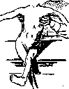
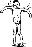
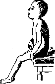
Figure 4. Cerebral diplegia. Position of the lower extremities when sitting. or sensory irritations). Pareses may be completely absent and are often simulated by spasms. True pareses show the usual type. However, there are cases where spasms are completely absent at rest and appear only with voluntary or sudden passive movements (intentional spasm). They occur most vividly when attempting to put children on their feet. A uniform round kyphosis is often observed, which (due to the spastic flexed position of the thighs) greatly hinders the ability to sit, stand, and walk. Spasms often interfere with the appearance of greatly increased tendon reflexes. Pathological reflexes may also be absent. The embracing reflex (Umklammerungsreflex), crossed adductor reflex, and in severe cases also the sucking reflex are often observed. Reflex synergistic movements of one limb during passive movement of the other are common. Hypertension of abdominal muscles can simulate the absence of skin reflexes. In rare atypical cases, decreased sensitivity is noted. The pelvic organs are not affected. It is necessary to note the extreme timidity of these children. Involuntary movements are observed less frequently than in hemiplegias. Tremor and ataxia occur. Among the lesions of cranial nerves, atrophy of the optic nerves and strabismus are sometimes observed. With lesions of facial muscles, an amimic mask-like face is noted, standing in sharp contrast to the excessive mimicry during laughter, crying, and fear. Dysarthria and bradylalia are often observed. Spasms of the pharyngeal muscles, making swallowing difficult, are also noted. In these cases, drooling is often present. Retardation in the growth of limbs is rarely observed, perhaps because it is less noticeable due to the bilateral nature of the lesion. Sometimes muscle hypertrophy is noted. Bones in these cases often show significant osteoporosis. To the abnormalities of body structure in these diseases belong changes in the skull. In some cases, hydrocephalic types are observed, in others microcephaly, flattening of the occiput and lateral parts. Among developmental defects are sometimes observed heart defects. Dislocations of the hip are also noted, which are sometimes congenital and sometimes a consequence of paralysis and muscle spasms. Seizures observed immediately after birth or after a few days much less frequently develop into epilepsy than in hemiplegic forms. As for the psyche, in these forms different degrees of impairment are noted - from profound idiocy to normal intelligence. It is necessary to point out that there are sometimes idioties which in the nature of the disease do not differ from cerebral diplegies and only do not show paralysis because the entire pathological process is concentrated in the so-called silent areas (cerebral paralysis without paralysis). Some cases of deaf-mutism may also belong here.
3. Less frequently than the indicated forms, pseudobulbar paralyses are encountered, under which is meant a symptom complex with disorders of the facial, speech, and chewing musculature, extremely similar to the phenomena observed with lesions of the nuclei of the corresponding cranial nerves. These disorders may be completely isolated; with more significant foci, however, both hemi- and diplegic phenomena may be observed. Two forms of this paralysis are distinguished: paralytic and spastic, between which transitional states exist (mixed forms). In the paralytic form, paralyses of muscles innervated by the lower and (mostly) middle branches of the facial nerve, rarely the upper, and paralyses of the tongue and chewing muscles are in the foreground. As a result, difficulties in chewing, swallowing, and movement of facial muscles, especially the muscles of the mouth, occur. Speech is always disturbed, monotonous, drawn out, with a nasal tint, sometimes completely impossible. Usually the mouth is open, the tongue is often protruded, and significant drooling is observed. Swallowing is to some extent disturbed. Food often gets under the tongue or between the cheek and teeth, from where it must be extracted with a finger and pushed to the back of the pharynx. Further swallowing occurs quite freely. Voluntary movements of the affected muscles are impossible, while mimetic and reflex movements are not disturbed. In this case, the sucking reflex of Oppenheim (Fressreflex), the reflex from the hard palate of Hennenberg, and the lip reflex of Toulouse and Würpe, normally observed in infants, are found. These patients are often timid and react extremely sharply to minor sound irritations. Sometimes forced laughter and crying are observed, often accompanied by athetosis of facial muscles. A disorder of respiratory muscles is also noted, which is often expressed during speech by short, jerky movements, with which involuntary sounds are produced. Besides the clearly expressed forms, formes frustes are observed, in which only certain phenomena are noted (e.g., speech disorder or more or less pronounced disorder of movements of facial muscles). Such children are often considered stutterers. In infants, it is striking that they cannot suck and swallow only when food is introduced dropwise deep into the mouth. In the spastic forms, the muscle lesions are more massive, and mimetic movements also become impossible. In this case, all muscles of the face may be affected, and then a mask-like face is observed, which does not change with emotion. Sometimes only the range of motion is affected, and in such cases every mimetic movement turns into inappropriate grimacing. Sometimes only the area of the mouth or the muscles of one half of the face are affected. In this form, formes frustes are also observed.
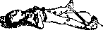
B. Cases with predominant lesions of extrapyramidal pathways. 1. Hemiplegic form. Choreic paresis represents such a form of cerebral hemiplegia in which, without a preceding spastic paralysis, a picture of choreiform movements develops. These forms are more often observed in children over 3 years old, with atrophy, epilepsy, and other mental defects usually not occurring. Infantile extrapyramidal hemipareses should also include cases of hemitremor, hemi-clonia, and cases of hemiparesis without pyramidal lesions. - 2. Diplegic extrapyramidal cerebral paralyses, a) Hypotonic form. Atonic-astatic type. In this symptom complex, described by Förster, general hypotonia or atonia of the entire musculature or significant areas of it is observed, with no resistance to passive movements, as a result of which excessive joint flexibility occurs. Static actions become completely impossible. Despite the fact that all muscles can be voluntarily innervated, children still cannot sit or stand. In the lying position, however, they are able to make all kinds of movements with their limbs. Coordination is disturbed, and ataxia is noted. Children cannot hold their head: it falls to different sides. Reflexes in pure cases are not increased, pathological reflexes disappear after the second year. Children are always mentally deficient, often epileptic. This symptom complex can be combined with various spastic phenomena. These conditions are always congenital, and there is a tendency to improvement. Forms described under the names of infantile cerebellar and cerebro-cerebellar diplegies also belong here. - b) Hypertonic forms: these include many cases of Little's disease, in which striatal symptoms are involved and predominate. In these cases, despite significant rigidity, the absence of pathological reflexes is often noted, and the rigidity often has an extrapyramidal character. Förster described a case of congenital pallidal syndrome, characterized mainly by an increase in plastic tone and passive resistance of muscles to stretching, poverty of movements, etc. Acquired cases of hypertonic forms include parkinsonism after epidemic encephalitis (see Parkinsonism and Encephalitides). - c) Hyperkinetic forms. These include bilateral athetosis (see Figure 5)
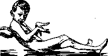
Figure 5. Bilateral athetosis.
and the general infantile chorea, which presents a picture very similar to bilateral athetosis, differing only in the nature of the movements. Combinations of both forms are often observed. This also includes cases of myoclonus that occur after encephalitis, in which there are constant lightning-like twitchings in different muscles (see Encephalitides). Course and outcome. In very early onset of the disease, in the first period there may be a complete absence of pathological phenomena or, after initial irritation phenomena, remain latent for 1½ years or more, as centers and pathways that normally reach their full development only in the first months of extrauterine life may be affected. For this reason, disorders caused by the absence or insufficient development of these centers and pathways manifest themselves only when, under normal conditions, they should begin to function. This probably explains, for example, the fact that the forced movements in congenital athetosis, as well as the phenomena caused by pyramidal lesions, appear after some time, whereas spastic phenomena caused by lesions of the pallidal system are already present from the first days of life. The developing brain copes well with minor disorders. Most hemi- and diplegias, as already indicated, have a tendency to improve. Sometimes such children learn to walk and talk at 8-10 years. In inflammatory (encephalitic) diseases leading to cerebral paralysis, the process usually begins with elevated temperature, severe cerebral symptoms (mainly convulsions) and often already at the onset of the disease with severe paralysis. Traumatic cases, according to the severity of the injury, initially show severe cerebral phenomena, which then partially subside. Vascular cases present the picture of an apoplectic stroke. Not all cases develop acutely; there are also cases with gradual development of pathological phenomena; in these cases, it is usually a matter of hereditary syphilis, but the same is observed in other progressive inflammatory processes. Often, epileptiform convulsions occur, after which at first mild pareses remain, and then severe paralysis. In many cases, epilepsy subsequently develops, in others the existing convulsions later disappear. Feeble-mindedness also often develops gradually. In most cases, however, the condition remains constant for many years. Contractures usually increase over time. Choreaic and athetotic movements show very little tendency to improve. Many patients, however, cope well with these movements and learn to use needles, various instruments, etc. These conditions do not pose a danger to life. Diagnosis. In hemorrhages during childbirth, convulsions, disorders of breathing, sucking and swallowing, paleness of the skin, twitching of facial muscles, and the phenomenon of Heibner (lightning-like raising of the upper and lower limbs when pressure is applied to the sternum) are often noted. Considerable restlessness and continuous crying, according to Seitz, indicate hemorrhage above the tentorium cerebelli, while considerable apathy indicates hemorrhage below it. In hemorrhages above the tentorium, which usually occur 24 hours after childbirth, gradually increasing symptoms of cerebral pressure often appear, as well as local irritation and deficit phenomena, especially from the facial nerves; in hemorrhages below the tentorium - severe phenomena from the centers of the medulla oblongata, directly or a few hours after childbirth, rigidity of the neck and opisthotonus. More subtle symptoms may be hemorrhage into the retina and disorders of the vestibular apparatus in the form of nystagmus, caloric unexcitability, and rotational decreased excitability. Blood in the spinal canal may also be a symptom of cerebral hemorrhage. In the chest age, mild cases of paresis are often difficult to diagnose, as the muscle hypertonia, characteristic of newborns, is intensified in various nutritional disorders. Tendon reflexes may be normally elevated and uneven. In this age, pathological reflexes may also be normal. Too early closure of the fontanelle, insufficient ability to hold the head, and signs of feeble-mindedness (delayed fixing and grasping), insufficient reactions to pricks may serve as important signs of the disease. In pronounced cases, it is sometimes necessary to exclude poliomyelitis and peripheral paralysis acquired during childbirth in differential diagnosis. It is important to distinguish contractures from spasms in diplegia. The spastic nature of the paralysis, increased reflexes, slight atrophy, intellectual defects, forced movements, and normal excitability speak for cerebral paralysis. These same symptoms make it possible to distinguish the atonic-atactic forms from congenital muscular atonia. Difference in pupils and absence of reaction to light speak for syphilis. New growths are distinguished by their progressive course, stagnant discs, headaches, and other symptoms of brain tumor. This also applies to hydrocephalus. Amaurotic idiocy is also distinguished by its progressive course and characteristic changes in the fundus of the eye. Family forms show atypical pictures (bradylalia, intention tremor, nystagmus) and are also distinguished by their progressive course. There are also family forms of diplegia that depend exclusively on the narrow pelvis of the mother. Therapy. In therapeutic treatment, it is necessary to distinguish acute cases from those that have already become chronic. In relation to the former, prevention of pregnancy and childbirth is important. In supratentorial hemorrhages with signs of brain compression, decompressive trepanation is performed, and in infratentorial hemorrhages - lumbar puncture or puncture of the posterior cistern (Brady), which, according to Sharpe, should be repeated until clear fluid appears under normal pressure. Punctures of the fontanelles and, in suspected hemorrhages into the ventricle, puncture of the latter have also been recommended. Based on the assumption of hemorrhagic diathesis, serum or blood injections are sometimes used in America. All these measures, however, have so far given insignificant results.
In acute inflammatory phenomena, cold, counter-irritants (flies, leeches) are applied. Massage and passive gymnastics, proper positioning of the affected limbs, and if necessary, splints, can prevent the development of undesirable contractures. In syphilitic etiology, specific treatment must be carried out, although in most cases improvement cannot be expected, as treatment does not restore the destroyed brain substance. In chronic cases, the most favorable forms for treatment are those without more severe disorders of brain functions and without significant pareses, with predominance of spasm of the limbs. Patients first of all need good general care. Too early and tiring attempts at walking should not be made. Muscle hypertonia responds well to long warm baths. Skillful massage of the par-
etic muscles and Fig 6 position of the lower, more an- weaker terior extremities after resection of the adductors. gonalists and pra-
ction weak, non-fatiguing gymnastic exercises (especially in lying and sitting positions) are also advisable. In speech disorders, systematic exercises in it are necessary. Intellectual defects often make it necessary to place the child in a closed institution. There are no internal remedies for affecting spasms. Bromide, belladonna, luminal, tetraphan, and cautious scopolamine are usually recommended. Often, intralumbar injections of narcotics have been used to reduce spasm. However, they are not safe and give only a temporary effect. Where physical therapy is ineffective, it is necessary to resort to surgical intervention. To correct the position of the legs, tenotomies of the corresponding muscles are performed (see Figure 6 and 7). Shortening and transplantation of tendons are also used. In recent times, nerve sections have also been resorted to. Lorenz recommended section of the n. obturatorius to reduce spasm of the adductors; Spitzy - transplantation of part of the nerve innervating the spastic muscles into the nerve supplying the weakened antagonist. Stoffel aims at the same goal by means of neurectomies, i.e., partial resection of motor nerves. It must be remembered that in all these interventions on the peripheral mixed nerve, pareses are produced instead of spasms, and that with regeneration of nerves, relapses can always be expected. In certain cases, the following
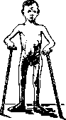
Fig. 7. Cerebral diplegia. Gait after tenotomy.
The operation of Förster.. He accepts two components of spastic paralysis—one paretic and the other spastic. Spastic symptoms occur only because the sensory irritations coming from the affected limbs through the posterior roots into the spinal cord can spread to the gray matter of the anterior horns and cause a state of constant increased irritation due to the absence of inhibitory fibers. The section of the corresponding posterior roots, i.e., the reduction of harmful excessive sensory irritations in the afferent part of the reflex arc, can result in a reduction of the spastic state that interferes with voluntary movements and simulates paresis. But even in true spastic pareses, this section, by reducing spasms, can to some extent reduce the phenomenon of paresis and thus cause an increase and expansion of the volume of active muscular movements. Thus, success of the operation can be expected only where spastic pseudopareses are in the foreground, i.e., in cases where there are no gross and extensive paralyses. To determine the remaining possible motor impulses, before the operation, lumbar or paravertebral conduction anesthesia can be performed. Purely spastic phenomena will disappear with this, and thus the remaining volume of voluntary movements can be discovered. It should be noted that this operation, like all surgical interventions in general, requires very long (1-2 years) sequential treatment in the form of various exercises in walking and standing, unthinkable without the active participation of the patient. Thus, operations are permissible only in cases of sufficiently developed intellect. Other motor disorders, such as cerebral ataxia or severe athetoses, significantly complicate operations. In severe cases of choreo-athetotic motor disorders, attempts have also been made to cause paresis instead of painful forced movements by removing the superficial layers of the motor centers. The operations are not without danger and do not always achieve their purpose. The treatment of epilepsy is carried out by conventional methods. In severe Jacksonian seizures, surgical intervention is indicated. Peripheral paralysis. These include lesions of the cells of the anterior horns (see Heine-Medina disease and Poliomyelitis), nuclei of the cranial nerves, plexuses, and peripheral nerves. The etiology of these lesions is diverse. A predisposing factor may be diseases of the parents (syphilis, alcoholism). In congenital motor disorders of the cranial nerves, the question is most often about developmental defects; sometimes the cause is intrauterine inflammatory processes or hemorrhages during childbirth. Paralysis of the facial nerves (see Figure 8) is often caused by pressure from forceps or pelvic bones on the trunk of the facial nerve.

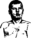
Figure 8. Paralysis of the right facial nerve due to trauma during childbirth. Pathological anatomy. Pathologically, aplasias of individual motor nuclei are observed, sometimes dysplastic disorders (infantiler Kernschwund Mobius'a), in which the nuclei, although laid down, are destroyed due to exogenous influences or endogenous inadequacy. There are hereditary familial forms of underdevelopment of nuclei. Lesions of the brachial plexuses develop due to excessive pressure on them or their strong stretching and are observed in severe, mostly artificial deliveries. They can occur in both head and breech positions (when turning on the leg, when freeing the arms, when extracting by the shoulders, etc.). They develop when the shoulder is pushed downward while the head is simultaneously bent in the opposite direction. In breech positions, this is observed when the shoulders have already passed but the head has not yet emerged. If in this position the trunk is lowered and raised while the head cannot follow these movements, then a pulling of the plexus occurs. In head positions, the same picture is observed if after the head passes the shoulders are held back and at the same time the head is bent downward while the anterior shoulder presses against the mother's symphysis. Paralysis in head positions occurs almost exclusively in large children with broad shoulders that hinder the passage of the trunk, especially with a narrow pelvis. Pathologically, in this case, lesions of the roots (from the 5th cervical to the 1st thoracic) are observed. At the same time, all roots or only some may be affected. In the so-called Erb's paralysis, the lesion concerns the 5th and 6th cervical roots or their plexus; it is caused by pressure on Erb's point (2-3 cm above the clavicle, at the posterior edge of the sternocleidomastoid muscle). In Klumpke's paralysis, the 8th cervical and 1st thoracic roots are subjected to stretching. At the sites of lesions, hematomas, neuritic phenomena, ruptures of nerves, and in older cases—connective tissue scars are found. Forms of paralysis. 1. Lesion of nuclei of cranial nerves. Most often, lesions in the area of the external eye muscles are encountered, mainly pareses of m. levatoris palpebrae (congenital ptosis, see Figure 9). Further, complete external ophthalmoplegias, paralysis of the external and superior rectus muscles of the facial nerve, masticatory muscles, and muscles of the tongue are observed. Pareses can be unilateral or bilateral, symmetrical or asymmetrical, can affect one or several muscles or be combined. Developmental defects in the area of the bulbar articulation center, the so-called congenital bulbar paralysis, are also described. Occasionally, in connection with such defects, disturbances of sensitivity— anesthesia of the conjunctiva and cornea—are also observed. This probably also includes hemispasm of the lower lip Variot. There is also a combination with other congenital defects of muscles, bones, and the visual apparatus. Clinically, it is interesting to note that in these congenital paralyses, the position of the eyeball remains normal, and even in paralysis of the abducens nerve, no contracture of antagonists develops; only occasionally a slight strabismus is observed; diplopia is absent. Sometimes peculiar associated movements in the masticatory muscles and the paretic upper eyelid are noted. In unilateral ptosis, a high position of the ipsilateral eyebrow often develops (long-term innervation of m. frontalis). A characteristic feature of congenital paralyses is their stationary state. The excitability of paretic muscles is reduced or absent, no reaction of degeneration is observed. 2. Plexitis. In plexitis, paralyses are usually unilateral, only in breech positions sometimes bilateral. In head positions, the leading side is always affected. When all roots are affected, the limb is completely immobile; in Erb's paralysis, according to the affected roots, lesions of nn. musculo-cutanei, axillaris, radialis, sometimes suprascapularis and paralysis of the muscles innervated by them (mm. biceps, brachialis, deltoideus, brachioradialis, sometimes also m. infraspinatus) are noted. The limb is rotated inward, the forearm is slightly bent, the palm due to excessive pronation is turned backward (see Figure 10). Raising the shoulder, flexion and supination of the forearm are severely limited or impossible. In Klumpke's paralysis, the muscles innervated by nn. ulnaris and medianus—small muscles of the hand, flexors of the arm and fingertips
are affected. In this case, the Horner's symptom complex is often noted, indicating a lesion of the first dorsal root before the departure of ramus communicans to the sympathetic nerve. Mixed forms are also observed. Paralysis of the lower extremities due to lesions of the lumbar plexus are extremely rare.-3. Lesion of peripheral nerves—see Polyneuritis. Diagnosis. In diseases of the cranial nerves, for differential diagnosis, in bilateral lesions, it is necessary to exclude progressive muscular dystrophy with involvement of the facial muscles. In it, however, mm. orbicularis are predominantly affected
mm. orbicularis are predominantly affected
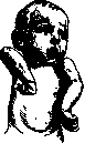
Figure 10. Erb's paralysis... oculi et oris; in the latter, hypertrophy is sometimes even observed. It is necessary to bear in mind also the phenomena of pseudobulbar paralysis (see above). Facial nerve paralysis due to birth trauma is sometimes indistinguishable from congenital paralysis (degeneration reaction in the former, sometimes changes in the temporal bone in the latter). Inplexititis, it is necessary to keep in mind fractures and dislocations of the humerus and clavicle, which can cause a similar picture and often complicate plexus lesions. With separation of the humeral epiphysis, positional anomalies and movement disorders reminiscent of Erb's paralysis are also observed. In these cases, it is important to establish crepitation or tenderness in the shoulder joint area. In doubtful cases, X-ray examination is necessary. Parrot's pseudoparalysis is easily excluded upon careful examination. It is also necessary to exclude the so-called painful paralysis of young children (paralysie douloureuse Chassaignac). Course and outcome. Paresis of the cranial nerves caused by congenital nuclear lesion can show some improvement. Facial nerve paralysis resulting from birth trauma usually has a good prognosis. Plexus lesions proceed variously depending on the degree. Mild lesions resolve in a few weeks; in other cases, especially when the entire plexus is affected, recovery is greatly delayed. In most cases, movements are fully restored or only minor pareses remain. In severe cases, movement disorders are often observed, which are explained, however, not so much by muscle paralysis as by secondary changes in the joints and ligaments that severely impede movement, as a result of which the corresponding muscles, despite normal electrical excitability, remain atrophic and paretic. The limb may lag in growth. In the shoulder joint, adductor contractures and inability to rotate outward are usually observed; in the elbow joint, pronation contracture and difficulty in supination. Therapy. In nuclear lesions, only plastic surgery is possible, such as transplantation of m. frontalis to levator palpebrae sup., etc. In plexitis, complete rest is necessary in the first days. Local dry heat is recommended (on the supraclavicular region in the form of warm cloths, hot-water bottles, bags of heated sand). After 1-2 weeks, besides warm baths, careful massage and passive movements of the paralyzed muscles are begun. From the beginning of the second month, electricity treatment is given for many months. It is extremely important to pay the most careful attention to the prevention of contractures by means of appropriate positioning of the paralyzed limb, etc. To induce active movements in the paralyzed limb, temporary binding of the healthy one is recommended. If function does not recover in 1/2 year or a year, Childhood Paralysis then surgical intervention is indicated, aimed at excising scar tissue and suturing the severed nerve ends. It is contraindicated if it is a question of root avulsion at the site of their exit (Horner's syndrome). Regarding childhood paralysis caused by Heine-Medin disease, see Heine-Medin disease. Prophylaxis. The prophylaxis of childhood paralysis, depending on its etiology, has a different character. One of the main conditions for healthy offspring is the satisfactory health of the parents. In view of this, a medical certificate of health for those entering into marriage should be a necessary prophylactic measure. Taking into account the enormous importance of hereditary factors in the transmission of many nervous and mental diseases, it is necessary to avoid marriages between knowingly sick and heavily burdened individuals in this regard (this is especially essential in relation to blood relatives). In the presence of clearly hereditary-familial diseases, having offspring can be permitted only after a thorough analysis of each case in terms of considering the inheritance pattern of the given disease and all eugenic rules stemming from the laws of heredity. Taking into account the adverse effect of a number of diseases and intoxications of parents on the embryo (e.g., syphilis, tbc, alcoholism, etc.), it is necessary to ensure that individuals with insufficiently treated syphilis, with open tbc, dipsomaniacs, etc., do not marry. The harmfulness of conception in a state of alcoholic or other intoxication, in a state of sharp nutritional decline, from very old parents, or with a very significant difference in their age should also be pointed out. As for the prophylaxis of pregnancy, the mother must avoid any moments that could adversely affect the embryo, such as physical and mental trauma, any diseases, excesses, excessive labor. In regard to childbirth, all possible harmful moments must be taken into account (e.g., narrow pelvis, abnormal fetal position, etc.) that can give cause to difficult labor and hemorrhages. When using forceps, versions, etc., extreme caution must be observed, as these measures can serve as the cause of facial nerve paralysis, brachial plexus paralysis, etc. In asphyxia, resuscitation methods that could cause or aggravate existing cerebral hemorrhage (e.g., Schultze's maneuver) should be avoided. To carry out preventive measures, widespread sanitary and educational propaganda is necessary, pointing out the dangers outlined above and the necessary measures. I. Prioman. Surgical treatment of childhood paralysis (mainly in Heine-Medin disease, from an orthopedic point of view). Orthopedic measures are necessary from the very first days of the end of the acute period of childhood paralysis. In the first period of the acute course and at the beginning of the second period of convalescence (recovery), the main attention should be directed to creating conditions of rest and preventing contractures and pathological postures of the limbs and spine. In the acute period, until the phenomena of irritation completely disappear, massage and electrification must under no circumstances be used. Rest is easily created with sandbags or light splints. In the second period (recovery), some muscles completely perish, in others strength decreases by 20-50% compared to normal, and only in exceptionally favorable cases does recovery reach normal. Optimistic orthopedists believe that muscle recovery is obtained in 80% of cases; others speak of 20% true recovery. This depends on the one hand on the nature of the epidemic; on the other hand, abortive cases must be taken into account. It should be considered that in those muscles where recovery occurred, there was no true paralysis, but only pseudoparalysis (Haglund) as a result of concomitant phenomena of edema and hemorrhage. Where actual destruction of spinal cord cells occurred, muscle recovery does not happen. Practically, it is important that even insignificant muscle damage can serve as the cause of secondary deformation under the influence of accompanying functional moments. This is frequently observed on the foot. Deformation usually develops gradually with the growth of the child. Therefore, already at the end of the first period, and especially in the second period of the disease (reparative period), measures are necessary to prevent and treat contractures if they have formed. The attending physician should direct main attention to the lower limbs, which are more frequently affected and most exposed to so-called static or functional influences. Preventive measures in the second period should be directed first of all to the lower limbs. A simple plaster posterior splint, preventing pes equinus and knee contracture, is quite satisfactory for a lying position, and when one leg is affected, it is also suitable for walking with the help of crutches. Most orthopedists consider a longer stay in bed desirable and in any case the elimination of harmful loading moments during the recovery period. In the stage of recovery, all conditions that worsen the recovery process must be excluded. Such conditions include: 1) overstretching of the muscle (for example, in pseudoparalysis or true paralysis of the deltoid muscle and the anterior group of foot muscles); 2) muscle fatigue, which significantly disrupts the reparative process; 3) the occurrence of deformation that reduces muscle strength; 4) walking in the absence of muscle force balance, leading to overstrain of stronger muscles rather than the development of weak ones. Therefore, under various body positions, it is necessary to take into account the state of dynamic forces, i.e., muscles that regulate not only the maintenance of balance, but also the development of the skeleton, especially in a growing organism. Thus, with weakness of the back and abdominal muscles, when the child is early given the opportunity to sit and stand, paralytic scoliosis-type deformation develops, and besides, the abdominal muscles are subjected to harmful overstretching. During crawling, the thigh muscle group sometimes develops, and the muscles of the lower leg and foot, especially the anterior ones, perish. Early standing without supporting apparatus leads to the development of a number of deformations on the lower limbs.
For the period of quiescence, in addition to prophylactic splints and appliances, physical-therapy procedures come into play: massage, therapeutic gymnastics, heat, electrization. In the acute period, massage and any attempts at electrization can bring only harm. In the second period, light, skillful massage and medical gymnastics are indicated. Rough massage leads to muscle atrophy. If significant improvement does not occur in the first 1-2 years, orthopedic measures are indicated; surgical treatment no earlier than in 4-5 years. After the acute period, improvement occurs in all patients. In 25% recovery occurs spontaneously within 4 years (Bowen). Heat has a favorable effect on muscles and joints. Water baths act worse than dry types of heat. The old view that splints and appliances are indicated only as a last resort must be abandoned. Appliances, splints, and corsets in poliomyelitis have their indications for all periods of the disease. They are used for the purpose of 1) enabling those who cannot walk to walk; 2) improving the gait of patients who walk; 3) preventing pathological positions; 4) correcting deformity. For the lower limb, the Thomas splint type, modified by Lowett, is very convenient. The design of this splint is based on three points of fixation of the knee: by means of a posterior loop above, a shoe below, and pressure on the knee from front to back by a fenestrated clasp (see figure 11). With a drop foot resulting from paralysis of the peroneal nerve, the Blencke boot in combination with a steel spring is very convenient (see figure 12). Conversely, with the loss of the muscular group of the triceps surae, an appliance with a spring producing plantar flexion is applicable. For lateral support of the foot (with pes calcaneo-valgus or varus) in childhood, an appliance is convenient which preserves the necessary position for the foot with a leather loop and allows free mobility in the ankle joint (see figure 18). With flattening of the arch of paralytic origin, as with other kinds of flat foot, individual inserts are indicated, passively correlating the skeleton of the foot, creating an arch. Conversely, with a slight tendency to pes varus, the so-called Taylor shoe finds application with leather padding on the outer part of the sole (see figures 15 and 17). For the upper limb, with frequent paralysis of the deltoid muscle, abductor splints or appliances are indicated, which serve to prevent overstretching of the deltoid muscle; they are also used in the postoperative period during muscle transplantation. Of serious practical importance is a timely given supporting corset for weakness of the spinal and abdominal muscles, since it prevents the development of persistent paralytic scoliosis (see figures 13 and 14). If paralysis of the oblique abdominal muscles predominates, a cloth corset with steel rods can be used. With simultaneous paralysis of the long muscles of the back (usually unilateral lesion), a supporting corset of the Biesalski type is indicated. Unfortunately, the majority of patients after suffering from childhood paralysis come to the orthopedist with already developed contractures and skeletal deformities. Treatment in such cases should be aimed at eliminating large contractures and skeletal deformities, at using dynamic factors in the form of well-preserved muscles, and at obtaining stability, mainly for the lower limbs and spine. Paralytic contractures most frequently occur in the form of adductor contractures on the upper limb, flexion contractures on the knee, flexor-adductor contractures on the hip joint (see figure 16), and equinus or varo-equinus type on the foot. In view of the fact that in the presence of marked contracture and deformity it is not always possible to judge the state of the muscles, it is recommended to preliminarily correct the contracture conservatively. For this purpose, serial bandages, tractions, and in individual cases the „tourniquet" are convenient. After the elimination of the deformity, it often turns out that muscles previously considered paralyzed show some viability. It is important with such serial bandages not to fix for a long time, but to combine the correction period with the period of physiotherapy. Sometimes the treatment period is shortened by combining conservative treatment with surgical treatment in the form of tenotomy-myotomy. Reducing the dynamic strength of a certain group of muscles sometimes appears advantageous for the correction of deformity. However, it should be emphasized that tenotomy in poliomyelitis must be treated critically. This operation in many cases proves to be very valuable for eliminating deformity, but sometimes it is useless and even harmful. Thus, with paralysis of the quadriceps muscle in combination with pes equinus, a mechanism is created that is very convenient for walking, not requiring a special appliance for the knee. Conversely, when eliminating equinus, the gait can be significantly impaired: there results
Figure 13.
Figure 14. Figure 1;
Figure 16.
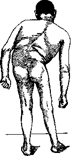
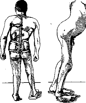
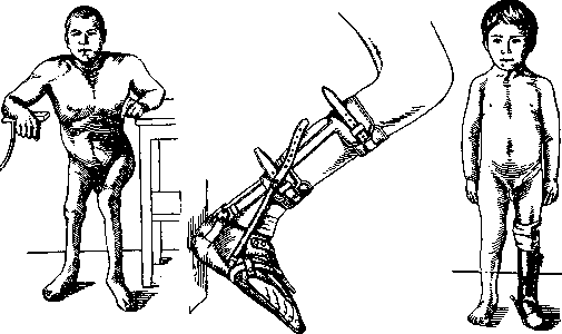
Figure 17. Figure 18. Instability in the paralytic knee joint, requiring an apparatus or plastic reinforcement of the quadriceps. Various kinds of tenotomies and fasciotomies occupy a prominent place in the system of surgical treatment for poliomyelitis. Even greater importance is attached to operations aimed at improving function. These include the transplantation of active tendons onto paralyzed ones. In addition to perfect technique, a strict selection of suitable cases and properly indicated indications for transplantation are necessary. If a muscle that is electrically excitable but with questionable clinical activity is transplanted, the success may be very insignificant. A prerequisite for successful transplantation is the preliminary correction of deformity. It is necessary to create the best conditions for the action of the transplanted muscle. The most common transplants are of the mm. sartorii and tensoris fasciae latae in paralysis of the quadriceps muscle; the mm. biceps and semitendinosus are less suitable for this purpose. In paralysis of the m. gastrocnemii, the transplantation of the tt. peronei long, and flex, hallucis (or tibialis postici) to the calcaneus is beneficial. In paralysis of the peroneal group, success is achieved by transplanting the m. tibialis ant. In paralysis of the m. glutei medii, Legg's operation is recommended: reinforcement of the tensoris fasciae on the thigh, 6-7 cm below the trochanter major. An even better effect is achieved by transplanting the m. sacrospinalis, with the missing tendon segment supplemented by silk ligatures, which are secured to the trochanter major during marked abduction of the thigh. In paralysis of the deltoid muscle, the use of the force of the m. trapezii in combination with fascial plastic surgery (W. Mayer's method) is indicated. For the success of transplantation, skillful subsequent orthopedic treatment and the patient's active will are necessary. The second type of operation to improve function is nerve plasticity. The transplantation of a healthy peripheral nerve onto the affected one has been proposed. Thus, in paralysis of the thigh muscles, the n. obturatorius is transplanted onto the n. cruralis. Nerve plasticity has not acquired wide clinical significance and is significantly inferior in practical results to tendon (rather, muscle) plasticity. The second large group of orthopedic operations for the sequelae of Childhood Paralysis consists of so-called stabilizing operations. Their aim is, in the absence of functioning muscles, to create passive stability of the limb for a short moment of loading. Among such operations, arthrodesis (see) occupies a central place. Its numerous modifications on the ankle joint pursue the main goal of achieving stability through a firm, painless ankylosis. However, not all orthopedists share the view that it is necessary to arthrodese the ankle joint. In the operations of Dunn and Hoke, the ankle joint itself remains free. In childhood, an apparatus that provides a more elastic gait should be preferred in any case. Arthrodesis in the knee joint is indicated in adults; in childhood, it should be avoided to prevent shortening of the limb due to injury to the epiphyseal line. In all cases where it is possible to achieve at least some stability in the knee joint without arthrodesis, it should be avoided. Such stability can be achieved - by means of tenodesis or by creating artificial recurvatum in the knee through supracondylal osteotomy of the thigh. In coxa laxata, as a consequence of the paralytic condition of the gluteal muscles, instability in the hip joint, subluxation, or even complete dislocation of the head is often present. Arthrodesis in such a joint is not always successful and is accompanied by prolonged pain. The surgical creation of a canopy from the edge of the acetabulum does not always provide firm stability. Jones proposes shortening and duplicating the capsule with subsequent prolonged fixation in the position of abduction and hyperextension. Arthrodesis in the shoulder joint is fully indicated if muscle plasticity is not possible. With the preservation of muscles moving the scapula, fairly good movement of the shoulder together with the scapula is obtained. In individual cases, satisfactory stability can be achieved by tenodesis and fasciodesis. This type of operation is especially indicated in drop foot and hand. Fixation of tendons in a bone canal ensures more reliable success for operations. The creation of artificial tendons with silk is also used, and such tendons in suitable cases are connected with an active muscle. For example, in drop foot and with an active m. gastrocnemii, silk threads sutured through this muscle are passed subcutaneously to the anterior part of the foot and secured to the dorsum of the foot medially and laterally, thereby suspending the foot. The technique of creating silk tendons has been developed in detail by Lange. Finally, to achieve good stability of the foot in various paralytic deformations, resection of the talus - astragalectomia according to Whitman - is widely used. In addition to eliminating deformity through astragalectomy, a completely stable position and practically satisfactory movement in the ankle joint are achieved. It is important in this case to shift the foot as far back as possible to avoid a sharp disproportion between the anterior and posterior parts of the foot. In individual cases, tenodesis of the extensors of the foot must be added to astragalectomy. In recent years, among most orthopedists, there is a tendency to avoid prolonged use of apparatuses in the sequelae of childhood paralysis, using surgical methods in all cases where there are no contraindications to this. Such a replacement of apparatuses by the art of surgical orthopedics is extremely valuable for socio-economic reasons. But unfortunately, there are a whole series of cases where the apparatus cannot be dispensed with. This especially applies to childhood and to severe cases of total paralysis of both lower extremities and back muscles in the presence of paralytic scoliosis in adults. Here, often only a well-fitted apparatus in combination with a corset gives the patient the possibility of moving. The condition of the back and abdominal muscles is of enormous importance in the mechanics of movements of the trunk and extremities. It is often necessary to be convinced that a significant disturbance of gait is associated with the paralytic condition of the mentioned muscles. In such cases, it is sufficient to give the patient a well-modeled corset to immediately change the type of gait. Paralytic scoliosis can be corrected, especially in young age, by repeated plaster corsets in combination with a period of bed rest and skillful massage. After eliminating the severe curvature, the patient can be provided with a permanent support corset, or the corrected position is maintained by surgical means. If the treatment of paralytic deformations in general presents a difficult task, then the treatment of paralytic scoliosis presents the most complex task. In the hands of the orthopedist, there are numerous conservative and surgical methods for treating the sequelae of Childhood Paralysis. Skillful use of all means (combining surgical methods with apparatus therapy) depends on the experience of the orthopedist, V. Chvclin.
Related articles
Mentioned in
Cite this page
“Childhood Paralysis.” Soviet Medical Encyclopedia. English translation of Bolshaya Meditsinskaya Entsiklopediya, 1st ed. (Moscow, 1928–1936), ed. N. A. Semashko. https://sovietmedicalencyclopedia.pages.dev/article/childhood-paralysis/