Heine-Medin Disease
Historical document, translated for reference. It reflects medical knowledge of the 1920s–30s and is not medical advice.
Summary
This 1930s encyclopedia article discusses Heine-Medin disease, commonly known as acute anterior poliomyelitis or infantile paralysis, detailing its epidemiology, pathology, and historical occurrence in epidemics across Europe and America.
Encyclopedia article (1928–1936)
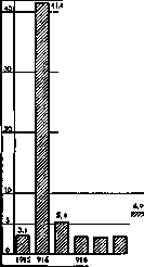
HEINE-MEDIN DISEASE (Heine, Medin), syn.: acute poliomyelitis (poliomyelitis anterior acuta), acute atrophic spinal paralysis, epidemic infantile paralysis, spinal infantile paralysis,—first described by Heine in 1840 (with the first detailed description given by Medin in 1890), is an acute specific infectious disease of the nervous system that appears both sporadically and epidemically. Since the beginning of the 20th century, this disease has appeared from time to time in Europe and America in the form of severe epidemics. Epidemiology and pathogenesis. Heine-Medin disease is a contagious illness, tends to spread along transport routes, and is sometimes transmitted directly. Schools may also play a role in the spread of the infection. The number of virus carriers is large. Healthy transmitters who have had abortive forms of the disease, as well as convalescents in whom the virus can persist for months—mainly on the mucous membrane of the nose, oral cavity, and pharynx (droplet infection)—apparently play a major role in spreading the disease. Transmission also occurs via feces; transmission is also possible through objects (handkerchiefs, linen, clothing, footwear), through dust, dirt, domestic animals (dogs, ducks, chickens, etc.), and apparently through insects (especially flies). However, a certain individual predisposition is necessary for the disease, which appears to be observed not very frequently, especially in adults and older children; it is possible that acquired immunity also plays a role here, and great significance is attributed to the body's defense mechanisms against the invasion of the pathogens. According to Flexner, Noguchi succeeded in cultivating the pathogen of poliomyelitis on a liquid medium and subsequently on a solid one. It belongs to the filterable viruses, is very resistant to external influences, weakens over time, does not die upon drying, but has low resistance to heat; at temperatures of 47-55°, it rapidly loses its activity. Of chemical reagents, it is rapidly destroyed by 1% hydrogen peroxide, menthol preparations, 0.2%-1% potassium permanganate, and formaldehyde, but is resistant to 0.5% carbolic acid, bile, and gastric juice. The pathogens of poliomyelitis are very small (0.15-0.3 µ) non-motile bodies, lying in pairs, short chains, or small clusters (staining well with Giemsa, Gram, and methylene blue). The upper respiratory tract and, perhaps, the gastrointestinal tract serve as the portal of entry for the virus. It enters the central nervous system via the lymphatic vessels of the cranial and spinal nerves, or, according to other authors, via the cerebrospinal fluid. Within the brain substance, it spreads mainly along the lymphatic vessels. Levaditi recently provided a very interesting and broad synthesis of clinical, epidemiological, histopathological, and experimental facts in connection with the study of acute poliomyelitis, encephalitis, and herpes, uniting these diseases under the concept of 'neurotropic ectodermoses.' The character of the epidemics naturally matters in terms of a large number of severe forms, but generally during the same epidemic the most diverse forms are encountered, while the number of abortive forms defies calculation, etc. Statistics. Observed sporadically or in the form of small epidemic outbreaks, acute poliomyelitis manifests from time to time as large and severe epidemics. Beginning in 1919, it has been registered in various countries (see Table 1): Table 1. Number of patients with acute poliomyelitis registered in various countries from 1919 to 1927 (absolute figures). Countries: Australia, Austria, England, Germany, Netherlands, Denmark, Italy, Canada, New Zealand, Norway (cities), Romania, USA, Finland, France, Switzerland, Sweden. 1919, 1920, 1921, 1922, 1923, 1924, 1925, 1926, 1927. Among European countries, the largest epidemics of acute poliomyelitis were observed in the Scandinavian Peninsula. In Norway, the first outbreak was registered in 1868, in Sweden in 1881 (Wernstedt). In 1905, the first major epidemic broke out, yielding 1,034 registered patients in Sweden and 981 in Norway for that year. The next (significantly larger) epidemic began in 1911. Over 3 years (1911-13), 9,447 patients were registered in Sweden and 2,788 in Norway. In 1919, a third epidemic was noted in Sweden, yielding far fewer cases, and in 1924-25 there was a new rise in acute poliomyelitis (see Table 1 and Fig. 1). Morbidity of acute poliomyelitis in Sweden from 1905 to 1927 (abs. figures). Figure 1. In other European countries, Heine-Medin disease is registered at significantly lower rates. In England, prior to the World War, the highest figures were registered in 1912 with 955 cases and 1913 with 881 cases. A new rise was observed in 1924 and 1926-27. In Germany, an epidemic of acute poliomyelitis was observed in 1909 with about 2,400 cases; the next epidemic was noted in 1926-27. Concurrently with Germany, an epidemic of Heine-Medin disease was also observed in Austria, where about 1,000 cases were registered in 1909. The largest epidemic of Heine-Medin disease in the current century was observed in 1916. Morbidity of acute poliomyelitis in the USA from 1915 to 1927 (per 100,000 population).
926
Figure 2. in the U.S.A., where about 27,000 cases were registered for this year (in previous epidemics in the U.S.A. there were registered: in 1907—about 2,800 cases, in 1909–10—about 12,000 cases). In New York City, from June 1 to November 1, 1916, there were registered (according to Vaughan) 8,928 patients with acute poliomyelitis, and in the entire state of New York—13,000 cases (in previous epidemics in New York, patients registered were: in 1907—about 2,500, in 1908—about 1,200). Subsequent rises in Heine-Medin disease, of significantly smaller dimensions, were observed in the U.S.A. in 1921, 1924–25, and 1927 (see Table 1 and Fig. 2). Of other countries besides those listed, an increase in Heine-Medin disease in recent years has been observed in Austria, Romania, Canada (1927), and New Zealand (1925; see Table 1). In the USSR, acute poliomyelitis is not subject to compulsory registration. According to very incomplete data, there were registered in the RSFSR in 1926 530 cases, of which 275 were in cities and 255 in rural areas. The largest number of cases was noted in Voronezh Governorate—47, Ivanovo-Voznesensk—79, Nizhny Novgorod—23, Samara—52, and the North Caucasus—116. In 1927, according to the RSFSR, also from incomplete data, 592 cases of acute poliomyelitis were registered, of which 35 were in Nizhny Novgorod Governorate, 154 in the Middle Volga Region, 101 in the Vyatka-Vetluga Region, 71 in the North Caucasus, and 75 in Tomsk Okrug. Morbidity in cities and rural areas is not identical. In the 1911–13 epidemic in Sweden, patients with Heine-Medin disease registered per 10,000 population were: Years 1911 1912 1913 Cities 6.3 5.1 1.8 Table 2. Rural areas 16.0 14.0 7.6. In rural areas, the incidence of Heine-Medin disease is registered much higher than in cities. In larger cities it is lower than in smaller ones. In the same epidemic, totaled over three years (1911–13), patients registered per 10,000 inhabitants in cities of various types were: Table 3. Urban population With paralysis Over 10,000 .... From 10,000 to 100,000 Less than 10,000 .... 6.3 14.0 76.3 Total 16.0 80.0. Morbidity by age and sex. Heine-Medin disease is observed chiefly in childhood. In cities, morbidity among young children is significantly higher than in rural areas. For individual epidemics, the following ratios in the morbidity of various age groups can be given (according to Wernstedt, see Table 4). Mortality and lethality. Table 5 shows the number of deaths from acute poliomyelitis in recent years. Mortality in nonepidemic years ranges from 0.1 (Germany) to 1–2 (Sweden, U.S.A.) per 100,000 population; in epidemic years it rises to 10 (U.S.A. in 1916) and 20 (Sweden in 1911–1913) per 100,000 population. Lethality Table 4. Number of acute poliomyelitis cases in cities and rural areas by age group (in % of total). Place and time of epidemic Norway, 1905. Sweden, 1905. New York, 1907. Vienna, 1908–09. Massachusetts, 1910. Warsaw, 1911. Sweden, 1911–13. New York, 1916. Cities 1–5 years Older than 5 years Total Rural areas 1–5 years Older than 5 years Total 78.1 90.5 75.2 78.7 90.7 61.8 85.5 21.9 9.5 24.8 21.3 9.3 38.2 14.5 39.9 40.3 63.8 37.5 60.1 59.7 36.2 62.5 525 868 525 5,142. Table 5. Mortality and lethality from acute poliomyelitis in selected countries for 1924–27. Countries Number of deaths (absolute figures) Australia ..... Austria ..... Germany ..... Holland ..... Denmark ..... New Zealand ..... Romania ..... U. S. A. ..... Sweden ..... 1924 g. 1925 g. 1926 g. 1927 g. in most countries constitutes from 10 to 20 percent in relation to registered patients. The same percentage of lethality is noted in individual epidemics. Thus, in New York in the 1916 epidemic, out of 8,928 patients, 2,407 deaths were registered, or 26.96%; in Brooklyn for that year, lethality from acute poliomyelitis was 21.6%, in Chicago—13.3%, and in 1917—33.6%. In Sweden in the 1905 epidemic, out of 100 paralyzed cases, 16.7 died (868 fell ill, 145 died), and in 1911–1913—19.8 (out of 6,754 cases, 1,337 died). For individual years of this epidemic, lethality was: in 1911—21.7%, in 1912—18.1%, in 1913—19.5%. Lethality by age groups. Lethality varies in different age groups. In Leipzig, from January 1 to October 20, 1927, 181 cases of acute poliomyelitis were registered, with 26 deaths (14.3%). For individual groups, the number of cases and deaths is distributed as follows: Table 6. Lethality from acute poliomyelitis by age group in Leipzig in 1927. Age groups Preschool children School-age children 9.7 14.1 58.3. Lethality increases with age. Wickman gives the following table of lethality by age group in the 1905 epidemic in Sweden (see Table 7). and 297 15 6 226 Lethality (per 100 patients) 1924 g. 10.9 21.0 20.8 50.0 20.5 30.1 21.2 25.4 1925 g. 22.2 20.6 22. 28. 20. 12. 19.5 1926 g. 21.8 21.3 12.7 32.6 17.2 20.7 19.8 1927 g. 7.3 10.8 34.9 13.9 10.4 Table 7. Lethality from acute poliomyelitis by age group in Sweden in 1905. Age years Total. 10.0 11.6 14.9 11.4 28.6 23.7 28.3 33.3 21.4 27.3 33.3 19.2 16.7. Based on these tables, it must be concluded that in older children and adults, the prognosis in acute poliomyelitis is significantly worse than in young children. Lethality in cities is generally lower than in rural areas. In Sweden in 1911–13 it constituted (per 100 cases with paralysis): Table 8. Years 1911 . . . 1912 . . . 1913 . . 1911–13 . Cities 19.2 19.8 13.5 18.0 Rural areas 22. 18. 19. 19. Time of onset of death. In the majority of cases, death occurs within the first week after the onset of the disease. Wickman collected the following data on the time of death in the 1905 epidemic in Sweden. Table 9. Died after onset of disease. on the 1st day in 1 case on the 9th day in 5 cases » 2 » » 6 cases » 10 » » 7 » » 3 » » 22 » » 11 » » 2 » » 4 » » 36 » » 12 » » 1 » » 5 » » 25 » » 13 » » 2 » » 6 » » 15 » » 14 » » 1 » » 7 » » 16 » » 15 » » 1 » » 8 » » 3. Out of 143 cases analyzed by the author, 121 died in the first week after the onset of the disease, or 84.6%, and in the second week—22, or 15.4%. Death most frequently occurs on the 3rd–5th day after the onset of the disease. Seasonality of acute poliomyelitis morbidity. The bulk of acute poliomyelitis cases fall in the summer-autumn period. According to Wickman, who collected information on 26 outbreaks of acute poliomyelitis (from 1881 to 1906), 14 of them were observed in summer, 5 in summer and autumn, 3 in autumn, and the remaining 4 captured, besides summer and autumn, partially winter (two outbreaks) or spring (two outbreaks). The monthly trend of some epidemics can be illustrated by the following table: Table 10. Monthly distribution of cases in individual epidemics (absolute figures). Sweden Romania Leipzig Months 1905 g. 1911–13 g. 1927 g. 1927 g. Jan.–June . July .... August . . . September . . 24 3 October . . . November . . - December . . . The height of the epidemic is observed mostly in August and September (see also Figures 3 and 4).
I. Dobreytser. Pathological anatomy. Macro- and microscopic pictures vary depending on the stage of the process. In the initial stage, the disease presents as an acute disseminated inflammatory process, predominantly in the anterior horns of the spinal cord (see Figure 5), not restricted to the area of the central arteries, but involving arteries and veins in general (their dilation and perivascular infiltrates). In the ganglion cells, disappearance of fibrils and various
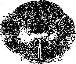
Figure 5. Cross-section of the spinal cord. Change in the anterior horns. Left—reduction of the anterior horn due to scar formation; right—disappearance of cells and fibers in the middle parts of the anterior horn.
degrees of chromatolysis up to complete degeneration and atrophy of the cells. In the degenerated cells there is a varying degree of neuronophagia. The disease almost always spreads to the gray matter of the posterior horns, as well as to the white matter and the pia mater. 89 Corresponding changes are also noted in the cerebrum, the pons varolii, and the medulla oblongata. Various degenerative phenomena are also observed in the nerve fibers: tortuosity, swelling, focal disintegration. Diffuse proliferation of nuclear glia is also noted. The cranial nerves, anterior and posterior roots, and spinal ganglia often also present phenomena of inflammation. With the final disappearance of nerve tissue, it is replaced by scar tissue, consisting chiefly of glial elements with a small number of thickened vessels. A severe degree of inflammation is accompanied by softening of the nerve tissue with the subsequent formation of a cyst. In addition to the indicated changes in the nervous system, it is necessary to note inflammatory phenomena in the lymph glands of the neck, upper respiratory tract, and intestinal tract. The kidneys, liver, and heart are often in a state of cloudy swelling. Symptomatology. The incubation period fluctuates between one to two days and 11/2 weeks. In the disease itself, three stages are distinguished: 1) the initial stage, characterized most often by elevated temperature and the development of paralyses, 2) the stage of partial or even complete recovery, and 3) the final stage with flaccid trophic paralyses. In the first stage, pre-paralytic phenomena of both a general and local character are often observed, consisting of influenza-like pictures (especially frequently the neuralgic form), disorders of the respiratory organs, gastrointestinal tract, and sometimes meningeal symptom complexes. Fever, almost always observed in the first days of the disease, is often overlooked in early childhood, but it may also be absent. Sometimes, a few days after the drop in temperature, a new elevation is noted. The pulse often does not correspond to the temperature. The acceleration of respiration is also not always a consequence of elevated temperature, but is sometimes a manifestation of initial bronchitis, bronchopneumonia, or paresis of the respiratory musculature. Regarding the psyche, children exhibit restlessness, tearfulness, caprices, anxious dreams, delirium, and motor excitability. Headaches are not particularly severe. A state of unconsciousness is rare. Sometimes a scarlatiniform rash is noted, rarely herpes labialis. Symptoms characteristic of poliomyelitis already during the pre-paralytic stage are general, sometimes local hyperesthesia, associated with significant pain upon passive movements, especially of the spine, as well as of the extremities that will subsequently become paralyzed, and spontaneous twitchings of a tonic or clonic character in them. Further, there is a significant tendency to sweating, local muscle hypotonia, and ataxic uncertainty during movements. The cerebrospinal fluid is transparent, sterile, increased in quantity, and secreted under increased pressure; cytologically, there is moderate lymphocytosis, chemically, a small increase in the amount of protein; the Wassermann reaction is negative; globulin reactions are positive. Changes in the cerebrospinal fluid depend on the intensity of the involvement of the meninges in the process. In the blood, according to some authors, there is leukopenia, according to others, leukocytosis. Typical for poliomyelitis is the development of paralyses directly following the initial phenomena or during this period. Usually, paralyses occur on the 1st–5th day (sometimes much later); most often the lower extremities are paralyzed (in 4/5 of all cases), and paralyses of the trunk muscles are in second place. The proximal areas of the extremities, both upper and lower, are usually more affected (exception: the peroneal group, and sometimes m. tibialis ant.). Respiratory muscles are also frequently paralyzed. Paralyses often do not occur simultaneously; they are asymmetrical and uneven. Their character is flaccid, with loss of tendon reflexes and lowering of tone. Due to the participation of the white matter in the process, sometimes in older children pathological reflexes (Rossolimo, Babinski, etc.) and an increase in tendon reflexes are observed. When the trunk muscles are paralyzed, abdominal reflexes disappear, as well as often the reflex from the cremaster. Sensitivity may be disturbed, but to an insignificant degree. Disruption of the pelvic organs is observed only in the initial stages. In the recovery stage, general phenomena, sensory disturbances, and pelvic organ disorders disappear first of all. But even paralyses of the extremities almost never remain in their original form. Along with the return of active movements, tone and reflexes usually return to normal. In severe and irreparable cases, muscle flaccidity persists, and atrophy begins to develop; at this time, the reaction of degeneration is discovered. The final stage is characterized by established atrophic paralyses, the forms of which are diverse, depending on the localization and spread of the paralyses and on their effect on the soft parts, skeleton, and articular apparatus. Muscle atrophy can reach the point of their complete disappearance and transformation into connective tissue (see Figure 6). Fibrillar twitchings are frequent. Compensatory hypertrophies can develop in non-paralyzed muscles. In severe paralyses, damage to bones and joints is constantly present. For the most part, abnormal shortening of the paralyzed extremities is observed, depending on bone hypoplasia and lag in growth. Ligaments become relaxed, joints become loose. In the formation of contractures, besides partial paralyses and the action of relatively well-preserved antagonists, the influences of gravity, external pressure, the tendency toward shrinkage of paralyzed muscles, and secondary shortenings of antagonists play a role. More frequent
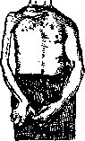
Figure 6. Marked
atrophy of the muscles of the right hand.
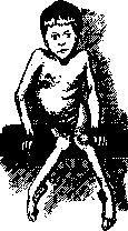
Figure 7.
Contractures develop mostly in the lower extremities (scoliosis or lordosis are observed; see figure 7). Vasomotor and trophic disorders are also noted: the skin becomes cyanotic, slightly edematous, smooth; anomalies in hair and nail growth are also observed. The psyche is usually not affected. Besides the spinal form, there are also distinguished the bulbar, pontine, encephalitic, neurotic, Landry's, meningeal, and abortive forms. In bulbar forms, peripheral facial nerve paralyses are most commonly observed, and pareses of the oculomotor nerves, hypoglossal nerve, and the motor branch of the trigeminal nerve, as well as swallowing and speech disorders, are also noted. Other forms present pictures characteristic of them (see Encephalitis, Meningitis, Landry's disease, Polyneuritis). Abortive forms are very frequent (15-20%), but their recognition is possible with a certain degree of probability only during epidemics. Contracting poliomyelitis confers lasting immunity. Immune bodies appear as early as the 6th day and can still be detected 20 and more years later. Differential diagnosis is particularly difficult in the initial stages of the disease, when poliomyelitis can be confused with various other infectious diseases, such as, for example, gastroenteritis, influenza, meningitis, typhus. Cases of poliomyelitis with initial symptoms from the respiratory organs may give rise to confusion with bronchitis, pneumonia, and even diphtheria. In acute infectious polyneuritis, paralyses in the distal parts predominate, sensory disturbances and a progressive course are noted. In small children, they are generally rare. From meningitis of another origin, the meningeal form of poliomyelitis differs in the composition of the cerebrospinal fluid and the presence of flaccid paralyses. In bulbar, pontine, and cerebral forms, the diagnosis can be established only in the presence of a poliomyelitis epidemic. An important test is the serodiagnostic test: neutralization of the poliomyelitis virus by the serum of convalescent patients or of immune animals. The technique, however, is difficult and expensive. Prognosis. Mortality, depending on the epidemic and locality, varies considerably; in Sweden it averages 10-15% (reaching up to 42.3%). Boys fall ill in greater numbers than girls (55.5% resp. 45.5%). At a mature age, the prognosis is particularly grave. Death usually occurs on the 3rd-7th day from bulbar paralysis of respiration, from spinal paralyses of the intercostal muscles and diaphragm, and pulmonary complications. Complete recovery is observed in approximately 15%. Severe paralyses remain in only 1/3 of the cases. An important criterion for prognosis is the state of electrical excitability. Muscles that have not lost faradic excitability during the first 2-3 weeks will, in all probability, become functional; those showing complete reaction of degeneration at the end of the first week, apparently, will not recover, although some improvement is not excluded here either. Prophylaxis, treatment. Effective immunization against acute poliomyelitis in humans does not yet exist. Treatment with serum from patients who have had poliomyelitis is still in the period of observation and experiment. Treatment with convalescent serum may be tried in the first days of the disease in the following form: intralumbally 10-25 cubic centimeters, subcutaneously or intravenously 30-120 cubic centimeters, depending on the severity of the case and the age of the patient. Prophylactically necessary are: isolation of the patient at home or in a hospital during the acute period. In suspicious or mild cases, precautions should be taken (sneezing, coughing, talking are dangerous); it is necessary to rinse the mouth and wash hands several times a day (avoid contact with children). Sick schoolchildren, as well as their brothers and sisters, must not be admitted to schools earlier than 6 weeks; disinfection of excreta and linen is necessary, at least in the acute stage. A record of diseases must be kept by public health authorities. In acute cases, physical and mental rest is necessary. Diaphoretics, urotropin internally and intravenously in the form of a 40% sterile solution are advisable. In very rapidly developing paralyses, as well as in severe spinal tenderness, especially in meningeal forms, lumbar puncture is indicated. It must not be forgotten that autohemotherapy can sometimes have a good effect. In phenomena of significant motor and sensory irritation—warm baths, diathermy, wraps, antineuralgic agents, luminal-sodium subcutaneously. For respiratory disorders—lobeline (with caution). During the first 14 days, during severe pain, the patient may be allowed to assume the most comfortable position for him. In the recovery period, the main concern is the prevention of contractures and incorrect limb positions, which are very often the consequence of inappropriate treatment. First of all, the correct position of the patient is necessary. If there is paralysis of the shoulder, it is necessary to abduct the upper limb at a right angle. In paralysis of the lower limbs, the patient must lie horizontally, with extended hip and knee joints and feet bent at right angles, which is achieved by a board placed under the mattress, splints. To avoid pressure of the blanket on the feet, it is placed on a bed cradle. Of the general measures in this stage, mercurial ointments, injections of novasural are recommended, contributing, mainly, to the resorption of edema. 3-4 weeks after the onset of the disease, faradization and careful active exercises, passive gymnastics, and massage can be gradually started. Rhythmic electrotherapy is especially recommended. For further treatment, it is necessary to be guided by the following principles: 6-8 weeks after the end of the acute stage, with great caution, children are begun to be put on their feet with the aid of appropriate devices in order that the still functional muscles begin to function. This is necessary also because only in a standing position, thanks to the compression of the joints and the physiological arrangement of the centers of gravity of individual parts of the body, do those sensory stimuli flow to the motor apparatus, without which the automatic centers cannot act and final muscle recovery cannot take place. If after 1-2 years, depending on the age and severity of the disease, no further success can be expected from the indicated measures, it is necessary to resort to surgical intervention. Orthopedic care, judiciously applied, is very useful in various stages of the disease. Surgical (incl. orthopedic) treatment—see Infantile Paralysis.
Related articles
Mentioned in
Cite this page
“Heine-Medin Disease.” Soviet Medical Encyclopedia. English translation of Bolshaya Meditsinskaya Entsiklopediya, 1st ed. (Moscow, 1928–1936), ed. N. A. Semashko. https://sovietmedicalencyclopedia.pages.dev/article/heine-medina-disease/