Motor Centers
Historical document, translated for reference. It reflects medical knowledge of the 1920s–30s and is not medical advice.
Summary
This article describes the motor centers of the cerebral cortex, their location, function, and associated pathways. It explains how stimulation or damage to these areas affects voluntary movement and discusses both pyramidal and extrapyramidal motor systems.
Encyclopedia article (1928–1936)
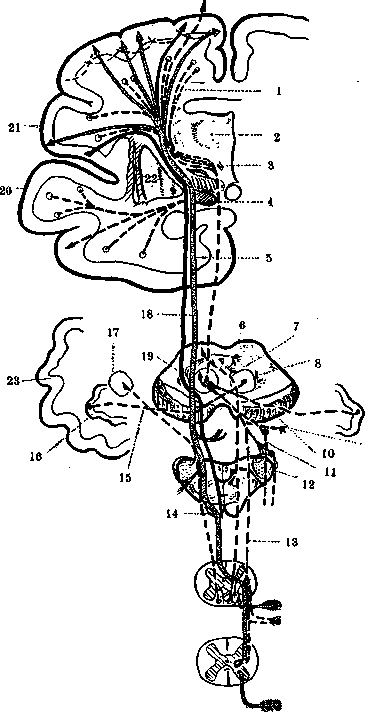
MOTOR CENTERS (and conducting pathways). Motor centers, or the motor area of the cerebral cortex can be defined as an area of the cortex, the lesion of which causes paralysis of half of the body, while irritation of this area with minimal electric current causes isolated contraction of individual muscles in the corresponding part of the body, resembling coordinated movement; with stronger current, widespread movements with the character of Jacksonian epilepsy are obtained. In the narrow sense of the word, these motor centers lie in front of the fissura Rolandi in the gyrus centralis anterior, lobus para-centralis, and in the posterior parts of gyri frontalis superioris et medii. According to some authors (Rothmann, Probst), the motor area of the cerebral cortex also extends to the posterior central gyrus, since its irritation also causes isolated movement of individual muscles, although such contraction can be explained by simultaneous irritation of the anterior central gyrus. The motor area of the cortex includes gyrus centralis anterior, lobus paracentralis (4th area according to Brodmann) and gyr. frontalis (6th area) [see Cerebral Cortex Architecture and Brain (anatomy)]; both these areas are combined into the precentral area, the main feature of which is the greater thickness of the cortex (on average 3.8 mm), the indistinctness of the boundaries between the layers, the comparative poverty of the cortex with small cells, and the absence of the grain layer. In addition, the 4th area is characterized by the presence in the 5th layer, in the layer of deep pyramids, of giant cells, or so-called Betz cells (see Betz cells). The largest of them are located in the upper parts. Their number in general is not particularly large: approximately in one hemisphere there are about 25,000 of them (Campbell). Due to the presence of such cells, this area of the cortex is distinguished under the name area giganto-pyramidalis, and the 6th area-area agranularis frontalis. Electrical investigation of the motor area in humans during surgical operations has shown that in this area there are about 40 motor centers, intended not only for the movement of various joints of the limbs, but also for various highly differentiated muscle groups, managing movements of flexion, extension, abduction, adduction, etc. In lobus paracentralis and in the upper quarter of gyrus centralis anterioris are centers for the movement of the lower limbs, located in reverse order relative to the segments of the limb; in the two middle quarters are centers for the movement of the upper limb in the corresponding sequence with its segments; in the lower quarter of the gyrus are centers for the face, tongue, pharynx, larynx; these last centers extend into the area of operculi. In the posterior part of gyrus frontalis medii are localized centers for the movement of the head and eyes; in gyrus frontalis superior-center for the movement of the trunk, although some authors place this center in gyr. centralis anterior, between the centers of the upper and lower limb [for details of the distribution of centers-see Brain, colored plate (Vol. VII, pp. 535-536), Fig. 1 and 2]. The centers are separated by non-excitable intervals; especially large intervals are found between the centers of the fingers of the hand and the centers of the face. In these non-excitable areas there are no characteristic motor cells, and pyramid fibers do not take their origin there. After unilateral destruction of cortical centers, loss of voluntary movements is observed on the opposite half of the body. Since the muscles of the larynx, pharynx, mastication, most of the muscles of the tongue, the upper muscles of the face, the muscles of the eyes, neck and trunk are innervated by both hemispheres, irritation of the centers of one side produces a bilateral effect in these muscle groups, and with destruction of the corresponding centers these muscles continue to function correctly. With destruction of motor centers, especially persistent paralysis is mainly subjected to isolated movements of the limbs, especially the upper, due to the presence in the cortex of numerous and differentiated centers for the movement of the hand. Combined movements, i.e., movements performed by several parts of the body at once or alternately, are less affected and recover more quickly. In addition to voluntary movements, the motor function of the cortex of this area also includes certain reflexes, so-called normal skin reflexes (plantar, from cremaster, gluteal and abdominal), which are of great importance in diagnosis; loss of these reflexes indicates cortical localization of the disease. Some authors suggest that irritation of the cortex of the motor area affects not only the striated muscles, but also the smooth muscles, without precisely determining the location of such centers; other authors (L. R. Muller) do not recognize cortical centers for internal organs, believing that, for example, for the bladder, the influence of the cortex is limited to the striated muscles, the contraction of which secondarily causes reflexes of smooth muscles. The cortex of the frontal lobe is related to maintaining equilibrium due to its connection with the cerebellum, and destruction of this area is characterized by the development of ataxia, similar to cerebellar (see Cerebellum).-In the precentral area of the cerebral cortex begin the motor conducting pathways, which are divided into cortico-nuclear, going to the cells of the motor nuclei of the cranial nerves, and pyramidal pathways, or cortico-spinal, directed to the cells of the anterior horns of the spinal cord (see figure). Some authors consider the entire system of these fibers as the pyramidal system (see). In addition to the pyramidal system, there are still systems of fibers related to movement and connecting the cerebral cortex with the cerebellum and with other subcortical formations located in various parts of the central nervous system, as well as these formations with the spinal cord. This system of fibers is distinguished under the name of the extrapyramidal system (see). For the proper functioning of the entire motor apparatus, it is necessary to preserve both the pyramidal and extrapyramidal systems. Motor conducting pathways: 1-fronto-pontine fibers; 2-thalamus; 3-tectal pathways; 4-optic nerve; 5-cortico-nuclear system; 6-superior colliculus; 7-tecto-bulbar fasciculus; 8-red nucleus; 9-rubrospinal tract (decussation); 10-base of the cerebral peduncle; 11-rubro-reticular fasciculus; 12-corpus restiform; 13-rubrospinal tract; 14-pyramid of the medulla oblongata; 15-brachium conjunctivum; 16-dentate nucleus; 17-fastigial nucleus; 18-pyramidal system; 19-tegmentum of the cerebral peduncle; 20-temporal gyrus; 21-operculum; 22-lenticular nucleus; 23-cerebellum. (From Monakow.)
"" 9
and extrapyramidal systems. Disease of the pyramidal system causes paralysis in the form of hemiplegias, paraplegias and monoplegias or convulsions, depending on whether there is a deficiency or irritation of the corresponding parts. Disease of the extrapyramidal system causes various kinds of hyperkinesias, akinesias, changes in body position and a number of other symptoms. Lit.-see lit. to article Brain. E. Kononova.
Related articles
Mentioned in
Cite this page
“Motor Centers.” Soviet Medical Encyclopedia. English translation of Bolshaya Meditsinskaya Entsiklopediya, 1st ed. (Moscow, 1928–1936), ed. N. A. Semashko. https://sovietmedicalencyclopedia.pages.dev/article/motor-centers/