Hemiplegia
Historical document, translated for reference. It reflects medical knowledge of the 1920s–30s and is not medical advice.
Summary
Hemiplegia is a condition characterized by paralysis affecting one side of the body, resulting from damage to the pyramidal motor system. The article describes various types including hemiparesis (incomplete hemiplegia), monoplegia, alternating hemiplegia, and crossed hemiplegia.
Encyclopedia article (1928–1936)
HEMIPLEGIA (from Greek hemi- half, and plesso- I strike), literally: injury to half the body. The term denotes impairment of voluntary motor function on one half of the body, i.e. unilateral paralysis of muscles. An incomplete degree of hemiplegia is called hemiparesis. In humans, voluntary motor function is carried out by means of the pyramidal motor system, beginning from the cortical cells of the anterior central gyri. The axons of these cells extend without interruption to the bulbar nuclei and motor cells of the anterior horns of the spinal cord (tractus cortico-bulbares and tractus cortico-spinales), constituting the so-called "central" paralysis. Dissociated hemiplegia (paralysis of one or more muscles of the opposite upper extremity). Monoplegia (paralysis of the opposite upper extremity). Hemiplegia (paralysis of the opposite half of the body, face, tongue, upper and lower extremities). Alternating hemiplegia (paralysis of the hypoglossal nerve on the side of the lesion, and of the upper and lower extremities on the opposite side of the BRAINSTEM). Crossed hemiplegia (paralysis of the homolateral arm and contralateral leg). Spinal hemiplegia (paralysis of the arm and leg on the same side of the focus).
Crossed paralysis.
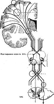
Crossed paralysis.
Crossed pyramidal path of the lateral column Uncrossed pyramidal l Figure 1. Diagram of hemiplegias with lesions of the pyramidal tract at different levels. The upper (voluntary) motor neuron; from the cells of the bulbar nuclei or the anterior horns of the spinal cord, axons extend through the roots and nerves to the muscles themselves, forming the so-called "peripheral" (lower, reflex) motor neuron. The latter, thus conducting impulses of voluntary movement, is at the same time the efferent part of the arc of the simplest segmental reflex (see Figure 1). Hemiplegia develops with lesions of the upper motor neurons, which conduct impulses of voluntary movement for the muscles of the opposite half of the body. In higher animals, the pyramidal path is less differentiated, and in the active innervation of their musculature, the short motor paths, originating from the subcortical ganglia, mainly the striatal system, play a greater role. This system of short motor paths (striatal, or extrapyramidal system) appears phylogenetically more ancient and in animals in which the neocortex is not yet developed, it is the main motor apparatus. With the gradual development of the mantle of the hemispheres (pallium), a powerful pyramidal tract develops, to which the functions of voluntary movement are also assigned, while the striatal system assumes the subordinate function of regulating muscle tone, as well as automatic and synergistic movements. Therefore, the destruction of pyramidal paths, for example, in a dog, leads only to a rapidly passing H., whereas in humans, the pyramidal tract being almost completely switched off, the person becomes almost completely immobile. But since the pyramidal path in humans begins to function relatively late (in the first year of extrauterine life), embryonic kinetics, as well as the mobility of the newborn, are still largely extrapyramidal. The pyramidal path, starting over a considerable length of the precentral gyrus, converges fanwise toward the center of the hemisphere and lies here in the form of a massive bundle, constituting a significant part of the internal capsule, located between the nucl. caud. and thalamus options on one side, and nucl. lenticularis on the other. In the internal capsule, due to rupture of arteries and hemorrhages, necrotic foci often develop, destroying the pyramidal bundle. This is how the so-called "capsular" H. is formed, which is the most common. The destroyed pyramidal bundle shows signs of descending degeneration, which does not extend to the peripheral motor neuron. However, with complete destruction of the pyramidal bundle in the area of the internal capsule on one side, not all the striated musculature of the opposite half of the body is paralyzed. Of the cranial nerves, the nerves moving the eyeball (III, IV and VI pairs) are preserved, the motor portion of the V nerve (masticatory musculature) is almost completely preserved, the X nerve (innervation of the corresponding half of the soft palate, pharyngeal musculature, vocal cord), innervation of m. sterno-cleido-mastoidei (from the XI nerve), and only in the area of innervation of two cranial nerves (VII and XII) are signs of paralysis observed, with the muscles innervated by the facial nerve being paralyzed dissociatively: in the lower half of the face, signs of paralysis are observed (flattening of the nasolabial fold, paralysis of the muscles retracting and elevating the angle of the mouth; similarly, platysma myoides is weakened), while the upper portions of the facial nerve (m. orbicularis oculi and especially m. frontalis) almost completely retain their function or are paralyzed only superficially and not for long; the hypoglossal nerve is paralyzed persistently, as a result of which the protruded tongue deviates to the side of paralysis (paralysis of m. genio-glossi). The muscles of the trunk (muscles of the spine, respiratory musculature, musculature of the anterior abdominal wall) are not paralyzed either. The musculature of the limbs undergoes paralysis, initially complete, which, however, later mostly undergoes more or less far-reaching regression, with the general rule that the function of the leg is restored much more completely than the function of the hand, and the mobility of the proximal parts of the limbs is restored significantly earlier and more completely than the mobility of the distal parts. Thus, the hand and fingers are the most severely affected segments, and often movements in them are not restored at all or are only partially restored (for example, flexion of the fingers becomes possible while their extension is impossible), which leads to a deep and persistent disturbance of the function of the upper extremities. On the contrary, walking becomes mostly possible in later stages of H. and is characterized by certain peculiar features: the paralyzed leg is swung forward in a semicircular movement, describing an arc similar to the movement of a scythe by a mower ("scissors gait", "demarche en fauchant"), with a straightened leg and foot fixed in the position of pes equinus. However, even movements restored after H. often require disproportionately large muscular effort and are possible only as summed, coarse, distorted movements due to the mass of synergistic movements. Thus, massive, interphalangeal flexion of all 5 fingers of the hand often becomes possible, but the patient is unable to isolate the flexion of any one finger. The preservation of certain muscles in lesions of the pyramidal system is usually explained by the fact that the non-paralyzing muscles in H. are to a much greater extent than the paralyzing muscles, innervated not only from the opposite, but also from the ipsilateral hemisphere. This ipsilateral innervation is sufficient to protect the muscle from paralysis. The physiological expression of such bilateral innervation is the difficulty of active contraction of the corresponding muscle or muscle group on one side: movements of the eyeballs, chewing, swallowing, phonation, respiration, etc. are always bilateral in normal conditions. This also explains why, if a focus of softening later develops in the other hemisphere of a hemiplegic, not only does a new H. of the usual type occur, but paralysis of chewing, swallowing, and articulation also joins (the so-called pseudobulbar paralysis). Ipsilateral H., however, is extremely rare, and is explained by compression of distant parts of the brain or by congenital absence of the crossing of the pyramidal paths. And even in ordinary H. on the side corresponding to the focus, small signs of pyramidal insufficiency can often be found, for example, some increase in tendon reflexes, even a slight decrease in muscle strength, mainly in the lower extremity. The paralyses in H. possess all the features of paralyses developing with lesions of the central motor neuron (so-called central paralyses, see). They are characterized by the absence of qualitative changes in excitability (absence of reaction of degeneration) and the preservation of nutrition of the paralyzed musculature. The reflex arcs in central paralysis are preserved and, even when isolated from central connections, come into a state of increased irritability, as a result of which the reflexes and muscle tone of the paralyzed muscles are increased (hyperreflexive, hypertonic paralyses); with preservation of subcortical impulses in the paralyzed limbs, synergistic involuntary movements are also preserved and even sharply intensified. However, upon more detailed consideration of this series of phenomena, the following is noted. The preservation of nutrition of the paralyzed muscles in H. turns out to be only relative: here, slight emaciation is often observed, for example, in the small muscles of the hand, especially the first interosseous, and in childhood H., the growth of the paralyzed limbs is also delayed; however, the matter never reaches such deep atrophy as in peripheral paralyses. Of the reflexes, immediately after the appearance of H., all normal reflexes from the skin and mucous membranes either disappear or decrease on the side of H. Tendon and periosteal reflexes are also sometimes decreased or even (in a comatose state) completely absent at first and only increase after a certain time; in later stages of H. (after several days or weeks) they are already sharply increased and are accompanied by clonus of the hand, patella, or more often the foot. In addition, a number of reflexes that are absent in normal conditions and appear only with the loss of function of the pyramidal bundle can be detected. Among the most important of these are: protective reflexes, Babinski's, Oppenheim's, Gordon's, Schaeffer's, Marie-Foix's, Rossolimo's and Mendel-Bechterev's reflexes. The last two reflexes, which have a rapid motor tempo and are obtained by rapid single irritation, can be detected in later stages of H. On the contrary, Babinski's symptom, as well as other tonic reflexes, are established immediately. Protective reflexes in their numerous clinical manifestations (palm scratch reflex, tonic movements of the upper limb when the muscular body of pectoralis major is pressed, Marie-Foix's symptom, etc.) are usually maximally developed immediately after the onset of H., in the further course they have a tendency to fade and in later stages of hemiplegia they often cannot be detected at all.
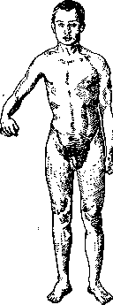
The normal 'forearm symptom' of Leri (automatic flexion of the forearm in response to passive flexion of the hand and fingers) is weakened or disappears on the side of H. The same applies to the normal symptom of Mayer (automatic opposition of the thumb of the hand during passive flexion of the proximal phalanx of the III or IV fingers) and to postural reflexes (see), which disappear on the side of hemiplegia. Associated movements (synkinesias) are absent at first and only later can be detected more and more clearly. In the late phases, they are often very pronounced, and any active forced muscle contraction (e.g., clenching the fist of the non-paralyzed hand) is accompanied by an involuntary tonic spasm of the paralyzed limbs. These synkinesias are observed in several different synergies, most often (see Figure 2) in the form of a shortening synergy for the upper limb (abduction of the shoulder + flexion of the forearm + pronation + flexion of the hand and fingers) and a lengthening synergy for the lower limb (extension of the thigh + extension of the leg + flexion of the foot). However, there are also opposite types: extension for the hand (especially often involuntary extension of the fingers during yawning) and flexion for the leg; the formula of synkinetic movement is influenced by the initial position of the limb, as well as various positions of the head: when the head is rotated toward the paralyzed limbs, associated
movements
in them may occur according to the extensor type, when the head is rotated to the opposite side - according to the flexor type (cervical reflexes of Magnus-Klein). To the associated movements, not uncommon in H., belongs also the symptom of combined flexion of the thigh and trunk of Babinski, as well as the symptom of combined abduction and adduction of the thigh of Raime, involuntary contraction of m. tib. anter. when flexing the leg (symptom of Strümpel), involuntary spreading of the fingers when raising the arm (symptom of Suk), flexion of the leg on the affected side when bending the trunk forward (symptom of Neri) and others. In other cases, tonic synkineses may be extremely sharply expressed when the actual phenomena of paralysis have completely or almost completely passed (the so-called hemi-tonia of Bekhterev). In the flexor synergy for the arm and in the extensor synergy for the leg, they want to see disinhibition of the automatic subcortical mechanisms underlying the grasping function and vertical standing. The muscle tone, like the tendon reflexes and synkineses, begins to increase only several weeks after the appearance of H., while at first (if there are no signs of so-called early contracture) the paralyzed limbs are hypotonic. Gradually increasing, hyper- Fig. 2. Traumatic right-sided hemiplegia. With strong compression into a fist of the left hand, the right hand performs automatic associated movements. tonia of the muscles eventually leads to the development of the so-called hemiplegic contracture, or late contracture of hemiplegics, when the muscles of the paralyzed limbs begin to increasingly spring during passive movements and may finally fix the limb in a stable pathological position. The muscles of the face rarely participate in the contracture. As for the nature of contracture in H., the most accepted view is that which connects it, like the formula of synkineses, with the liberation of subcortical mechanisms from the influence of inhibiting central impulses: contracture - as it were, 'frozen associated movement'. In favor of this, one can cite both the consideration that occasionally hypotonic H. without synkineses are encountered, and the circumstance that the motor formula of late hemiplegic contracture, in general, coincides with the nature of synkineses: the arm is fixed in the position of internal rotation of the shoulder pressed to the chest, flexion of the forearm, pronation of the hand and clenched fist position of the fingers, while in the leg typical extensor positions predominate (the so-called 'predilection type of contracture' - Wernicke-Mann). The muscles affected by contracture, as well as the joint-ligamentous apparatus, gradually undergo fibrous retraction. Arthritides develop comparatively early (especially in the shoulder joint), which often make attempts at passive movement very painful. The contracture increases with cooling, with sharp skin irritations, with rapid passive movements, with excitement; it decreases with rest, with warming of the limb (e.g., in a warm bath), during sleep. Often it is extremely variable and, exacerbating with various external factors (e.g., with excitement), almost disappears with complete rest of the patient ('latent contracture').-A quite different picture is presented by the so-called early contracture of hemiplegics, appearing immediately after the development of paralysis and, if the disease does not lead to death, regressing in the future. This is a strong tonic spasm of the paralyzed limbs, now increasing, now weakening, sometimes interrupted by temporary states of hypotonia, and at the moment of setting the spasm the paralyzed limb usually performs involuntary movements in space. Early contracture is closely connected with the development of protective reflexes, always strongly expressed in this condition, and its motor formula usually coincides with the motor formula of the protective reflex, which is why it can be considered as an analog of the so-called spontaneous protective movements and protective contracture (see Protective reflexes). Early contracture develops with very extensive shutdowns of centers (very extensive necrotic foci, hemorrhages into the ventricles of the brain, etc.), usually in a comatose state, and is therefore always a symptom dangerous to life. In favorable cases, early contracture can gradually turn into late contracture in the further course. If H. did not appear suddenly (as in vascular foci), but gradually increases (as, e.g., in brain tumors), the phenomena of this so-called 'early' contracture, on the contrary, can characterize the late, terminal phases of the disease. On the side of H., various vasomotor and trophic disorders are not uncommon: arthropathies, cyanosis, swelling, cooling of the limbs, asymmetry of blood pressure, skin t°, sweating, pilomotor reflex, etc. (vegetative syndrome in hemiplegia). If the pyramidal tract is not completely destroyed, but only damaged comparatively superficially or only temporarily, paralyses can undergo reverse development to complete restoration of function, without passing through the above-described phases of late contracture, associated movements, pathological synergy, etc. Partial restoration of function in H. is possible within the first year of the disease. With destruction of the entire pyramidal tract, the so-called hemiplegia facio-linguo-brachio-cruralis develops. If only part of it is destroyed, the development of partial H. is possible - hemiplegia facio-linguo brachialis or hemiplegia brachio-cruralis, etc. Monoplegia is called paralysis of only one segment, e.g., of the arm or leg. With even more fractional localization of the lesion, development of dissociated monoplegia is possible, in which only a small area of the limb is paralyzed, for example, only the small muscles of the hand or further only some of these muscles. The closer the necrotic focus is located to the cortex, the greater the chances for the formation of incomplete H. or monoplegia. However, sufficiently extensive cortical foci can lead to the development of true hemiplegia facio-linguo-brachio-cruralis.-Right-sided H. (in left-handers - left-sided) is often accompanied by symptoms of aphasia. In addition, with cerebral H., a more or less deep degradation of psychomechanics is often observed. With localization of necrotic foci in the brain stem, the so-called alternating H. (hemiplegia alternans, see Alternating syndromes) may occur, characterized by the fact that the focus simultaneously destroys the pyramidal tract and one or several nuclei of cranial nerves or their root fibers, as a result of which a paralysis of the same name of the cranial nerve and contralateral H. develop. Depending on which of the cranial nerves are involved in the process, very precise topographic diagnosis is possible here. With localization of the focus in the region of the decussation of the pyramids, the so-called crossed H. (hemiplegia cruciata) is occasionally encountered, in which the upper limb on the side of the focus and the lower limb on the opposite side are paralyzed. Finally, with localization of the focus in the lateral columns of the spinal cord, the so-called spinal H. develops, which in combination with contralateral thermoanesthesia and analgesia is a component of the so-called Brown-Séquard syndrome (see). S19 The cause of hemiplegia is most often the development of necrotic foci due to disturbance of cerebral circulation (cerebral and meningeal hemorrhages, thrombosis and embolism of arteries, thrombosis of veins). Usually H. develops suddenly, often accompanied by symptoms of cerebral apoplexy, and later reveals the partial regression of symptoms described above. H. can also develop as a result of inflammatory (encephalitis, abscesses, meningitis) and traumatic processes, in tumors, animal parasites and syphilis of the brain. More rarely, H. is encountered in disseminated sclerosis. Temporary and very rapidly passing H., or hemiparesis, often remains after Jacksonian epileptic seizure as a sign of exhaustion of the motor centers. Spasm of cerebral vessels (e.g., the so-called vascular crises of Raynaud) in certain forms of migraine can cause a short-lasting hemiparesis, which completely recovers each time. Recognition of fresh H. may present some difficulty in the state of coma after apoplexy. Here important are: unilateral extinction of reflexes (corneal, tickling reflex of the lips, abdominal and cremaster reflex), passive bulging of the cheek during expiration (the 'sails'), flaccid tone of the corresponding limbs and absence in them of automatic gesticulation, if the latter is expressed on the healthy side; often there is associated deviation of the head and eyes toward the cerebral focus. The distinction between organic H. and hysterical H. does not present great difficulty, thanks to the typical changes in tone, reflexes and associated movements in H., - all these functions in hysterical H. do not appear changed. In addition, in organic H., the motor functions of the distal parts of the limbs are more affected, in contrast to the distribution of paralyses in hysterical H.
A typical gait, often observed in hysterical hemiplegia: the patient steps forward with the healthy leg and passively drags the paralyzed leg behind them across the floor, as if dragging a log ('demarche en draguant' of the French, Todd's gait). Of course, it is completely different from organic hemiplegia, and also the general setting in which hysterical paralyses appear and disappear. The therapy of hemiplegia consists in treating the underlying disease (e.g., specific treatment of syphilitic arteritis), as well as measures to prevent contracture, which most hinders subsequent active movement. Warm baths, passive gymnastics, light and careful massage are appropriate, as well as - in later phases - active gymnastics. Any irritating procedures, such as electrization, are contraindicated in cases with a tendency to contracture and are permissible only in hemiplegia with low muscle tone. Hemiplegia spastica infantilis, see Childhood paralyses.
The structure of blood circulation in the brain (Course of Nervous Diseases, edited by G. I. Rossolimo, M.-L., 1929); Astvatsaturov M., Diseases of the Brain (Private Pathology and Therapy of Internal Diseases, edited by G. Lang and D. Pletnev, vol. IV, issue 2, M.-L., 1928); Strümpell A., Private Pathology and Therapy of Internal Diseases, vol. III, M.-L., 1929; Klippel M. et Monier-Vinard B., Syndrome pyramidal, hemiplegie (Nouveau traite de medicine, sous la direction de G. Roger, F. Widal et P. Teissier, fasc. 19, P., 1927); see also literature for the article Hemianesthesia.
Related articles
Mentioned in
Cite this page
“Hemiplegia.” Soviet Medical Encyclopedia. English translation of Bolshaya Meditsinskaya Entsiklopediya, 1st ed. (Moscow, 1928–1936), ed. N. A. Semashko. https://sovietmedicalencyclopedia.pages.dev/article/hemiplegia/