Internal Capsule
Historical document, translated for reference. It reflects medical knowledge of the 1920s–30s and is not medical advice.
Summary
This article provides a detailed anatomical and pathological description of the internal capsule of the brain, covering its structure, fiber pathways, and clinical significance in the context of 1930s neurology.
Encyclopedia article (1928–1936)
CAPSULA INTERNA (internal capsule), a band of white brain matter lying between the basal ganglia of the cerebral hemispheres. The internal capsule is studied on sections of the cerebral hemispheres made in various directions—frontal, horizontal, and sagittal.
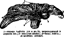
(Figs. 1, 2, and 3), and depending on the direction of the section, it has a different appearance. The internal capsule consists of 4 parts: 1) pars anterior, s. lenticulo-caudata, or the anterior limb of the internal capsule, lies between the nucl. caudatus (medially) and the nucl. lenticularis (laterally); 2) pars posterior, s. lenticulo-optica, or the posterior limb of the internal capsule, connecting with the anterior limb at an obtuse angle, forming the genu (knee) of the internal capsule (Fig. 2); 3) pars retrolenticularis—a section of white matter lying posterior to the nucl. lenticularis, and 4) pars sublenticularis—a section lying under the nucl. lenticularis. Upward, anteriorly, and posteriorly, the fibers of the internal capsule diverge fan-like into various parts of the cerebral cortex, forming the corona radiata (radiant crown, see figure); downward, the majority of fibers pass into the basis of the cerebral peduncle (see figure), and a part terminates in the thalamus opticus and in the basal ganglia (many authors deny such a termination). The anterior limb of the internal capsule is shorter; in the lower sections, it is penetrated by strips of gray matter (the connection of the nuclei caudati and nuclei lenticularis); the fibers in this part of the internal capsule run in a horizontal direction from the cortex of the frontal lobe, partly to the thalamus opticus, forming its anterior peduncle (pedunculus anterior), and partly to the pons Varolii through the cerebral peduncle; the horizontal fibers are intersected by numerous
transverse bundles running from the nucl. lenticularis to the nucl. caudatus.—The posterior limb of the internal capsule, which is longer, is formed by vertical fibers, as is the genu of the internal capsule. In the genu of the internal capsule, fibers run from the operculum and the lower sections of the anterior central gyrus to the motor nuclei of the cranial nerves—fibrae cortico-nucleares. The anterior 2/3 of the posterior limb are occupied by pyramidal fibers running from the gyr. centralis anterior, lob. paracentralis, and the posterior sections of the gyri frontalis super. et medii to the spinal cord—fibrae cortico-spinales. Fibrae cortico-nucleares and cortico-spinales are motor in their function and are distributed in the internal capsule in a known sequence. Throughout the entire length, fibers running to the optic thalamus, to the nucl. ruber, substantia nigra, and corpus Luysi are mixed with the pyramidal bundle. In the posterior third of the posterior limb, fibers pass from the thal. opticus to the gyr. centralis posterior; they carry sensory impulses, as a result of which Charcot called this part of the internal capsule the carrefour sensitif; many authors disagree with the doctrine of such an isolated position of the sensory bundle, but assume that its fibers are mixed with the motor ones; finally, there is a third opinion, according to which in the upper sections of the internal capsule, motor and sensory fibers are mixed, and in the lower ones, the sensory bundle becomes isolated and occupies the posterior part of the posterior limb. In the posterior section, fibers also run from the cortex to the thalamus opticus.—Fibers included in the pars retrolenticularis of the internal capsule mainly belong to the visual pathway and run to the occipital lobe; a small part of the fibers runs to the gyr. angularis and supramarginalis and from the cortex of the occipital lobe to the primary visual centers.—In the pars sublenticularis of the internal capsule, fibers are arranged in 2 layers: in the upper one, auditory fibers pass from the corpus geniculatum mediale to the gyr. tempor. sup. and gyr. transversus of Heschl; these fibers are mixed with the bundle of Türck (temporopontine system); in the lower layer, the fascic. temporo-thalamicus of Arnold and fibers belonging to the peduncul. inferior thalami optici run, connecting the thal. optic. with the anterior sections of the temporal lobe and with the insula Reili.—The internal capsule receives blood from the art. cerebri media (art. capsularis, lenticulo-optica, lenticulo-striata). Physiology of the internal capsule is clear from its anatomical structure: many systems, very important in their function—motor, sensory, visual, auditory, corticopontine—and fibers to some vegetative centers pass through it. The arrangement of bundles in the internal capsule, especially for motor function, was established not only on the basis of pathological-anatomical studies but also experimentally on monkeys by stimulating the internal capsule with an electric current. These experiments allowed conclusions to be drawn not only about the isolated position of fibers for the lower and upper limbs but even for parts of the limbs and for some muscle groups. According to these experiments, in front of all motor conductors (in the genu of the internal capsule) run bundles for combined eye movements, then fibers for the movement of the mouth, tongue, and head; prepared from paper, starch, or gelatin. Gelatin and starch capsules are swallowed together with the medicine contained in them; the purpose of their use is to hide the taste or smell of the medicine (about glutol capsules—see below). 1. Paper capsules (Figs. 6 and 7) are applicable only for powders or generally for solid medicinal forms (individual pills, tablets). Weighed in advance (on capsule trays), doses of powders are poured into the capsules, stacking them and folding them at one end (Fig. 9). To open the capsules, an unhygienic method of blowing with the mouth is often used; this has caused the appearance of capsules with a glued bent strip of cardboard, capsules with folds, etc., which open when pressed. Another method was introduced by the Oberländer factory: capsules in stacks of several pieces or (in boxes) hundreds are weakly glued at one end. Due to the composition of the gluing starch paste, it is easy to tear off an individual capsule or the required quantity, but the adhesion strength is sufficient to keep the rest glued together when one capsule is pulled slightly; at the same time, the capsules stretch fan-like and open. For opening ordinary capsules, capsule blowers with bellows or electric ones (Typhoon system, etc.) were also proposed. These devices did not prove themselves in practice. Previously, capsules made of Japanese paper were also prepared, swallowed together with the powder. For fatty powders, capsules are made of parchment, paraffin, or waxed paper—charta cerata, wax capsules; powders with volatile substances (ammonium carbonate, essential oils) or hygroscopic powders are enclosed in such capsules.— 2. Gelatin capsules (capsulae gelatinosae, Ph VII) are hard, consisting of two halves, closing like boxes (capsulae operculatae; Figs. 1 and 2), or soft (elastic), closed after filling with a drop of gelatin mass (Fig. 5). Composition: gelatin jelly with the addition of sugar
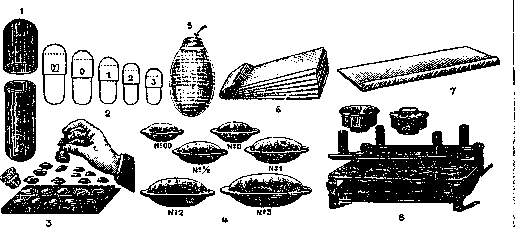
Figure 1. Shape of a hard gelatin capsule. Figure 2. Hard gelatin capsules (numbers; reduced approx. 1.5 times). Figure 3. Machine for filling Fasser wafers. Figure 4. Glued Fasser wafers (reduced approx. 1.5 times). Figure 5. Soft gelatin capsule filled with oily liquid; the arrow shows the opening closed by a drop of solidifying mass. Figure 6. Folded paper capsules. Figure 7. Paper capsule for powders. Figure 8. Morstadt wafers and a machine for their filling and closing.
in the posterior limb run fibers for the movement of the upper limb, posterior to them—for the movement of the trunk and then for the lower limbs.—Pathology of the internal capsule. Disease of the internal capsule is one of the frequent diseases of the brain; most often observed are arteriosclerosis with hemorrhages, softenings (in arteriosclerosis of the cerebral arteries); embolisms in endocarditis, cysticercosis, and less often—tumors. The anterior limb gives unclear focal symptoms, more likely related to the compression of adjacent parts; paresis and transient speech disorders are observed. More definite symptoms are given by disease of the knee and posterior limb, where motor and sensory conductors pass. Lesion of the anterior section of the posterior limb and the knee gives paralysis or paresis of the opposite part of the body in the form of hemiplegia with involvement of the face; the paralysis, initially flaccid, becomes spastic, pathological reflexes appear, cutaneous ones disappear; sometimes a sensory disorder is observed (in the opinion of some authors, only with simultaneous lesion of the thal. optic.) (see Hemiplegia and Brain). With bilateral lesion of the internal capsule, paralysis of 4 limbs, facial muscles, and disorder of swallowing and phonation develop, the so-called pseudobulbar paralysis. With lesion of the partis retrolenticularis of the internal capsule, there are visual disorders in the form of hemianopsia (see), and with disease of the partis sublenticularis—disorder of hearing. Lit.—see lit. to art. Brain. K. Kononova.
Related articles
Mentioned in
Cite this page
“Internal Capsule.” Soviet Medical Encyclopedia. English translation of Bolshaya Meditsinskaya Entsiklopediya, 1st ed. (Moscow, 1928–1936), ed. N. A. Semashko. https://sovietmedicalencyclopedia.pages.dev/article/internal-capsule/