Subthalamic Region
Historical document, translated for reference. It reflects medical knowledge of the 1920s–30s and is not medical advice.
Summary
This article from the 1928–1936 Soviet Medical Encyclopedia describes the anatomy of the subthalamic region (regio subthalamica or hypothalamus), detailing its boundaries, structures forming the floor of the third ventricle, and internal nuclear formations such as Luys' body and the mamillary bodies.
Encyclopedia article (1928–1936)
REGIO SUBTHALAMICA (subthalamic region; synonyms: regio hypothalamica, hypothalamus), the lower part of the diencephalon (diencephalon), located inferior to the thalamus and forming the lower part of the wall of the third ventricle and its floor. It develops from the anterior primary brain vesicle, the cavity of which narrows due to the thickening of the walls and turns into the third ventricle, and posteriorly into the cerebral aqueduct of Sylvius. The third ventricle communicates with the cavities of the lateral ventricles through the interventricular foramina of Monro. From the cerebral aqueduct of Sylvius to the lower edge of the foramen of Monro extends the hypothalamic sulcus
VI VII
VIII
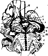
Figure 1. Base of the brain. Lower surface of the regio subthalamica and pons Varolii: 1 - hypophyseal sulcus, surrounded by the diaphragm of the sella turcica; 2 - optic chiasm; 3 - tuber cinereum; 4 - lateral geniculate bodies; 5 - pulvinar; 6 - mamillary bodies; 7 - pons Varolii; 8 - Gasserian ganglion; 9 - posterior perforated substance; 10 - optic tract; 11 - anterior perforated substance; 12 - olfactory tract; II, III, IV, V, VI, VII, VIII - cranial nerves. (sulcus hypothalamicus), dividing, together with the lateral wall of the ventricle, the diencephalon into the upper section—the thalamus, the region of the geniculate bodies—and the lower—the subthalamic region (regio subthalamica). The anterior part of the latter in the lower third is formed by the terminal lamina (lamina terminalis). Higher up, it borders on the anterior columns of the fornix, surrounding the foramen of Monro in front. Departing from this foramen, the columns of the fornix plunge into the lateral wall of the third ventricle. Anteriorly, the anterior commissure (commissura alba anterior) adjoins the columns of the fornix, belonging together with the fornix to the cerebrum. Posteriorly, the subthalamic region passes into the midbrain, and laterally into the hemispheres. The lower parts of the subthalamic region, which are part of the floor of the third ventricle (Fig. 1), are formed by the optic chiasm and the tuber cinereum, passing downwards into the infundibulum, at the end of which is the hypophysis; posterior to the tuber cinereum lie the mamillary bodies, the posterior perforated substance, and the interpeduncular fossa; in the higher layers of the subthalamic region and laterally (Fig. 2) lies Luys' body, or the subthalamic nucleus (see Corpus Luysi); next to it is the zona incerta (see Zona), and upwards is the nucleus of Forel's field. The mamillary bodies are formed by two round gray nodes covered by a white peripheral layer. In each mamillary body, two nuclei are distinguished—an internal one, consisting of small cells, and an external one, of smaller size, formed by larger cells 18 12
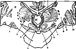
Figure 2. Frontal section of the brain through the thalamus opticus and regio subthalamica: 1 - lateral geniculate body; 2 - thalamus; 3 - red nucleus; 4 - Meynert's bundle; 5 - posterior perforated substance; 6 - substantia nigra; 7 - Luys' body; 8 - internal capsule; 9 - globus pallidus; 10 - putamen; 11 - caudate nucleus; 12 - lateral ventricle; 13 - habenula; 14 - fornix; III - third ventricle.
(see Olfaction, Fornix). The tuber cinereum consists of small cells with a narrow protoplasm and a large vesicular nucleus. The grey matter dorsally embraces the convexity of the corpus mamillaris and here passes without a sharp boundary into the substantia reticularis hypothalami. Among the grey matter thus constructed, isolated nuclei and pathways are scattered. Along the entire dorsal edge of the optic tract, closer to the center, is located an arched, orally, dorsally, and caudally lying group of rather large, polygonal ganglion cells that stain strongly with Nissl's method and impregnate weakly with Bielschowsky's method: the nucleus supraopticus, s. ganglion opticum basale Meynerti. This nucleus can be established in all mammals; only in Aplacentalia does it lose its compact general form and, penetrated by the fibers of the basal ganglia, divide into separate small groups. On sagittal sections, this nucleus can be differentiated into 3 groups: 1) lying in front of the optic tract—nucl. supraopticus ant., 2) located dorsally to the first—nucl. supraopticus dorsalis, and 3) lying between the tuber and the optic tract—nucl. supraopticus post. Near the edge of the III ventricle, in its wall, is a vertical group of cells—nucl. paraventricularis (Malone, Friedemann, Spiegel, and Zweig), consisting of cells morphologically identical to the cells of the nucl. supraoptici; reaching its largest dimensions toward the middle of the subthalamic region and forming a continuous cellular column, this nucleus extends to the region of the optic chiasm. The fibers of the fornix divide the nucl. paraventricularis into two parts, of which the ventral part—nucl. paraventricularis accessorius—rapidly disappears on a series of frontal sections. This nucleus is found in all mammals, although its dorsoventral expansion undergoes various adaptations in different species. Connections with other cell groups have not yet been established. Greving suggests that the conductors of this nucleus join the nearby fasciculus thalamo-infundibularis. In the middle part of the tuber cinereum, at its very base, lie three groups of triangular cells, smaller than the preceding ones, with peripheral nuclei, staining pale with Nissl's method—nucleus tuberis. Phylogenetically, a progressive development of this nucleus can be established. In rabbits, it is represented only in the form of lateral groups; in carnivorous animals, it can be ascertained in the form of a continuous, narrow cell group at the ventral periphery of the tuber, medio-dorsally from the optic tract. In primates, it appears to disintegrate into numerous groups, of which the lateral ones have the greatest sagittal distribution. However, this division into groups is only apparent, as can be verified on sagittal sections. From all sides, the nucl. tuberis is surrounded by fine, delicate afferent and efferent nerves penetrating into it, forming a kind of capsule at the reticulated base of these groups. Here also belongs the pathway running in the dorsal direction, named by Greving tractus tuberis. According to Greving, this pathway extends to the n. tuberis in a horizontal direction from the corpus striatum (tractus striohypothalamicus) or from the lobulus paraolfactorius. The remaining part of the tuber cinereum, located around the III ventricle and designated as the grey matter of the tuber, is composed of diffusely scattered, small accumulations of small cells with a small protoplasm that stain pale with Nissl's method. These accumulations are somewhat denser around the fornix and dorsally from the optic chiasm. Spiegel described them as nucl. perifornicatus or interfornicatus or nucl. suprachiasmaticus. Especially clearly these cell accumulations are expressed in most animals, in rabbits and in Perameles. The precise delimitation of the tuber region and its nuclei is complicated by numerous pathways passing here. Orally and laterally from the tuber is an accumulation of cells located at the base of the lenticular nucleus above the substantia perforata ant., between the fibers of the ansa lenticularis (nucl. ansae lenticularis). The cells of this accumulation penetrating into the tuber are bipolar or multipolar with intensely staining tigroid substance with Nissl's method, larger than the cells of the nucl. supraoptici, and in humans, especially elderly ones, heavily pigmented. They are separated (especially sharply in the calf) from the cell groups of the globus pallidus located between the optic nerve and the peduncle—this formation was described by Wagner as the nucleus of Meynert's commissure (commissura Meynerti). The latter can be well distinguished from the region of the tuber on the basis of its characteristic location between the optic nerve and the peduncle, as well as its connection with the globus pallidus (extremely demonstrative on a series of sections). According to Greving, this nucleus bears the name nucl. pallido-infundibularis. In the circumference of the corpus mamillaris, orally and outside of it, are scattered groups of triangular, multipolar, pigmented cells, slightly smaller than the cells of the nucl. supraoptici, weakly contoured and less intensely stained with Nissl's method, described by Malone as nucl. mamillo-infundibularis and later as nucl. tubero-mamillaris. To this nucleus Friedemann and Malone attach the nucl. parvocellularis corporis mamillaris. The nucl. mamillo-infundibularis is poorly demarcated from the nucl. perifornicatus and can be connected with the very similar cells of the nucl. campi Foreli (Cajal); Levy and Dresel (Levy, Dresel), connecting these cell accumulations into a single nucleus, call it nucl. periventricularis. The nerve pathways of the latter are still unknown. Approaching the above-described nuclei by the character of its cells is the nucleus (nucleus reuniens) embedded in the wall of the III ventricle in the internal nucleus of the thalami optici (see Thalamus opticus). Concepts regarding the pathways of the subthalamic region are still very unclear, since they are almost all unmyelinated and were only recently discovered for the first time by Greving. Brugsch, Dresel, and Levy, who obtained degenerative changes in the ganglion cells of the nuclei periventricularis after an injection into the dorsal nucleus of the vagus nerve, consider their observations proof of the existence of a pathway connecting the subthalamic region in a centrifugal direction with the medulla oblongata. However, this view has not received general recognition. The same can be said regarding Dresel's report on finding degeneration in the nucl. periventricularis after transection of the thoracic part of the spinal cord. It is apparently fully proven that there exists a pathway running from the nucl. supraopticus to the posterior part of the hypophysis—tractus hypophyseo-supraopticus. For the first time, Ramón y Cajal noted fibers in the latter running from a nucleus lying behind the chiasm; after an injection into the hypophysis and damage to the fibers passing into it from the diencephalon, degeneration is observed in the ganglion cells of the nucl. supraoptici. Mogilnitsky and Podlyashchuk definitively confirmed the existence of these connections; upon X-ray irradiation of the hypophysis with subsequent atrophy of it, they obtained retrograde degeneration of nerve bundles from the hypophysis to the n. supraopticus and small ganglion cells of the tuber cinereum, as well as degenerative changes in the latter. Stengel proved that these fibers originate not only in the nucl. supraopticus and in the cells of the floor of the III ventricle, but also from the cells of the paired lateral, anterior part of the tuber cinereum (part of the nucl. tuberis). In addition, from the nucl. supraopticus for some distance run fibers along the outer edge of the tractus optici—the residual bundle of Marie and Leri or the angular bundle of Moeli. In the same region (Greving), fibers of the tractus paraventricularis cinerei pass from the nucl. paraventricularis to the nucl. supraopticus and partly to the hypophysis. Laterally in the tuber cinereum pass fibers coming from the base of the globus pallidus to the nucl. tuberis. Apparently these bundles originate from the ansa peduncularis and are described by Greving as tractus fronto-tuberis. The subthalamic region is connected with the optic thalamus by fibers coming from the tuber cinereum—fasc. thalamo-infundibularis (Greving), and from the corp. mamillaris—tr. mamillo-thalamicus. In addition to these pathways, the subthalamic region includes fibers from sensory pathways. According to Wallenberg, fibers from the medial lemniscus pass through the pedunculus corporis mamillaris into the lateral nuclei of the corp. mamillaris, into the zona incerta and into neighboring regions, ending approximately in Forel's field. Thanks to such a connection, the latter receive excitation coming from the last sacral segments through the bundle and nucleus of Goll. As regards the connections of the subthalamic region with the underlying section, it must be noted that they have been studied extremely poorly up to now, with the exception of the hypothalamo-hypophysial bundles. Recently, based on the study of preparations impregnated with silver on sagittal sections, Greving distinguishes in the subthalamic region: 1) tractus substantiae griseae tuberis, running from the medial part of the central grey matter of the tuber to the central grey matter of the midbrain; 2) tractus reticularis hypothalamicus, beginning in the dorsal part of the subst. reticularis Malone; 3) tractus tuberis, beginning on the dorsal side from the nucl. tuberis. However, the terminal ramifications of the latter two pathways have not yet been found.
The caudal connection of the nuclei tubero-mamillares (n. mamillo-infundibularis) with the nuclei of the corpus mamillare (especially with its lateral nuclei) gives reason to assume that the latter transmit excitation from the region of the tuber cinereum into the corpus mamillare. From the corpus mamillare, the following fibers emerge in a descending direction: 1) the fasc. mamillo-tegmentalis, which begins from its medial nucleus and goes to the nucl. tegmenti (of Gudden) in the tegmentum of the pons and to the substantia reticularis of the region of the corpora quadrigemina posteriora; 2) the pedunculus corporis mamillaris from the lateral ganglion of the corpus mamillare to Gudden's ganglion tegmenti dorsale, and perhaps to the ganglion tegmenti profundum. The existence of these pathways gives reason to assume that through the nucl. tubero-mamillaram, as well as the corpus mamillare and its fibers, the transmission of excitation from the region of the tuber cinereum to the substantia reticularis and to the central gray substance of the diencephalon is possible; apparently, as yet unknown neurons to the nuclei of the midbrain, medulla oblongata, and spinal cord also arise here. Physiology. At the present time, certain centers of metabolism and other vegetative functions are localized in the subthalamic region. To study questions related to the latter, methods of destruction, punctures, irritation, and the analysis of various pathological processes of this region are used. However, the observed effects are far from being attributed with sufficient grounds to the function of specific centers (vegetative nuclei), and many of the existing concepts are frequently not only hypothetical, but also purely fantastic products of abstract thought. The reason for this state of affairs becomes completely understandable if one takes into account that one has to operate in a region whose anatomy and especially physiology are still in the stage of development. Although the cytoarchitecture of the centers has been studied to a greater or lesser extent, some of them are nevertheless differentiated into nuclei very arbitrarily. Furthermore, the concepts regarding the pathways of these cell aggregates are in most cases very unclear. Physical and pathophysiological data are insufficient, imprecise, and frequently contradictory, and contribute little to diagnostics, contrary to the optimistic views of Dresel, Lewy, and Löwy. Great difficulties in pathomorphological and histological investigations are presented by the presence in the subthalamic region of unmyelinated pathways, which as yet offer no technical possibilities and prospects other than impregnation. The application of the staining methods of Schulz, Bielschowsky, and Ramón y Cajal has made it possible to discover certain conduction pathways (Ramón y Cajal, Greving, Pines, see above). It is quite natural that this anatomical substrate (cell aggregates and pathways) is insufficient both for morphological-clinical parallels and for pathohistological localizations. At the present time, the data on experimental lesions and diseases of the subthalamic region appear as follows. Experiments on the corpus Luysi suggest that in humans the corpus Luysi has some relation to vasomotor functions, perspiration, lacrimation, and the innervation of the urinary bladder; however, the data of these studies do not yet provide sufficient grounds to consider this nucleus as the "center" of the aforementioned functions. It is impossible to assume that a nucleus with a uniform morphological substrate, such as the corpus Luysi, would control such diverse functions without any internal connection among them (pupillary innervation, perspiration, innervation of the bladder). In any case, the close proximity of the pathways passing here suggests the possibility of their simultaneous irritation and damage along with this nucleus, which indeed causes these diverse phenomena in the periphery. Based on the experiments of a number of authors, it has long been accepted that the anterior and medial parts of the optic thalamus play a significant role in thermoregulation. Bechterew believes that this part is difficult to distinguish from the tuber cinereum. Ott also ascribes great regulatory-temperature significance to the latter. The cessation of heat regulation and its complete disruption were observed by Isenschmidt and Krehl upon a transverse section of the brain behind the thalamus; Isenschmidt and Schnitzler obtained the phenomenon of hyperthermia upon removal of the tuber cinereum, and Jacoby and Römer upon the introduction of metallic mercury into the infundibulum. This phenomenon, however, in the opinion of the author, can be explained along with the irritation of the tuber cinereum also by an alteration in the outflow of the pituitary hormone. From Boyers' experiments on pigeons, it can be concluded that the diencephalic part of the brain in birds also plays a major role in central heat regulation. Fever induced by the administration of tetrahydronaphthylamine or NaCl is not eliminated by punctures into the tuber cinereum. Animals become, as it were, poikilothermic. Pathological processes in the subthalamic region and the hypophysis can only to a certain extent indicate the heat-regulating role of the cell aggregates and conductors located near the third ventricle, since questions constantly arise concerning the possibility of pressure on the optic thalamus, as well as the influence of other factors: thus, upon damage to the subthalamic region or the pituitary gland, the factor of hormonization of the pituitary secretion of these areas is lost. Of great interest are cases of expansion of the third ventricle or irritation of its walls (hemorrhages). At the beginning of irritation, fever was observed; with more prolonged irritation (e.g., childhood hydrocephalus observed by Stettner), a drop in temperature and poikilothermia were noted. The special interest of this case lies in the fact that the relief of pressure by puncture of the third ventricle led to an increase in temperature, i.e., to the elimination of inhibitory factors in the region of the tuber cinereum. As early as 1885, Claude Bernard found that a puncture (sugar puncture) into a specific point of the floor of the fourth ventricle—the vegetative nucleus of the vagus nerve—leads to hyperglycemia and glycosuria. Upon histological examination of the brain of animals with a sugar puncture, Dresel and Lewy established retrograde degeneration in the nerve bundle connecting the nucleus of the vagus nerve with the paraventricular nucleus, and changes in the cells in this latter bundle, on the basis of which these authors put forward the hypothesis that this nucleus is one of the centers regulating carbohydrate metabolism. Asher and later Camus showed that hyperglycemia and glycosuria can also be obtained by punctures in the region of the tuber cinereum. There are indications in the literature regarding the role of the subthalamic region of the brain in fat metabolism. Clinicians have long known that various pathological processes in this area are accompanied by obesity and sexual dystrophy. Dora Göring and Müller, based on experiments, suggest that specific obesity and emaciation centers—centers for the synthesis and analysis of fats—exist on the floor of the third ventricle. Toennissen joins their opinion. Müller thinks that impulses for obesity are directed along parasympathetic pathways. Observations by Camus and Roussy indicate that Frohlich's syndrome and cachexia can be caused by damage to the tuber cinereum, but the mechanism of the processes underlying fat metabolism is still insufficiently known, and therefore there are no real grounds to assume the existence of specific centers for fat metabolism. Based on the localization in the subthalamic region of various pathological processes accompanied by disorders of water metabolism, Camus and Roussy, Löwy, Erdheim, and others consider the diencephalon to be the site of its regulation. Satec, Roussy, Bailey, and Bremer, who established an increase in water excretion upon removal of the hypophysis, showed that damage to the frontal sections of the tuber cinereum is accompanied by persistent polyuria. Based on pathohistological studies of cases of diabetes insipidus, Lewy links the polyuric symptom complex with damage to the supraoptic nucleus. Polydipsia, which is usual in acute experimental and traumatic cases of damage to the tuber cinereum, and the preceding polyuria give proponents of a specific chemical action on vegetative centers reason to emphasize the importance of the latter in the genesis of thirst. The increase in the osmotic pressure of the blood due to an increase in the concentration of crystalloids acts, in their opinion, as a stimulus on the centers of the musculature of the pharynx and esophagus. Tonic contractions of the latter are perceived by the cerebral cortex as the sensation of thirst. However, Kappers suggests that the latter arises in the tuber cinereum without the participation of the cortex. Not to mention the fact that these "specific" centers are hypothetical on the basis of the considerations given above in relation to other types of metabolism, we allow ourselves to express the supposition that the disorder of water and salt metabolism is the result of 1) changes in the function of peripheral organs due to their damage, 2) disruption of isotonicity and isooionic state, 3) damage to the pathways and the center of these organs or the hypophysis. There are no indications in the literature regarding the participation of the subthalamic region in protein metabolism.
Related articles
Mentioned in
Cite this page
“Subthalamic Region.” Soviet Medical Encyclopedia. English translation of Bolshaya Meditsinskaya Entsiklopediya, 1st ed. (Moscow, 1928–1936), ed. N. A. Semashko. https://sovietmedicalencyclopedia.pages.dev/article/subthalamic-region/