Basal Ganglia
Historical document, translated for reference. It reflects medical knowledge of the 1920s–30s and is not medical advice.
Summary
The basal ganglia form central gray matter in the brain with significant physiological importance, including the caudate nucleus, lenticular nucleus, claustrum, and amygdaloid nucleus. These structures differ in their anatomy, development, and function, with the corpus striatum being a phylogenetically newer formation compared to the pallidum.
Encyclopedia article (1928–1936)
BASAL GANGLIA (subcortical nuclei) form central gray matter in the brain of considerable volume and great physiological importance. These nuclei include the nucleus caudatus, nucleus lenticularis, claustrum, and nucleus amygdalae (see figure); some authors also include the thalamus opticus among them, but it is more correct to describe it separately. Connected as if into a single whole under the name of basal ganglia, all these formations differ from each other in their structure, development, and function. -Nucleus caudatus (caudate body) has a pear-shaped curved form; its anterior part, or head (caput), almost connects with the posterior part, or tail (cauda), in general describing a circle, open downward and forward. -Nucleus lenticularis, or lentiform body, has the shape of a wedge; the apex of the wedge is turned inward and downward toward the subthalamic area, while the base is turned outward toward the insula. Internally, this nucleus is bounded by the capsula interna (see), the anterior part of which separates it from nucleus caudatus, and the posterior part from thalamus opticus; the inferior surface lies on pars sublenticularis of the internal capsule, on the crus inferius of thalamus optici (see Visual thalamus) and on the commissura alba anterior. To the base of nucleus lenticularis lies the capsula externa (see). Nucleus lenticularis consists of three segments, separated from each other by white plates - laminae medullares nuclei lenticularis; two inner segments are distinguished under the name of globus pallidus, and the outer one is known under the name of putamen. -Outward and parallel to nucleus lenticularis, along the external capsule, extends a narrow plate of gray matter - claustrum, or fence, belonging to the insula, from which it is separated by the capsula externa (see). -Nucleus amygdalae (amygdaloid body) is a collection of gray matter lying in the thickness of the anterior part of the gyrus hippocampi, in the uncus. Phylogenetic, ontogenetic, and histological research has shown that nucleus caudatus and the outer segment of nucleus lenticularis - putamen - represent a single nucleus, which in higher mammals is divided into two by the appearing fibers of the internal capsule; these two nuclei bear the name corpus striatum, to which is opposed the other part of nucleus lenticularis - globus pallidus, or simply pallidum. According to comparative anatomical data by Kappers, corpus striatum is a phylogenetically later formation and belongs to the neostriatum; it develops as the forebrain develops; pallidum is an older formation and belongs to the palaeostriatum; nucleus amygdalae belongs to the archistriatum. Corpus striatum reaches its greatest development in monkeys; in humans it does not decrease or increase in its development, only its connections with other formations change. Corpus striatum develops from the inferior wall of the lateral vesicle (hemisphere), forming a thickening in the form of an elongated protrusion.
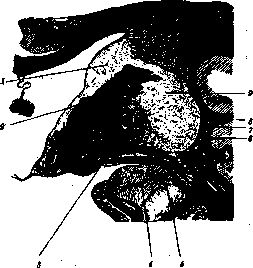
1-nucleus caudatus; 2-capsula interna; 3-globus pallidus; 4-nucleus amygdalae; 5-commissura alba anterior; 6-capsula externa; 7-claustrum; 8-capsula externa; 9-putamen. In humans it does not decrease or increase in its development, only its connections with other formations change. Corpus striatum develops from the inferior wall of the lateral vesicle (hemisphere), forming a thickening in the form of an elongated protrusion. Diagram of the main centers of the extrapyramidal system (according to S. and O. Vogi) ncF+mv+t (Tub. cm) v"'"':-V"aLP' /** ^ y к *Ж//*/ > S^^rJeruss./wrii {Patlid. и Corp. /-ujvs)
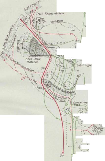
LEGEND: Connections of corpus striatum with pallidum: black - bundles from corpus striatum to pallidum; red (thick lines) - pyramidal cells (Py); red (thin lines) - tractus frontopontinus et cerebellaris; red dotted - fibers going from central gyri to thalamus opticus and to substantia nigra. Nucleus ruber Nucleus ruber Substantia nigra. (comm. post) Nucleus interstitialis. Brachium conjunctivum. (Tr.tectospinalis) Cerebellum Dentatum Cerebellum ad pontem ir-4jVm7.B«hf. V--' \.'"' '^clrCKl. Fasc. rubro-spinalis (Monakow) and from hypothalamus-
Black and to pallidum: bundles of Forel - H, //:); // - large cells of corpus striatum; III - fibers from corpus striatum and pallidum; IV - fibers from pallidum to thalamus opticus; V - fiber from pallidum to tuber cinereum; VI - fibers from pallidum to corpus striatum of the other side; VII - fibers from pallidum to nucleus ruber; VIII - fibers from pallidum to 'bundle of Forel' to nucleus interstitialis; IX - fibers from pallidus to nucleus interstitialis pedunculi; X - fibers from pallidum to nucleus subthalamicus Luys; XI - fibers from pallidum to substantia nigra; XII - fibers from substantia nigra to pedunculus cerebri; XIII - fibers from pallidum to nucleus campi Foreli; XIV - nucleus medialis anterior thalami optici; XV - nucleus medialis ventralis thalami optici; XVI - nucleus ventralis thalami optici; XVII - nucleus lateralis thalami optici; XVIII - nucleus medialis thalami optici; t - tuber cinereum. 3,
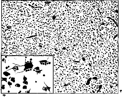
Figure 1. Putamen. Two types of cells - large and small (at weak and strong magnifications).
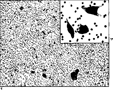
Figure 2. Globus pallidus. One type of cells - large (at weak and strong magnifications).
along the floor of the lateral ventricle (see Brain). Initially, as is evident from comparative-anatomical study, it represents a single continuous mass of gray matter, which by branches of the internal capsule is divided into two nuclei: nucleus caudatus and nucleus lenticularis. Regarding the ontogenetic development of the pallidum, it is not yet fully established from which parts it develops; more hypotheses have been expressed for its development from the intermediate brain (diencephalon). Nucleus amygdalae, on the basis of its anatomical connections and comparative anatomy data, is referred to as corpus striatum; on the other hand, the lack of isolation of nucl. amygdalae from the cortex of the uncus and histogenetic data lead other researchers to classify it as part of the archipallium cortex. Claustrum is considered as a separated part of the insula cortex. The microscopic structure of corp. striatum brings it closer to the cortex; according to data from Vogt, in it one can find hints of the layered structure of the cortex. In nucl. caudatus, under the ependyma, there is a zone poor in myelin fibers; the outer part of this zone is also poor in glial cells, while the inner part is richer; further there is a layer of tangential fibers containing a small number of ganglion cells, and then follow the layers of ganglion cells. Corpus striatum has two types of ganglion cells: a large number of small ones of various shapes, and scattered among them in a small number of large ones (see table II, fig. 1). The small cells are bubble-like, have delicate granulation and a large nucleus with a nucleolus. The large cells are triangular, polygonal, their nucleus is often eccentric, in the protoplasm there are chromatin granules and a large amount of yellow lipoid pigment, which collects near the nucleus; these cells are normally surrounded by satellites, giving a picture similar to neuronophagy. The small cells have a short axon cylinder, which branches without leaving the corpus striati; they belong to the second type of Golgi cells. The large cells belong to the first type, have a long axon cylinder, branching outside the corpus striati. On this basis, Vogt established the conductivity mechanism in corpus striatum; striopetal fibers end around small cells, closely connected with each other and with large cells, from which striofugal fibers already begin. In small cells, fibrils are not differentiated, in large ones they are distributed fascicularly. There are few myelin fibers in corpus striatum, most of them arise in the striatum itself and serve to connect with the pallidum; between the bundles of myelin fibers there is a dense network of non-myelinated fibers. A rich glial reticulum surrounds the nerve cells and nerve fibers. In the pallidum there is only one type of cell, very large, of various shapes - pyramidal, spindle-shaped, multipolar with long dendrites (see table II, fig. 2). In the protoplasm there are many chromophilic clumps; the surface of the cells is covered with loop-shaped terminal bodies - endings of non-myelinated fibers surrounding the cells and myelin fibers; there are many more myelin fibers than gray matter; this explains the white appearance of the nucleus (globus pallidus - pale nucleus). Chemically, the pallidum also differs from corpus striatum; it contains fat, iron, calcium deposits, which are either very scarce or completely absent in corpus striatum. There is a very rich connection between the basal ganglia and thalamus opticus (see table I); from it they receive all their afferent pathways; part of the fibers from thalamus opt. goes directly to the pallidum, and part - through corpus striatum; through the commissura Meynert, fibers from thal. opt. go to the opposite nucl. lenticularis; almost all fibers from corpus striatum end in the pallidum. Efferent fibers of the basal ganglia depart from glob. pallidus and are directed: 1) to formations playing a role in the motor system of extrapyramidal pathways - to nucl. ruber, to the nucleus of Darksevich, to the nucleus of the quadrigemina, to the inferior olive, from which the peripheral motor pathways begin - rubrospinalis, tectospinalis, posterior longitudinalis; through these same formations, the basal ganglia also have connections with the cerebellar and vestibular systems; 2) to formations connected with the autonomic system - regio subthalamic, substantia nigra, corp. Luysi, tuber cinereum. The direct connection of the basal ganglia with the cortex is denied by most authors, and this connection is carried out through the thalami opt. Claustrum forms a polymorphic layer of the Reilii insula cortex. In nucl. amygdalae, the main nucleus and the lateral nucleus are distinguished, which merges with the cortex of the g. hippocampi. Detailed studies on the connection with claustrum and nucl. amygdalae have not yet been conducted; it is only known that claustrum sends fibers to the insula cortex, and nucl. amygdalae, in addition to the cortex of g. hippocampi, connects with gl. pallidus. The basal ganglia have blood supply, mainly from art. cerebri media - art. lenticulares (aa. lenticulo-optica and lenticulo-striata); art. cerebri anterior also participates in their blood supply. The basal ganglia are very rich in capillaries, especially corpus str., in which their distribution resembles the distribution in the cortex; in vascular diseases of the brain, corpus str. is especially a favorite site for softening foci. The histological picture shows that the pallidum has a rather primitive structure - one type of cell; a large number of fibers carrying irritation from the outside enter it; in its cells, switching of these irritations in the centrifugal direction occurs, and from here impulses, after several switches, reach the periphery. The function of the pallidum is subordinate to the striatum, which is a very complex structure formation, analogous to the cortex. The reflex activity of the pallidum is moderated by corpus str., which thus plays the role of a regulator for the extrapyramidal motor function. The physiological role of the basal ganglia has been clarified only recently, thanks to anatomical-clinical studies; experimental data on this question are almost absent. The basal ganglia, due to their close connection with thal. opt., react to every impulse reaching thal. opt.; this reaction is motor (for which numerous connections with motor subcortical organs speak), and since at the same time there is no direct connection with the cortex, one can think that in the motor sphere the basal ganglia play the role of a central organ of automatic movements; all complex synergies that make up each motor act are carried out through the mediation of these same nodes; under their control is the setting, fixation of the system of the entire peripheral apparatus for dynamics or kinetics of any movement, their participation achieves the smoothness of movements. Especially they participate in animals in combined movements manifested in running, climbing; in humans, the innervation mechanism formed in the basal ganglia is not abolished, but only subordinated to the cortex, which acts on it through the mediation of thal. opt. Therefore, diseases of the basal ganglia and the descending pathways from them do not lead to paralysis, but to significant motor disorders of another nature - immobility, absence of facial expression, muscular rigidity, various hyperkineses. Some authors assume that the basal ganglia have different centers for different parts of the body. The connection that the basal ganglia have with the region of the subthalamic region, substantia nigra, corp. Luysi, tuber cinereum, parasympathetic and sympathetic systems in the medulla oblongata and spinal cord, indicates that the basal ganglia, mainly glob. pallidus, are a regulator of all autonomic functions (see Autonomic Nervous System). The basal ganglia system plays a role in affective life. (For a more precise acquaintance with the pathophysiology of the basal ganglia, see Hyperkinesia, Extrapyramidal system, Amiostatic symptom complex, Chorea, Athetosis, Paralysis agitans, Amiotaxia.)
Related articles
Mentioned in
- Ansa
- Atony
- Basal
- Brain (551 General diagnostics of brain diseases)
- Brain (cerebrum, a comprehensive term for the entire)
- Claustrum
- Commissural Fibers
- Constantin von Economo
- Corpus Luysi
- Extrapyramidal System
- Fever (a complex of phenomena in the organism,)
- Hyperkinesis
- Hypobulic Mechanisms
- Internal Capsule
- Mobile Spasm
- Paralysis Agitans
- Pilomotor Fibers
- Pseudobulbar Paralysis
- Sieve State
- Subcortical Functions
Cite this page
“Basal Ganglia.” Soviet Medical Encyclopedia. English translation of Bolshaya Meditsinskaya Entsiklopediya, 1st ed. (Moscow, 1928–1936), ed. N. A. Semashko. https://sovietmedicalencyclopedia.pages.dev/article/basal-ganglia/