Paralysis Agitans
Historical document, translated for reference. It reflects medical knowledge of the 1920s–30s and is not medical advice.
Summary
Paralysis agitans is a chronic progressive brain disease characterized by tremors, muscle rigidity, and movement difficulties. It typically begins in middle age and affects men more frequently than women.
Encyclopedia article (1928–1936)
PARALYSIS AGITANS, paralysis agitans (syn. disease of Parkinson after the author, who first described this disease in 1817 and gave it the name "shaking palsy"), a chronic progressive disease of the brain. Charcot considered P. a. a neurosis, since the technique of that time did not reveal anatomical changes in autopsy cases. At present they are found in the basal ganglia (see below), why paralysis agitans belongs to the group of diseases of the extrapyramidal nervous system. P. a. most often begins in the pre-senile age (from 50-60 years), more rarely in the senile age. Cases of so-called juvenile P. a. at present are questioned as to their belonging to epidemic encephalitis (see Parkinsonism), or to the disease of Wilson, or to a special hereditary form, not identical with the present paralysis agitans. Among men P. a. is almost twice as common as among women. P. a. is more common in the central part of Europe than in the southern part. Among Negroes P. a. occurs exceptionally rarely. Some authors note that sometimes the onset of the disease is preceded by mental, especially prolonged trauma, much more rarely physical, in which the psychological factor must also be taken into account. In cases of physical trauma the disease itself at first seems to be localized on the injured limb. Guillain described cases of Parkinson's disease after contusions in war. P. a. usually begins insidiously, with slight tremor in one limb, more often the hand (especially the right) than the foot, or even in a small segment of it, for example in one finger. At first the tremor is intermittent. It appears only during excitement, fatigue of the patient or when moving this limb. With time the tremor becomes persistent and passes to the homolateral lower limb according to the hemiplegic type. Later (the period is counted in many months and years) the tremor also affects the other hand, and in severe cases it spreads to all four limbs. The muscles of the trunk, shoulder and pelvic girdle do not participate in the tremor. The lower jaw, lips and tongue are involved in the tremor far from rarely (together or separately). More rarely the tremor spreads according to the crossed type, i.e. from the hand to the opposite leg. The tremor is rhythmic, of medium frequency; it is observed at rest and somewhat decreases during active movements. During sleep it disappears. Following the tremor, and sometimes before it, constant muscular tension develops, which does not necessarily exactly follow the tremor in territory. Usually this tension appears earlier in the muscles of the neck, face, back, and then asynchronously passes to the muscles of the limbs. Against the background of these two main symptoms, a number of fine motor changes develop, which are part of the hypokinetic-hypertonic syndrome (see Parkinsonism). The external picture of a patient with paralysis agitans is extremely characteristic. The face becomes frozen, immobile, mask-like, which is due to tension of the facial muscles, absence of normal play of muscular musculature and rarity of blinking. Mimic movements, with difficulty called forth, have a tendency to freeze. These disorders of facial play are called dysmimia. The skin of the face is usually thickened. Sometimes there is tremor of the lips, chin. The head and trunk are bent forward, so that sometimes the chin touches the sternum, the back is curved backward in an arch. The forearms are slightly bent at the elbow joints, the hands are somewhat extended, the fingers are bent at all phalanges or only at the base and extended in the middle and nail. This is the most common type of P. a., called by Charcot the flexion type. The extension type is less common, in which the head and spine are thrown backward and straightened like a stick (Fig. 1). The position and configuration of the fingers of the hands and to a lesser degree of the feet change sharply. On the hands the fingers are sometimes bent so strongly that they dig into the palm with the nails and cannot be passively extended. The thumb is usually adducted and opposed close to the index finger, creating the so-called "writer's hand" position (Fig. 2). Sometimes at this time it makes a series of small rotational movements around the pulp of the second finger—the pill-rolling symptom. In later stages of the disease the configuration of the fingers changes noticeably, they become unevenly curved and become very similar to the fingers in deforming arthritis (Fig. 3). On the foot the fingers in P. a. do not undergo such regular changes as on the hand. Here they are unevenly bent or extended. The gait of patients changes noticeably. The first steps are especially difficult. Before starting to walk, the patient seems to overcome some internal obstacle, his first steps are usually small—the so-called demarche a petits pas. Then, when the patient gets going, on the contrary, he shows a tendency to move forward quickly, sometimes almost to running. This disorder of gait is called propulsion (propulsio). It becomes especially clear if the patient is slightly pushed from behind. Retropulsion (retropulsio) is less common, consisting in a tendency to walk quickly backward, if the first step is made in this direction. And this disorder of gait is easier to detect if the patient is slightly pushed in the chest or abdomen. (For the pathophysiology of these disorders—see Parkinsonism.) All active movements are performed in P. a. extremely slowly, without the lightness and grace inherent in a healthy person. The small number of movements, their impoverishment, is striking. Brissaud says that parkinsonians "are stingy with movements". Automatic synergies (e.g. swinging of the arms when walking) are absent. The strength of the muscles is usually preserved for a long time, only comparatively late it begins to suffer damage, and under

active movements are performed with little force, while resistance to passive movements is quite sufficient. The excitability of the muscles is normal or shows in some cases myotonic, in others myasthenic (Dekhterev) reaction. Atrophy of the muscles does not occur. Tendon reflexes are usually unchanged or slightly increased, pyramidal signs
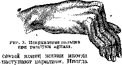
Fig. 1. Extension type paralysis agitans. Fig. 2. Positions of fingers in paralysis agitans (left—"writer's hand"). (Fig. 3). On the foot the fingers in P. a. do not undergo such regular changes as on the hand. Here they are unevenly bent or extended. The gait of patients changes noticeably. The first steps are especially difficult. Before starting to walk, the patient seems to overcome some internal obstacle, his first steps are usually small—the so-called demarche a petits pas. Then, when the patient gets going, on the contrary, he shows a tendency to move forward quickly, sometimes almost to running. This disorder of gait is called propulsion (propulsio). It becomes especially clear if the patient is slightly pushed from behind. Retropulsion (retropulsio) is less common, consisting in a tendency to walk quickly backward, if the first step is made in this direction. And this disorder of gait is easier to detect if the patient is slightly pushed in the chest or abdomen. (For the pathophysiology of these disorders—see Parkinsonism.) All active movements are performed in P. a. extremely slowly, without the lightness and grace inherent in a healthy person. The small number of movements, their impoverishment, is striking. Brissaud says that parkinsonians "are stingy with movements". Automatic synergies (e.g. swinging of the arms when walking) are absent. The strength of the muscles is usually preserved for a long time, only comparatively late it begins to suffer damage, and under active movements are performed with little force, while resistance to passive movements is quite sufficient. The excitability of the muscles is normal or shows in some cases myotonic, in others myasthenic (Dekhterev) reaction. Atrophy of the muscles does not occur. Tendon reflexes are usually unchanged or slightly increased, pyramidal signs
are absent. When testing muscle tone through passive movements, a sharp tension of the musculature is noted, sometimes approaching cadaveric rigidity. The tone during passive movements is either constantly elevated to the same sharp degree or increases in steps, forming the cogwheel symptom (signe de roue-dentee). Among sensory disturbances, pulling pains in the joints and muscles are sometimes noted at the beginning or during the course of the disease. All types of sensitivity are usually preserved in uncomplicated cases. Occasionally, hypesthesia in the form of patches has been observed in the trembling extremities. Changes in the autonomic nervous system are significantly more pronounced. Profuse sweating is common, and sometimes increased salivation occurs. The skin, especially on the face, becomes coarse. Attacks of a feeling of heat are common, which sometimes localize in the hypochondriac region, in the abdomen, and sometimes affect the entire body. A characteristic feature of paralysis agitans is its peculiar excitement. The patient cannot tolerate any one position for long, especially when in bed. He often turns from side to side, raises himself, turns again, and, as it is said, "cannot find his place." The speech of patients sometimes becomes poorly modulated and monotonous even at the beginning of the disease. The voice is mostly quiet. Writing, if possible at all, bears the imprint of tremor and is characterized by small letters (micrographia). On the side of the mental sphere, a depressive state is usually noted, in some individual cases reaching the point of suicide. In other cases, mental disorders in the form of hallucinatory and paranoid states have been observed. In the picture just drawn of R. a., both basic symptoms are combined: tremor and muscle tension. There are cases when one of these components, namely tremor, is absent - the so-called tremorous paralysis without tremor (paralysis agitans sine agitation); the absence of muscle tension is incomparably rarer, and the entire symptom complex is reduced to tremor alone. The duration of the disease is usually counted in many years (5-15 years). Sometimes sufferers from R. a. exhibit longevity. Gunther described a family where R. a. was transmitted directly from grandfather to son to grandson in three generations among seven of its members. R. a. itself is rarely the direct cause of death. The latter usually occurs in paralysis agitans due to senile debility, arteriosclerosis, or intercurrent diseases. The treatment of tremulous paralysis has only symptomatic significance. It consists in prescribing scopolamine, bromine, arsenic, warm baths, and massage. Special attention should be paid to maintaining the patient's mental calm. While the clinical picture of tremulous paralysis has been well studied since Charcot, who gave classic descriptions of the entire condition and its individual components, the details of its pathological anatomy constitute the achievement of the last two decades. The most difficult aspect in studying the pathological anatomy of R. a. is distinguishing the changes specific to it from the usual senile changes found in the brain of every person who has died from this disease. The distinguishing feature is not the very nature of the pathological process; on the contrary, both in R. a. and in old age it has the same basic type - a degenerative process, but with different localization. In old age, the cortex is mainly affected, while in tremulous paralysis, the subcortical ganglia are affected. However, the exact localization of the process within the ganglia themselves has not yet been fully established. Most authors see the cause of R. a. in disease of the globi pallidi (see Basal ganglia). On the basis of extensive pathological material, Lewy concluded that in the pallidum in R. a., a large part of the ganglion cells perish, and the remaining ones are in a state of deep destruction. However, according to Lewy, a number of other parts of the brain are also involved in the process: the cortex, cerebellum, nuclei of the medulla oblongata, and the gray matter of the third ventricle. According to his concept, R. a. is a diffuse presenile disease of the entire brain with emphasis on the pallidum. C. and O. Vogt see the cause of R. a. in a special lesion of the striopallidal system - in the so-called status desintegrationis. The latter process consists of widespread death of ganglion cells and fibers, thinning and rarefaction of perivascular glia, and the formation of small lacunae (status lacunaris). This lacunar degeneration, called etat precrible by French authors, without sharp boundaries, merges into etat crible, long ago noted in R. a. The French school (Brissaud, Souques, Tret'yakov) sees the center of gravity in R. a. in the lesion of the substantia nigra. Foix and Nicolesco take a reconciling position, accepting a lenticulo-subthalamic localization for R. a. The correlational connection between the anatomical process and the symptoms of R. a. is conceived in the following variants. Tret'yakov attributes all symptoms of Parkinson's disease to the lesion of the substantia nigra. This opinion stands somewhat apart. Most researchers, following C. and O. Vogt, recognize that the muscle tension in R. a. depends on the disease of the globi pallidi. The tremor, however, does not yet appear to be so strictly connected with the lesion of any one part of the subcortical system. Attempts have been made to show that within the striopallidal system itself, not all its cellular elements suffer to the same degree in R. a. Thus, Hunt, Jacob, and Lewy think that the large cells are more affected, while the small ones remain spared. Previous theories about the origin of R. a., namely the myogenous theory, which saw the cause of the disease in primary suffering of the musculature, and the endocrine theory, which connected R. a. with changes in the thyroid and parathyroid glands, now have only historical significance. In the light of recent work, R. a. appears as a degenerative, localized, and systemic disease of the basal ganglia.
Related articles
Mentioned in
Cite this page
“Paralysis Agitans.” Soviet Medical Encyclopedia. English translation of Bolshaya Meditsinskaya Entsiklopediya, 1st ed. (Moscow, 1928–1936), ed. N. A. Semashko. https://sovietmedicalencyclopedia.pages.dev/article/paralysis-agitans/