Brain (551 General diagnostics of brain diseases)
Historical document, translated for reference. It reflects medical knowledge of the 1920s–30s and is not medical advice.
Summary
This article from the 1928–1936 Soviet Great Medical Encyclopedia provides an overview of the anatomy and study methods of the human brain. It details the brain's structural divisions and the historical development of neuroanatomical research techniques.
Encyclopedia article (1928–1936)
551 General diagnostics of brain diseases...
595 The brain is the name given to that part of the central nervous system which is enclosed in the cranial cavity. The brain does not constitute a solid mass, but consists of a series of sections, which, although connected to each other, differ in their
own. There are considered to be six such sections of the brain: 1) the cerebrum, or hemispheres, 2) the diencephalon (diencephalon) - thalamus opticus, regio subthalamica, 3) the midbrain (mesencephalon) - pedunculus cerebri, corpora quadrigemina
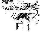
Figure 1. Brain of a human embryo at the end of the first month of intrauterine life: (right half): a-telencephalon; b-prosencephalon;
c-diencephalon; d-mesencephalon; e-isthmus rhombencephali; f-metencephalon;
g-rhombencephalon; h-myelencephalon; i-rhinencephalon: 1-pars mamillaris hypothalami; 2-pars optica hypothalami; 3-corpus striatum; 4-pallium; 5-thalamus; 6-corpora quadrigemina; 7-pedunculus cerebri; 8-brachium conjunctivum et velum medullare ant.; 9-cerebellum; 10-pars dorsalis; 11-sulcus limitans; 12-pars ventralis; 13-pons Varolii. (According to Spalteholz.)
4) isthmus (isthmus cerebri) - brachium conjunctivum, velum medullare anterius; 5) hindbrain (metencephalon) - pons Varolii, cerebellum; 6) medulla oblongata (myelencephalon) (see fig. 1-3).
The last of these sections (except for the cerebellum) are connected into the general concept of the brainstem (truncus cerebri); thus, by brain, one understands the cerebrum (hemispheres), the brainstem, and the cerebellum.*
* In this article, the anatomy, physiology, and pathology of only the cerebral hemispheres are set forth, while the description of all other parts of the brain is relegated to separate articles (see Basal ganglia, Pons Varolii, Thalamus opticus, Cerebellum, etc.).
Even at the beginning of the 19th century, the external appearance of the brain was sufficiently well studied, but the situation was different (due to the lack of correct and precise methods) regarding the internal structure of the brain and its physiology.
At that time, to study the brain, it was cut in all directions and dissected with the help of a scalpel and tweezers (Reil, Vicq d'Azyr); they worked with both fresh and fixed brains.
From 1830, they began to study the brain histologically, and in 1833, the nerve cell (Remak) and the nerve fiber (Ehrenberg) were discovered.
Then, new, more precise methods were gradually developed for studying both the fine structure of the brain and the connections that exist between the various sections of the brain, as well as its diverse functions and their localization in the cortex.
Methods of studying the brain. The methods of studying the brain that were and are applied are as follows.
1. The anatomical method, which includes the study of the normal brain using serial sections (thinly sliced and specially stained, allowing one to trace the course of fibers from beginning to end); it was introduced by Schilling, and later developed by Darkshevich, Bekhterev, Meynert, Gudden, Dejerine, and others.
This method of studying the brain is especially valuable in combination with the method of secondary degeneration, based on Waller's law—'every fiber separated from the cell degenerates'; the combination of these two methods for studying the brain allows one to very accurately trace the course of fibers, and thanks to this, the connections that exist between the various sections of the nervous system.
The method of secondary degeneration is used in pathological cases on the human brain and in experiments on the brains of animals. With its help, fiber systems were isolated (Türck, Charcot, Vulpian).
- The architectonic method, which also falls under the anatomical, is based on the different structure of individual sections of the cerebral cortex, which allowed it to be divided into a known number of areas (or fields), differing from each other in the number of layers and their structure (see Architectonics of the cerebral cortex).
The progenitor of this doctrine was Meynert, who was the first to suggest that if areas of the cortex are constructed differently, then they must have a different function, which confirms the general biological position: in the process of organ development, the differentiation of form corresponds to the differentiation of function.
2. The comparative-anatomical method, which consists of studying the gradual development of the brain in various classes of animals (in ascending order), and in the brain itself—the complication of the structure of the cortex by layering onto the oldest part of the cortex, the archipallium, a new one, the neopallium, which reaches enormous development in higher animals (Vogt, Rose, Jan).
In connection with the development of the brain, and in particular the cortex, the intellect of animals also develops, which manifests itself in their behavior and reaction to the environment.
Figure 2. Brain of a human embryo (left - 5 weeks, right - 3 months of intrauterine life): 1-hypophysis; 2-chiasma opticum; 3-hypothalamus; 4-corpus striatum; 5-pallium; 6-fiss. chorioidea; 7-thalamus; 8-sulcus hypothalamicus (Monroi); 9-epithalamus (corpus pineale); 10-metathalamus (corpora geniculata); 11-corpora quadrigemina; 12-pedunculus cerebri; 13-cerebellum; 14-fossa rhomboidea; 15-pons Varolii. (According to Spalteholz.)
The brain is connected into the general concept of the brainstem (truncus cerebri); thus, by brain, one understands the cerebrum (hemispheres), the brainstem, and the cerebellum.* Even at the beginning of the 19th century, the external appearance of the brain was sufficiently well studied, but the situation was different (due to the lack of correct and precise methods) regarding the internal structure of the brain and its physiology. In that time, to study the brain, it was cut in all directions and dissected with the help of a scalpel and tweezers (Reil, Vicq d'Azyr); they worked with both fresh and fixed brains. From 1830, they began to study the brain histologically, and in
* In this article, the anatomy, physiology, and pathology of only the cerebral hemispheres are set forth, while the description of all other parts of the brain is relegated to separate articles (see Basal ganglia, Pons Varolii, Thalamus opticus, Cerebellum, etc.). 1833, the nerve cell (Remak) and the nerve fiber (Ehrenberg) were discovered. Then, new, more
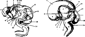
Figure 2. Brain of a human embryo (left - 5 weeks, right - 3 months of intrauterine life): 1-hypophysis; 2-chiasma opticum; 3-hypothalamus; 4-corpus striatum; 5-pallium; 6-fiss. chorioidea; 7-thalamus; 8-sulcus hypothalamicus (Monroi); 9-epithalamus (corpus pineale); 10-metathalamus (corpora geniculata); 11-corpora quadrigemina; 12-pedunculus cerebri; 13-cerebellum; 14-fossa rhomboidea; 15-pons Varolii. (According to Spalteholz.)
precise methods were gradually developed for studying both the fine structure of the brain and the connections that exist between the various sections of the brain, as well as its diverse functions and their localization in the cortex. Methods of studying the brain. The methods of studying the brain that were and are applied are as follows. 1. The anatomical method, which includes the study of the normal brain using serial sections (thinly sliced and specially stained, allowing one to trace the course of fibers from beginning to end); it was introduced by Schilling, and later developed by Darkshevich, Bekhterev, Meynert, Gudden, Dejerine, and others. Especially valuable is this method of studying the brain in combination with the method of secondary degeneration, based on Waller's law—'every fiber separated from the cell degenerates'; the combination of these two methods for studying the brain allows one to very accurately trace the course of fibers, and thanks to this, the connections that exist between the various sections of the nervous system. The method of secondary degeneration is used in pathological cases on the human brain and in experiments on the brains of animals. With its help, fiber systems were isolated (Türck, Charcot, Vulpian). - The architectonic method, which also falls under the anatomical, is based on the different structure of individual sections of the cerebral cortex, which allowed it to be divided into a known number of areas (or fields), differing from each other in the number of layers and their structure (see Architectonics of the cerebral cortex). The progenitor of this doctrine was Meynert, who was the first to suggest that if
areas of the cortex are constructed differently, then they must have a different function, which confirms the general biological position: in the process of organ development, the differentiation of form corresponds to the differentiation of function. 2. The comparative-anatomical method, which consists of studying the gradual development of the brain in various classes of animals (in ascending order), and in the brain itself—the complication of the structure of the cortex by layering onto the oldest part of the cortex, the archipallium, a new one, the neopallium, which reaches enormous development in higher animals (Vogt, Rose, Jan). In connection with the development of the brain, and in particular the cortex, the intellect of animals also develops, which manifests itself in their behavior and reaction to the environment.
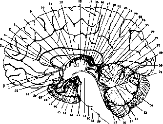
Figure 3. Sagittal section of the brainstem (right half): schematically; 1-commissura anterior (cerebri); 2-sulcus parolfactorius post.; 3-gyrus subcallosus (pedunculus corporis callosi); 4-genu corporis callosi; 5-rostrum corporis callosi; 6-lamina rostralis; 7-columna fornicis; 8-septum pellucidum; 9-foramen interventriculare; 10-corpus fornicis; 11-truncus corporis callosi; 12-massa intermedia; 13-thalamus; 14-tela chorioidea ventriculi III; 15-ventriculus III; 16-aditus ad aquaeductum cerebri; 17-commissura posterior (cerebri); 18-recessus pinealis; 19-commissura habenularum; 20-recessus suprapinealis; 21-corpus pineale; 22-splenium corporis callosi; 23-lamina quadrigemina; 24-aquaeductus Sylvii; 25-velum medullare anterius; 26-lobulus centralis; 27-monticulus (culmen); 28-ventriculus IV; 29-monticulus (declive); 30-laminae medullares; 31-fastigium; 32-tuber vermis; 33-pyramis (vermis); 34-corpus medullare; 35-uvula (vermis); 36-tela chorioidea ventriculi IV; 37-nodulus; 38-medulla spinalis; 39-foramen caecum; 40-pons Varolii; 41-decussatio brachii conjunctivi; 42-recessus posterior fossae interpeduncularis; 43-sulcus nervi oculomotorii; 44-nervus oculomotorius; 45-fossa interpeduncularis; 46-corpus mamillare; 47-recessus anterior fossae interpeduncularis; 48-hypophysis (lobus anterior et posterior); 49-chiasma opticum; 50-recessus infundibuli; 51-recessus optici; 52-lamina terminalis. 3. Embryological method, based on the study of the gradual development of various parts of the brain in human embryos of different ages. 4. Myelination method, or Flechsig's method, based on the fact that different parts of the brain do not myelinate simultaneously, but in a certain regular order; according to Flechsig, parts that myelinate simultaneously must also have the same degree of functional complexity; the higher the function, the later the fiber develops, i.e., myelinates. 5. Teratological method, based on the study of congenital deformities in humans resulting from either an arrest in development or insufficient, incorrect development of one or another part of the brain, which causes the absence of corresponding fibers and is clinically manifested as a defect in the corresponding function (Flechsig, Forel, Kaufmann). 6. Experimental-physiological method, which breaks down into the stimulation method and the ablation method. - The stimulation method consists of stimulating individual, strictly localized areas of the cerebral cortex with an electric (faradic) current of very low voltage, in order to avoid its spread to neighboring areas and to the white subcortical substance. - The ablation method consists of switching off the corresponding areas of the cortex and in a precise analysis of the symptoms observed during this. Switching off the cortex is achieved either by extirpation or by poisoning, the so-called toxicological method. Poisoning can be local or of the entire organism. A surgical-toxicological approach to the study of the physiology of the nervous system was proposed by G. I. Rossolimo in 1893 and consisted of the preliminary destruction of those parts of the cortex whose function was to be studied, and the subsequent subcutaneous injection of atropine or cocaine. In 1901, a method of local poisoning of the cortex (Beglioni) with various stimulating or paralyzing poisons was proposed. Fursikov and Voskresensky combined this method of local poisoning of the cortex with the method of conditioned reflexes, which makes it possible to study altered functions most objectively. The ablation method is the primary method allowing for the switching off of desired parts of the brain, and thus gives the researcher, unlike the clinical method, complete freedom in setting one task or another; the conclusions obtained from experiments on animals can be transferred (albeit with some reservations) to humans. This method is for the most part combined with the anatomical study of secondary degenerations resulting from the removal of one or another part of the brain. The disadvantages of this method lie in the development of postoperative shock, diaschisis (see), which obscures the clinical picture in the first days after the operation; in a later stage, the formation of a scar is possible, which acts as an irritating factor leading to convulsions, and sometimes to death. 7. Clinico-anatomical method boils down to careful observation of a given clinical disorder in a human and to the study after death of the changes in the brain that caused this disorder; it is necessary in each case to prove that it is precisely the given anatomical substrate that causes a specific disorder; precise microscopic examination, excluding the presence of other local diseases, acquires special importance in this regard. The clinical method was first proposed for solving specific questions of cerebral localization in France by Broca and in England by Jackson. The disadvantage of this method is that the choice of the clinical question is predetermined by the existing material. 8. The method of conditioned reflexes was introduced 25 years ago by I. P. Pavlov. It allows for the study of the nervous activity of higher animals in an exclusively objective way, i.e., to study it from a purely external, factual side, without resorting to considerations about what the animal might be experiencing and thinking under certain conditions by analogy with humans. When studying the function of the nervous system by this method, the researcher first becomes acquainted with the basic behavior of the animal, with the reaction it manifests in relation to its surroundings, i.e., with its innate (or so-called unconditioned) reflexes. As an objective indicator, the food reflex is taken (its secretory component, since with secretion a very precise measurement is possible; for observing the secretion, the duct of the salivary gland is brought to the outside). The test animal is strictly isolated from all external influences, and the experimenter, at his own discretion, introduces certain stimuli or various combinations of them, thanks to which he gains the possibility to study their influence on the animal one after another, to follow how new associations are formed in the animal, and how habits and reactions to the surroundings change. These new reflexes, which are formed in the animal under new conditions, Pavlov called conditioned (see Conditioned reflexes). When combining this method with the experimental-physiological method, observations were made on how these conditioned reflexes changed in connection with the removal of one or another part of the brain. The method of conditioned reflexes is very valuable, as it allows the researcher to remain an objective observer dealing exclusively only with external manifestations. As applied to humans, the reflexological method was developed by V. M. Bekhterev and is being continued at the present time by his school (see Reflexology). 9. The method of objective-psychological research is also used for studying the functions of the brain; this method is based on observations of human behavior, manifested in gestures, facial expressions, speech, and then in the registration and description of all the material obtained. When studying individual reactions, it is possible to combine the method of objective simple observation with tests or experiments, i.e., to artificially induce the phenomena that one wishes to study.
E. Kononova. Phylogenetic and ontogenetic development of the brain. Between the embryonic development of an individual and the biological evolution of the animal world, there exists a regular connection, which was first pointed out by Meckel (1811), and later pointed out by Fritz Müller (1864) and E. Haeckel (1866); the latter gave the following formulation of this connection: "The development of the embryo (ontogeny) is a compressed and abbreviated repetition of the development of the species (phylogeny)"; this generalization is known as the "fundamental biogenetic law." Although the relations between phylogeny and ontogeny are very complex, in principle the biogenetic law as a brilliant generalization allows one to understand the sequence observed in the embryonic development of the whole embryo and its individual organs, including the brain. The doctrine of the development of the brain assumes a sequential presentation of its phylogenetic evolution, then ontogenetic development, and finally the pathology of the latter. Phylogenetic evolution of the brain. In the brain of teleost fish (Teleostei), the significant development of the cerebellum is striking first of all
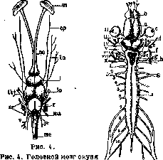
from the dorsal side: (- transmitted; io-middle; c-posterior]
Figure 5. pin; ta - medulla oblongata and spinal cord; sn - nasal capsule; no - olfactory nerve with olfactory lobes; or - orbital branch of the trigeminal nerve; tn - trochlear, thj - trigeminal, ac - auditory, and v - vagus nerves. Figure 5. Brain and spinal cord of a frog: a - hemisphere; o - olfactory lobe; c - eye; d - thalamencephalon; e - optic lobe; f - cerebellum; g - medulla oblongata; h - fourth ventricle; r - spinal cord; I - olfactory nerve; II - optic nerve; III - oculomotor nerve; IV - trochlear nerve; V - trigeminal nerve; VII - facial nerve; VIII - auditory nerve; IX - glossopharyngeal nerve; X - vagus nerve; 1-10 - spinal nerves (2 and 3, joining, form the brachial plexus; 7 and 8 - sciatic). (metencephalon) and midbrain (mesencephalon), with a weakly developed forebrain (prosencephalon, see figure 4). The cerebral hemispheres are absent; of the parts of the forebrain, only the olfactory lobe (lobus olfactorius) is developed, serving as the termination point for the olfactory nerve, and the striatum (corpus striatum) adjacent to it on the inside. Instead of hemispheres (cerebral mantle, pallium), the upper wall of the forebrain ventricle is formed by a thin epithelial membrane, on which, posteriorly, in the region of the diencephalon, the pineal gland (epiphysis) is located. In the diencephalon, the lateral wall of the ventricle is formed by the thalamus and hypothalamus; fibers from the olfactory lobe and from the striatum go to the latter; a series of bundles from the cerebellum, from the medulla oblongata, and from the spinal cord are directed into the thalamus. The floor of the ventricle passes here into the infundibulum, which plunges into the pituitary gland (hypophysis). The midbrain is a vesicle covered by a longitudinally curved, arch-like plate (tectum opticum), which has a layered structure. To this tectum, in addition to the fibers of the optic nerve after their decussation, go pathways from the cerebellum, from the spinal cord, and from the nuclei of the auditory and trigeminal nerves; from the tectum, bundles go to the nuclei of the thalamus and medulla oblongata and to the spinal cord. At the base of the midbrain is the interpeduncular body (corp. interpedunculare); the oculomotor nerve (III) and (more caudally) the trochlear nerve (IV) emerge here. The cerebellum is a thick-walled sac with a narrow lumen; in the wall of the cerebellum, one can distinguish an inner granular layer, then a layer of Purkinje cells, and an outer molecular layer; from the hindbrain, the restiform bodies (corp. restiformia) are directed into the cerebellum; from the cerebellum to the tegmentum of the midbrain go the superior cerebellar peduncles (brachia conjunctiva). The rhomboid fossa is not completely covered by the cerebellum. The medulla oblongata (myelencephalon) gives rise to the so-called lateral nerve and, in addition, to the vagus (X), glossopharyngeal (IX), auditory (VIII), facial (VII), abducens (VI), and trigeminal (V) nerves. From the experiments of Steiner, Bethe, and Loeb, it follows that motor disorders appear in fish only after the destruction of the midbrain and hindbrain; for impressions related to feeding, the thalamus and hypothalamus, according to Kappers, are a correlation area. Brain of amphibians (Amphibia; see figure 5). The cerebral hemispheres (telencephalon) already exist but are still poorly developed and diverge in their posterior part. A longitudinal fissure separates them only from above; on the ventral side, they are fused. Inside the hemispheres, there are lateral ventricles. Histological study of the mantle reveals the existence of the following layers in it: the inner layer, facing the ventricle, consists of cylindrical ependymal cells; outward is located a zone of pear-shaped cells; the outer layer is formed by a molecular zone. The diencephalon (diencephalon) is not covered by the cerebral hemispheres; on a section of the diencephalon, one can see that its dorsal part is formed by the thalamus, the ventral part by the hypothalamus; both parts are separated by the suprainfundibular commissure (commissura suprainfundibularis); the thalami form the lateral wall of the diencephalic ventricle, while the infundibulum is contained within the hypothalamus. The hemispheres of the midbrain diverge significantly in the posterior part; ventrally from the midbrain ventricle, as in fish, the nucleus of the oculomotor nerve (III) is located, and more caudally the nucleus of the trochlear nerve (IV), the roots of which decussate in the anterior medullary velum and emerge ventrally from the tectum opticum. In sections of the medulla oblongata, many nerve nuclei, especially of the lateral and vagus nerves, turn out to be significantly less developed than in Teleostei. In terms of cerebellar development, most amphibians are also inferior to fish, especially those that swim well. Decerebration experiments with an intact thalamus (Schrader, Munk, and others) show that the behavior of such an amphibian differs little from the behavior of a normal animal, although, according to Burne, a decerebrated frog is still not as agile in its movements as a normal one; if the diencephalon is also removed, significant and persistent movement disorders occur. Brain of reptiles (Reptilia). The cerebral hemispheres pass into the olfactory lobes without a noticeable boundary; posteriorly, they do not yet cover the optic tectum of the midbrain. The cerebellum is weakly developed (see figure 6). The cerebral hemispheres (telencephalon) are separated on the dorsal surface by a median fissure (fiss. mediana); in the depths, they are connected by means of the lamina terminalis, which contains fibers of the anterior commissure (commissura anterior). One part of the fibers of the anterior commissure serves to connect the striata, the other contains olfactory conductors. Inside each hemisphere is a ventricle, extending from the posterior pole to the olfactory lobe; by means of the interventricular foramen (Monro), the cerebral ventricles communicate with the ventricle of the diencephalon; a part of the striatum, the so-called epistriatum, protrudes into the ventricle. In the cerebral cortex, according to Krause, one can see: a layer of ependymal cells facing the ventricle cavity; outward from the ependyma is the medullary layer, consisting of myelinated centripetal fibers, then a deep molecular layer; outward from the latter lies a layer formed by pyramidal cells, and finally an outer molecular layer; parallel to the surface, at the very periphery, is a tangential layer consisting of thin, non-myelinated fibers. The diencephalon
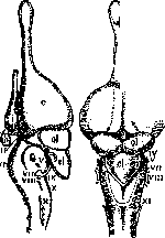
Figure 6. Brain of a young alligator: a - cerebral hemispheres; cl - cerebellum; e - pineal gland formations; h - pituitary gland; g - optic chiasm; ol - olfactory lobes; III-XI - cranial nerves. (After Gerin.)
is covered by the cerebral hemispheres; on the ventral side, the optic chiasm (chiasma) of the optic nerves adheres to it. The ventricle of the diencephalon, passing downward into the infundibulum, narrows in the caudal direction and passes into the cerebral aqueduct (aquaeductus). In the diencephalon, one can distinguish the epithalamus (epithalamus), the thalamus (thalamus), and the hypothalamus (hypothalamus); the epithalamus is formed by the habenular ganglion (gangl. habenulae), in which fibers decussate (taenia thalami); from the lateral side, fibers of the optic tract (tractus opticus) approach the thalamus, heading to the lateral geniculate body (corp. genic. laterale). In many reptiles, the so-called parietal organ ("parietal eye") is connected to the pineal gland by means of a long stalk. The tectum opticum s. lobus opticus forms two dome-shaped hemispheres, not completely separated by a median fissure. Below the cerebral aqueduct, downward and outward from the nucleus of the oculomotor nerve, is the red nucleus (nucleus ruber) of the tegmentum, to which fibers of the superior cerebellar peduncle approach and from which begins a bundle, according to Kappers, homologous to the Monakow's bundle of mammals; this bundle decussates. In the medulla oblongata, the abducens, facial, auditory, glossopharyngeal, vagus, accessory, and hypoglossal nerves originate. The cerebellum is a semicircular plate connected to the midbrain by means of the anterior medullary velum; the cerebellar cortex has a typical three-layered structure. With suprathalamic decerebration in reptiles, a state of stupor develops while retaining the ability to respond to external stimuli; with thalamic decerebration, the animal shows no motor activity. Brain of birds (Aves). The egg-shaped cerebral hemispheres border directly on the cerebellum posteriorly; the midbrain is covered by the cerebrum and cerebellum; the rhomboid fossa is for the most part covered by the cerebellum. The smooth (non-convoluted) cerebral hemispheres pass into the olfactory lobes (lobi olfactorii) at the anterior end; only on the lateral surface of the hemispheres
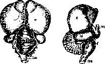
Figure 7. Brain of an eagle: c - cerebral hemispheres; c.l - cerebellum; f - flocculus; mo - medulla oblongata; ol - optic lobe; on - optic nerve. (After Herrick.)
One can note a shallow groove (fovea limbica), starting near the olfactory lobe and soon smoothing out. The hemispheres are separated by a narrow longitudinal fissure (fissura longitudinalis); only in their middle part are they connected by a short transverse bridge (commissura anterior). Inside the hemispheres are ventricles, which have the shape of a narrow slit (see figure 7). The mantle (pallium) reaches significant thickness only along the inner dorsal edge of the hemisphere; on the lateral surface it is thinned. In the cortex (cortex cerebri), according to Krause, one can distinguish three layers of cells: under the superficial molecular layer is a layer of stellate cells; medial to the latter is located a layer of pyramidal cells, and even more medially lies a layer of internal stellate cells, bordering on the cuboidal cells of the ventricle's ependyma. The axons of the cortical cells, having given off a significant number of collaterals, are directed toward the thalamus. Inside the cerebral hemispheres are the striate bodies, which with their medial and upper surface strongly protrude into the ventricles of the brain. The enormous development of the striate bodies with a comparatively poorly developed mantle constitutes a peculiarity of the bird brain. In the striate body, it is customary to distinguish four formations: hyperstriatum, mesostriatum, ectostriatum, and epistriatum, separated from each other by medullary laminae (laminae medullares); the most significant in size is the hyperstriatum; the epistriatum forms the caudal part of the striate body. The striate body is connected to the thalamus by means of the tractus striothalamicus; a part of the striatal fibers, having passed the thalamus, reaches the subthalamus and the midbrain. The diencephalon is covered by the cerebral hemispheres; between the latter and the cerebellum one can see the pineal gland (epiphysis); on the ventral side under the diencephalon is the optic chiasm, behind which lies the pituitary gland (hypophysis). The floor of the diencephalon forms the tuber cinereum (tub. cinereum). The lateral wall of the diencephalon is formed by the optic thalamus, in which one can distinguish several nuclei. Fibers from the medulla oblongata and spinal cord, from the cerebral cortex, from the striate body, from the midbrain, and from the subthalamus go to the nuclei of the thalamus. Between the thalamus and the optic tract is the lateral geniculate body (corpus geniculatum laterale), to which the fibers of the optic tract are directed; medial to the lateral one lies the medial geniculate body (corpus geniculatum mediale). Behind and above the thalamus is the habenular ganglion (ganglion habenulae), which is part of the epithalamus (epithalamus); the subthalamus (hypothalamus) in birds is weakly developed; behind the chiasm are the mammillary bodies (corpora mamillaria). The midbrain protrudes from under the cerebellum with its optic lobes. In a cross-section of the midbrain, one can see the layered structure of the lobi optici; superficially is located the visual zone, in which the fibers of the optic nerve terminate; medial to it is the intermediate zone, built of pyramidal-shaped cells, and finally the deep brain zone. Ventral to the cerebral aqueduct, in the thickness of the midbrain, are located the nuclei of the oculomotor (III) and trochlear (IV) nerves; even more ventrally one can see the medial longitudinal fasciculus and the red nucleus. In front of the lobi optici passes the posterior commissure (commissura posterior), the fibers of which arc around the cerebral aqueduct. The ventricle of the hindbrain (rhombencephalon) communicates with the ventricle of the diencephalon by means of the cerebral aqueduct; it narrows in its lower part and passes into the central canal. The cerebellum in birds is a powerful organ; as noted above, it covers a large part of the midbrain. In it, one can distinguish three lobes: the anterior, the middle, and


Figure 8. A—brain of a domestic dog; B—brain of a baboon; C—brain of a rabbit. (According to Gegenbaur.) I—hemispheres; III—midbrain (corpora quadrigemina); IV—cerebellum; V—medulla oblongata (medulla oblongata); r—spinal cord; lo—lobus olfactorius; vm—vermis. (According to Wiedersheim.)
posterior [lobes], separated by the anterior and posterior sulcus, and in addition the flocculus. The surface of the cerebellum is covered with a multitude of transverse sulci; on a cross-section, one can see the ventricle of the cerebellum, communicating with the ventricle of the hindbrain. In the thickness of the cerebellum, its center and nuclei are visible: the median, or roof [fastigial], and the lateral, or dentate; the cortex consists of an outer molecular layer, a layer of Purkinje cells, and a granular layer. Through the posterior cerebellar peduncles, fibers from the spinal cord are directed, and through the anterior peduncles, pathways of the cerebellum go to the midbrain and diencephalon. With suprathalamic decerebration in birds, the ability to react to gross stimuli is preserved, while the ability to independently find food is lost. When the cerebral hemispheres and the optic thalami are removed, the bird appears to be plunged into a deep sleep; it is incapable of feeding itself and requires artificial feeding.
Brain of mammals (Mammalia). Depending on whether the surface of the cerebral hemispheres is smooth, i.e., having no sulci, or, conversely, covered with sulci, mammals form two groups: the first includes those whose cerebrum has no sulci (these are the so-called lissencephalic animals), and the second includes those whose cerebrum is covered with sulci (gyrencephalic animals); the first group includes Monotremata (especially the platypus) and Marsupialia (marsupials); but even in some monkeys (Tarsiidae, Hapale), the surface of the cerebral hemispheres appears almost smooth. However, in all mammals, even lissencephalic ones, there exist such sulci as fiss. rhinalis and fiss. hyppocampi, which are conventionally called primary sulci (see Figure 8). The olfactory sulcus (fiss. rhinalis) separates from the hemispheres their olfactory part, the so-called rhinencephalon, consisting of the olfactory tract (tr. olfactorius) and the piriform lobe (lob. piriformis). In the depth of the longitudinal fissure separating both hemispheres is the great commissure of the brain, or the so-called corpus callosum (corp. callosum), in which one can distinguish the anterior, downward-curved end (or knee), the middle part, and the posterior, somewhat thickened end. Adjoining the ventral surface of the thickening of the corpus callosum is the fornix (fornix), in which horizontal and descending parts are distinguished. The space between the fornix and the knee of the corpus callosum is filled with the septum pellucidum (septum pellucidum); near the descending part of the fornix is the anterior commissure (commiss. anterior). On sections through the cerebral hemispheres, one can see the lateral ventricles, starting in the frontal lobe and arching around the caudate nucleus (nucl. caudat.) and the hippocampus (cornu Ammonis); in the ventricles are the choroid plexuses. On sections, it is also visible how the fibers of the internal capsule (capsula interna) separate the caudate nucleus from the lenticular nucleus (nucl. lenticularis); the lenticular nucleus breaks down into the inner segment, penetrated by a multitude of fibers—the globus pallidus (globus pallidus)—and the outer segment, the so-called putamen (putamen); outside the putamen is the external capsule (capsula externa), to which the claustrum (claustrum) adjoins. The internal capsule includes the corticothalamic, corticopontine, corticobulbar, and pyramidal (corticospinal) tracts, and finally, fibers of the optic and auditory radiations (radiatio optica, rad. auditiva). The striatum is connected to the thalamus, the subthalamic body (corpus Luysii), the red nucleus, and the substantia nigra (subst. nigra). In the cerebral cortex, one can see the following layers: the superficial molecular, consisting of small multipolar cells, below it three layers of pyramidal cells—small, medium, and large pyramids—and internal to them a layer of polymorphic cells—pyramidal, spindle-shaped, etc.; of the fibers, one can see a system of tangential fibers and deeper, under the large pyramids, the striae of Baillarger, outer and inner. The diencephalon is completely covered by the cerebral hemispheres, and its parts can be seen only at the base of the brain, where the mammillary bodies, the infundibulum, the pituitary gland (hypophysis), and the optic chiasm (chiasma) are distinguished. The ventricle of the diencephalon (third ventricle) is covered by the fornix; in the lower part, it passes into the infundibulum, and in the posterior part, into the cerebral aqueduct. The diencephalon includes the epithalamus, the thalamus, and the hypothalamus. The epithalamus includes the habenular nucleus (gangl. habenulae), from which the so-called retroflex bundle (fasc. retroflexus) goes to the interpeduncular nucleus (gangl. interpedunculare). In the optic thalamus, anterior, lateral, median, and ventral nuclei are distinguished, separated by medullary laminae (lam. medullares). Near the lateral nucleus is located the lateral geniculate body, which is the main place of termination of the optic fibers; from here begins the optic pathway, directed through the retrolenticular part of the internal capsule to the cortex of the occipital lobe of the hemisphere. On the border of the diencephalon and midbrain lies the medial geniculate body, serving as the main intermediate station of the auditory pathway, directed to the temporal lobe. The midbrain is covered by the cerebral hemispheres; upon removal of the latter, it is visible that the upper surface of the midbrain is formed by the corpora quadrigemina, with the anterior pair of colliculi being significantly larger than the posterior. The superior colliculus has a typical seven-layered structure; in it terminate fibers of the optic nerve, fibers from the spinal cord, and part of the fibers from the cerebral cortex; from it also originate bundles to the spinal cord, to the medulla oblongata, and to the cerebellum. In the central gray matter of the brain, at the level of the superior colliculus, under the cerebral aqueduct, are located the nuclei of the oculomotor nerve: lateral and median (unpaired). Ventral to the central gray matter, below the posterior longitudinal fasciculus, is visible Meynert's decussation, formed by fibers going from the superior colliculus to the spinal cord (tr. tectospinalis); still lower is Forel's decussation, formed by Monakow's bundles (tr. rubrospinalis), originating in the red nuclei. Ventral to this decussation is located the interpeduncular nucleus, into which the aforementioned Meynert's bundle (fasc. retroflexus) is directed. Dorsolateral to the red nucleus pass the fibers of the medial lemniscus, and ventral to it is the substantia nigra; in the base of the cerebral peduncle run the fibers of the pyramidal tract. The inferior colliculus is covered on the outside by a layer of fibers (stratum zonale); inside it contains a nucleus (nucl. corp. bigemini post.); to the inferior colliculus approach fibers of the lateral lemniscus, among which is located the nucleus of the lemniscus (nucl. lemn. lat.); internal to the lateral lemniscus are the fibers of the superior cerebellar peduncle, forming a decussation along the midline (the so-called Wernekinck's commissure). The cerebellum does not cover only the lower part of the IV ventricle. In the cerebellum, one can distinguish the middle part (vermis) and the hemispheres; by means of three pairs of peduncles, it is connected with different parts of the brain and spinal cord: the superior peduncles are directed to the midbrain, the inferior to the spinal cord, and the middle form the base of the pons Varolii, which in most mammals is still short and leaves the trapezoid body (a layer of auditory fibers in the depth of the pons) open. Inside the cerebellum, between the dentate and fastigial nuclei, is a gray mass, which bears the name nucleus interpositus; the cerebellar cortex consists of an outer molecular layer, Purkinje cells, and an inner granular layer. On sections of the pons and medulla oblongata, one can see the location of the nuclei of the exiting nerves (V-XII pairs). Numerous experiments with suprathalamic decerebration in mammals show that after it, the animal, such as a cat in the experiments of Dusser de Barenne (1919), despite the impairment of the functions of the sense organs, is capable of finding food, avoiding obstacles, and refusing beef soaked in a quinine solution. A dog in similar experiments by Rothmann (1923) gave energetic reactions to painful stimuli, barked, and made attempts to bite; all vegetative functions in it turned out to be preserved. From the experiments of Goltz on a dog, in which the cerebral hemispheres were removed together with the striatum, the Brain
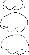
Figure 9. Brain of a gorilla (1), primitive man (2), and modern man (3). ...tive functions in it turned out to be preserved. From the experiments of Goltz on a dog, in which the cerebral hemispheres were removed together with the striatum, the thal-...
and partly the quadrigeminal plate, it turned out that it still retained the alternation of sleep and wakefulness, the ability to maintain balance, and the ability to run on three limbs when the fourth was injured; the dog refused food moistened with a quinine solution and turned its head toward light. Thus, the phylogenetic development of the brain is expressed in the complication of the structure, mainly of the forebrain and especially the cerebral mantle (pallium), which is therefore often called the "new brain" (neoencephalon)—in contrast to the olfactory lobe, the striatum, and other, older parts (paleoencephalon). Corresponding to the structural development of the forebrain, there is a shift of higher correlation-associative functions into it (Kappers, Monakow). Brain of primitive man (Homo primigenius). Regarding primitive man, it is known that he, like modern man (Homo recens), possessed speech and that his psychology was quite complex and differentiated. "Piltdown Man" is recognized as the most ancient type of man whose brain size is known; his antiquity is approximately 100,000–300,000 years. An endocranial cast of Piltdown Man shows that his brain was asymmetrical; it had developed those sections that are functionally connected in modern man with the speech function. "Neanderthal man," whose antiquity is 25,000–40,000 years, also possessed an asymmetrical brain; the left hemisphere of the cerebrum was distinguished by its significant size compared to the right hemisphere; the frontal lobes were of relatively insignificant size. "Cro-Magnon man" belongs to the modern type; his antiquity is determined at 25,000 years; the study of Cro-Magnon art shows that their mental abilities were close to ours; an endocranial cast shows that Cro-Magnons possessed a large-sized brain; the anterior lobes of the brain were well developed (see figure 9). Anthony, Klaatsch, Osborne, and others invariably recognize that the evolution of the brain consisted of progressive development—in height and width—of its anterior region, which, according to Anthony, in prehistoric times was the region of intellectual functions (see figure 10). Figure 10. Cross-section of the brain of a gorilla (1), primitive man (2), and modern man (3).
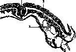
Ontogenetic development of the brain. The central nervous system of all vertebrate animals develops from the outer germ layer, the so-called ectoderm (see figure 11); in a two-week-old human embryo, the edges of the neural plate are noticeably delimited from the rest of the ectoderm. At this time, the primary cranial part of the tube is divided into three vesicles: anterior, middle, and posterior, of which the largest is the posterior. The tube itself appears bent almost to a right angle; the bend occurs in the region of the midbrain. In the third week, this bend changes from a right angle to an acute angle, so that the axes of the forebrain and hindbrain appear parallel; at this time, two more bends appear; besides the parietal one, the pontine and occipital bends are formed, which result from the uneven growth of various parts of the primary neural tube. In the mature brain, the parietal and partly the occipital bends remain from these; the pontine bend smooths out. Subsequently, the cerebral hemispheres and also the basal ganglia are formed from the anterior vesicle (prosencephalon); the quadrigeminal plate and cerebral peduncles develop from the middle vesicle (mesencephalon); the cerebellum, pons, and medulla oblongata develop from the posterior (rhombencephalon). By the end of the first month (see figure 1) of embryonic development, the hindbrain is better developed, not counting the spinal cord; in the forebrain, no differentiation is noticeable; the forebrain at this time represents a simple, thin-walled tube.
From the hindbrain, on the ventral side, emerge the root fibers of the hypoglossal, accessory, vagus, glossopharyngeal, vestibulocochlear, facial, and trigeminal nerves. The olive of the medulla oblongata, the pons Varolii, and the superior olive appear in the third month; with the appearance of the pons, the middle cerebellar peduncles are formed. At the end of the 2nd month, the cerebellum consists of two thickened plates not connected to each other; closer to the midline, a thickening is noticeable in both plates, corresponding to the primordium of the vermis. In the 3rd month (see figure 2), the cerebellar hemispheres represent two convex masses connected by means of the middle peduncles; the connection on the dorsal side occurs quite slowly along the midline; the vermis is covered with grooves at this time; at the end of the third month, the dentate and roof nuclei appear inside the cerebellar hemispheres, with the superior peduncles extending from the former and fibers of the vestibular nerve heading to the latter. In the fourth month, the formation of the external shape of the cerebellum is completed; the grooves
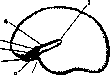
Figure 11. Part of a cross-section of a 20-day-old human embryo: 1-neural groove; 2-mesoderm; 3-primordium of vessels. (According to Tperet.)
appear on the cerebellar hemispheres in the fifth month; part of these grooves is a simple continuation of the grooves of the vermis. Regarding the midbrain, it is known that the red nucleus is well delimited in the third month; at this time, fibers of the superior cerebellar peduncle, coming from the dentate nucleus, approach it; at the same time, the fibers of the main and lateral lemniscus also become noticeable. Regarding the diencephalon, it has been established that the pituitary gland is already a solid organ in the third month. The pineal gland becomes noticeable in the second month. In the second month, according to Streeter, bundles of fibers from the mammillary body, the anterior column of the fornix, and the fasciculus retroflexus appear in the hypothalamus. By the end of the second month, the fibers of the lateral lemniscus reach the medial geniculate body, and the fibers of the optic tract reach the lateral geniculate body. Regarding the cerebrum (telencephalon), it is known that in a four-month-old embryo, the surface of the hemispheres is smooth; in sections through the forebrain, one can see a protruding ganglionic mass covered with ependyma (the primordium of the caudate nucleus) on the floor of the lateral ventricle; the lentiform nucleus forms in its depth. The subthalamic (Luysian) body is already found to be developed. In a five-month-old embryo, besides the already existing Sylvian fissure (see figure 12), the calcarine, parieto-occipital, callosomarginal, and central sulci appear. The
corpus callosum is formed later than all other parts of the brain; its development, according to Blumenau, is not finished even at the 5th month. In a six-month-old embryo, the presence of the main cortical sulci is noted; in the cortex, a six-layered type of structure can be distinguished; in the fifth layer of the precentral gyrus, giant pyramidal cells are encountered. In later months, the further development of sulci and gyri continues (see figures 13 and 14). Myelination of the fibers of the cerebral hemispheres occurs to a significant extent after birth; in a newborn
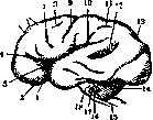
Figure 13. External surface of the brain of an embryo at the beginning of the 8th month of intrauterine life: 1 and 10- Sylvian fissure; 2- olfactory gyrus; 3- insula; 4- horizontal ramus of the Sylvian fissure; 5- frontal sulcus; 6- frontal gyrus; 7 and 8- precentral sulcus; 9- Rolandic fissure; 11- interparietal sulcus; 12- superior temporal sulcus; 13- parieto-occipital fissure; 14- cerebellum; 15- medulla oblongata; 16- inferior olive; 17- trigeminal nerve; 18- pons Varolii. (According to Dejerine.)
Many centripetal pathways of the hemispheres turn out to be myelinated, whereas the pyramidal tracts receive myelin later; commissural and association fibers become myelinated even later. The centrifugal pathways of the corpus striatum (gl. pallidi) develop earlier than the pyramidal tracts. Thus, it must be recognized that phylogenetically older formations of the brain develop first, and their development proceeds in a shorter period compared to such late products of biological evolution as the cerebral mantle, its commissure (corpus callosum), and the association fibers of the hemispheres. The structural relationships observed in the central nervous system in the process of its phylogenetic and ontogenetic development indicate the existence of a certain regularity; Ramón y Cajal sees in this a manifestation of chemotaxis, caused by the secretion of special substances and the reaction of cells to these substances; Kappers sees in the regularity of structural relationships the action of galvanotaxis and galvanotropism, which are connected with bio-electric phenomena observed in living protoplasm. Kappers was the first to notice that the nuclear masses of the medulla oblongata are formed depending on the position of the conductors through which the greatest number of excitations flow; he named this phenomenon neurobiotaxis. Bok established the dependence of the direction of the growing axis cylinder on the direction of the excitation current (e.g., the centrifugal direction of the axis cylinder). Ingvar, working with nerve tissue culture by the Harrison method, i.e., in vitro, proved the dependence of the direction of growth of the nerve cell and its processes on the lines of force of a galvanic field. A. Gurwitsch discovered special rays, which he named mitogenetic (i.e., producing mitoses), emitted by dividing cells; these rays are radiant energy with a short wavelength (in terms of wavelength, they lie between ultraviolet and X-rays); mitogenetic radiation also occurs from the neural plate (Anikin). Based on the studies of Ambronn, Held, and others, it must be recognized that the development of myelination to a significant extent depends on the excitations flowing along the conductor. The stages of brain morphogenesis are fundamentally the result of a very complex physico-chemical process, which depends on the external and internal conditions of the embryo's life. Westphal, Soltmann, Minkowski, and others proved the inexcitability of the cerebral cortex in the fetus and in the newborn in the first
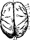
Figure 14. Upper surface of the fetal brain at the beginning of the 8th month of intrauterine life: 1 and 11 - fiss. sagittalis; 2, 3 and 4 - fiss. frontales; 5 and 6 - fiss. praecentralis; 7 - fissura Rolandi; 8 - fiss. Sylvii; 9 - fissura interparietalis; 10 - fissura parieto-occipitalis. (According to Dejerine.) days of extrauterine life; the movements of the fetus are reflex and completely independent of the cerebral cortex; these movements are identical to the movements of an anencephalic. During the first months, all movements in an infant are awkward and passive; at the same time, reflexes whose center is in the spinal cord, e.g., the knee jerk, are intensified; subsequently, the individual development of the infant proceeds in parallel with the maturation of the central nervous system. Using the methodology of Academician Pavlov, N. Krasnogorsky was able to verify that the mechanism of conditioned reflexes reaches its functional perfection only during the second year of life; V. Osipova established that associative (conditioned) reflexes are developed more quickly in children than in adults. Pathology of brain development. Anomalies of brain development in most cases are defects of its formation, or agenesis (aplasia), less often defects of growth, or hypoplasia; sometimes they consist of the development of some part, e.g., the cortex, in an unusual place, which is called heterotopia; often development occurs at an unusual time, i.e., heterochronia is observed; a significant displacement of the brain or only a part of it outward from the cranial cavity may also occur, which is called ectopia. The viability of the fetus after its birth in such cases is determined by the peculiarities of the brain structure in each individual case; it is known that anencephalics (see figure in the article Acrania), pseudencephalics, and exencephalics can live for several days; thus, the anencephalics described by Solovtsov lived from 3 to 15 days, the one described by Leonova lived only 17 hours and 20 minutes; but in the observation of Edinger and Fischer, an anencephalic lived for 4 years. Regarding the heredity of brain developmental defects, it can only be said that the chances for the transmission of grosser forms generally exist very little; it is only known that both the mother and the father can be predisposed to the reproduction of deformed offspring without themselves being burdened by the same anomalies; often among children of the same family, only one particular anomaly is reproduced; thus, in one woman who had 14 children, the 4th, 12th, and 14th child were anencephalics; in another who had 6 children, all children were born anencephalics. In the origin of brain developmental anomalies during the period of intrauterine life, embryonic hydrocephalus is of great importance; Virchow, Foerster, Ahlfeld, Morgagni, Solovtsov, and others saw in it the basis of various deformities of the central nervous system. The occurrence of anomalies is also facilitated by a strong curvature of the cerebrospinal axis of the embryo, due to which the transformation of the neural plate into the neural groove does not occur (Lebedev, Solovtsov); in some cases, the cause of the deformity was meningoencephalitis (Rabeau, Schob). Traumatic injuries to the brain during the act of birth are also significant. Meningeal hemorrhages are observed especially often; according to Neurath, they arise from ruptures of the intracranial venous sinuses, the longitudinal, transverse, and straight, or the veins flowing into them. As etiological factors, alcoholism (Forel, Babonneix, and others), congenital syphilis (Babonneix, Fournier, Gaucher, Solovtsov, V. Gilyarovsky, and others), and tuberculosis are also significant; "in fairness," says Lesbre, "alcoholism, tuberculosis, and syphilis are considered as powerful teratogenic factors"; in such cases, so-called blastophthoria (Forel), or a persistent change in germ cells, occurs.
A. Kapustin. Weight of the brain. Extensive works by very many researchers (such as Broca, Bischoff, Huschke, Weisbach, Meynert, Gilchenko, Chernyshev, and many others) are devoted to the study of brain weight. Based on numerous calculations, the average weight of the brain of an adult human is equal to 1,360 g. It can be said with certainty that the brain weight of all animals, with very rare exceptions, is less than the weight of the human brain; for example, the brain weight of a horse is 650 g, an ox is 500 g, and a gorilla is 400 g. If one compares the ratio of brain weight to total body weight of even such huge animals as the elephant, whose brain weight is 4,600 g, and the whale (brain weight = 2,800 g), one can be convinced that the human brain weighs relatively more than that of other mammals. The predominance of the development of the human brain over other animals is even more noticeable if one compares the figures indicating the ratio of brain weight to spinal cord weight: Turtle 1.0, rooster 5.0, pigeon 1.5, sheep 2.5, ox 7.0, cat 2.5, chimpanzee 10.0, horse 2.5, human 49.0. According to Meynert's research, the ratio of the weight of the cerebral mantle to other parts of the brain in humans is also significantly greater than in other animals. If the weight of the total mass of brain substance is taken as 1,000, the following values are obtained: In an adult human 780:220, monkey 708:292, elephant 630:370, horse 696:304, seal 673:227, bear 644:356, dog 728:272, cat 614:386. The ratio of the weight of individual lobes of the hemispheres to each other, according to Meynert, is as follows: Lobes of the hemispheres: Frontal, Parietal, Occipital. Human, Monkey, Bear, Dog. According to the same author, the weight of the frontal lobes of a human is 450 g, parietal 251 g, temporal and occipital 383 g, brain stem without cerebellum 148 g, cerebellum 148 g; the spinal cord weighs on average 30 g. The average weight of the brain of an adult man, according to Chernyshev, is 1,368 g, and of an adult woman 1,227 g (the brains belonged predominantly to natives of the central provinces of Russia). These figures were obtained based on the weighing of 1,979 brains, of which 1,310 were men and 669 were women. Race undoubtedly has an influence on brain weight. Based on numerous studies, the average weight of the brain is: Caucasian race 1,358 g, Chinese 1,332 g, inhabitants of the Sandwich Islands 1,302 g, Malays and Indians 1,266 g, Negroes 1,244 g, Australians 1,185 g. And among European peoples, a certain difference in brain weight is found. Thus, Bischoff, Huschke, Handmann, and Chernyshev, who operated with relatively large material, provide figures indicating that Germans have slightly less voluminous brains than Russians. The brain weight of the English and Japanese approaches the brain weight of Russians. The French, as well as Italians, have a slightly lighter brain than Russians. The influence of sex on brain weight is firmly established by science and confirmed by all authors without exception. According to Chernyshev's data, the female brain is 137 g, or 10.3%, smaller than the male. This reduction in the average weight of the woman's brain compared to the weight of the man's brain does not depend at all on the difference in body length. An explanation for this can be the following table, indicating the amount of brain substance per 1 cm of body length: Height Men | Women 130 cm... 8.0, 8.4, 9.9, 8.1, 7.7, 7.5. Body length undoubtedly also influences brain size. The taller a person is, the greater the absolute weight of the brain, and vice versa. This rule applies to both sexes. People with short body length possess a greater amount of brain substance per unit of their height than tall people. A numerous series of researchers asserts that age also undoubtedly influences brain weight. According to Handmann, the average brain weight of a newborn boy is 400 g, and of a girl 380 g. During the first year, brain growth is very rapid, and by the end of the year, the brain weight doubles; by the end of 4-5 years, it triples; then the increase in brain weight proceeds slowly. Based on Chernyshev's weighings, the male brain reaches its greatest development in terms of weight around the age of 25. From 25 to 50 years, the man's brain decreases very little in weight. Starting from the age of 50, the weight of the male brain gradually falls. The weight of the woman's brain reaches its maximum slightly before 20 years. After 20 years, a gradual fall in brain weight begins in women. In the period from 40 to 50 years, a new small increase in brain weight is observed in them. This or that influence on brain weight is also exerted by diseases causing the patient's death. Besides hyperemia and anemia observed in the brain during various diseases, there are many other conditions causing an increase or decrease in brain weight (for example, tumors, hydrocephalus, brain abscesses). Many chronic diseases exhausting the organism lead to a decrease in brain weight. In such a pathological form as progressive paralysis of the insane, in which there is always hyperemia in the brain and, alongside it, atrophic changes, a decrease in brain weight occurs. For a long time, attempts have been made to draw a parallel between the spiritual activity of a person and the weight of their brain. Regarding this, one of the most authoritative anthropologists, Broca, says the following: "It cannot occur to an enlightened person to measure the degree of mental development by weighing the brain." It must be assumed that, besides its weight, the brain's working capacity is influenced by its structural peculiarities, the chemistry of nerve elements, blood supply, etc. If there are known examples of outstanding people with a very large brain weight, such as Turgenev (2,012 g), Cuvier (1,861 g), Cromwell (2,000 g), then, on the other hand, many richly gifted individuals are known who had a brain weight below the norm. The heaviest brain of all described so far turned out to be that of an idiot (it weighed 2,850 g). Nevertheless, according to the investigations of Obersteiner, the limit below which brain weight cannot drop without being associated with a lowering of mental abilities (idiocy) can be established for the male brain at 1,000 g, and for the female brain at 900 g.
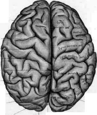
S. Chernyshev. Anatomy of the brain. Macroscopic and microscopic structure of the brain (morphology of the brain). The cerebrum (or cerebral mantle), pallium, is divided by a longitudinal fissure (fissura pallii s. fiss. longitudinalis, see separate table, fig. 1) into two hemispheres, right and left, connected in the middle part by means of the corpus callosum. Each hemisphere has three surfaces—outer, inner, and lower, two poles—anterior (or frontal) and posterior (or occipital), and three borders—upper, lower-outer, and lower-inner. The upper border is rounded and separates the outer surface from the inner; on this border, Pacchionian granulations are especially developed; throughout its entire length, it is in contact with the sinus falciformis. Three surfaces are clearly distinguishable at the anterior pole, whereas at the posterior pole, the boundaries between the lower surface and the inner one are blurred. The outer surface of the brain, convex in all directions, corresponds to the bones of the skull—frontal, parietal, occipital, and temporal, and therefore is divided into corresponding lobes (see figure 15). The inner surface of the brain, flat, corresponds to the inner surface of the other hemisphere, from which it is separated in the upper sections by the falx cerebri major of the dura mater. The lower surface...
The lower surface of the brain, which is complex in its relief, rests on the base of the skull, specifically on the anterior and middle cranial fossae. It is divided into the orbital part (corresponding to the orbital plate of the frontal bone) and the temporal part (corresponding to the middle cranial fossa). The lower surface of the frontal lobe is separated from the temporal lobe by the deep Sylvian fissure (fissura Sylvii). The brain is covered by three membranes: the dura mater (dura mater), the arachnoid (arachnoidea), and the pia mater (pia mater).
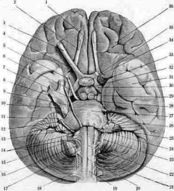
Fig. 1. Base of the brain (after Rauber). Fig. 2. Lateral surface of the brain (after Rauber): 1-olfactory bulb; 2-olfactory tract; 3-optic chiasm; 4-optic nerve; 5-oculomotor nerve; 6-trochlear nerve; 7-trigeminal nerve; 8-abducens nerve; 9-facial nerve; 10-vestibulocochlear nerve; 11-glossopharyngeal nerve; 12-vagus nerve; 13-accessory nerve; 14-hypoglossal nerve; 15-cerebral peduncle; 16-pons; 17-medulla oblongata; 18-pyramid; 19-cerebellar hemisphere; 20-flocculus; 21-cerebellum; 22-inferior olive; 23-temporal lobe; 24-frontal lobe; 25-occipital lobe. Base of the brain: 1-frontal lobe; 2-temporal lobe; 3-cerebellum; 4-medulla oblongata; 5-pons; 6-cerebral peduncle; 7-optic chiasm; 8-olfactory bulb.
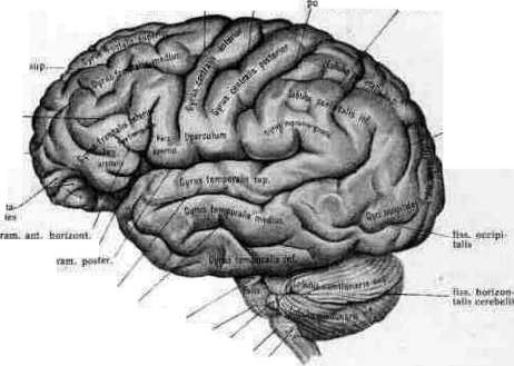
Fig. 3.
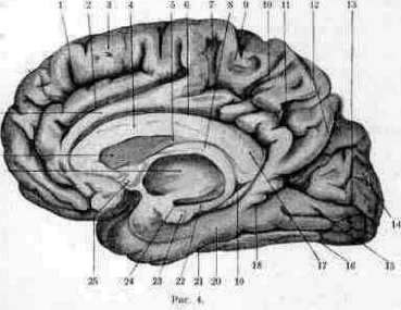
Fig. 4. Medial surface of the cerebral hemisphere (after Rauber): 1-corpus callosum; 2-fissura cinguli; 3-gyrus frontalis sup.; 4-gyrus cinguli; 5-fissura parieto-occipitalis; 6-lobus parietalis; 7-lobus occipitalis; 8-subparietal sulcus; 9-paracentral lobule; 10-sulcus subparietalis; 11-sulcus parieto-occipitalis; 12-cuneus; 13-fissura calcarina; 14-gyrus lingualis; 15-gyrus fusiformis; 16-gyrus hippocampi; 17-uncus; 18-thalamus; 19-commissura ant.; 20-fornix; 21-septum pellucidum; 22-ventriculus tertius; 23-thalamus; 24-colliculus superior; 25-colliculus inferior; 26-cerebellum; 27-medulla oblongata; 28-pons; 29-cerebral peduncle; 30-hypophysis.
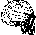
Figure 15. Relationship between the lobes and gyri of the brain and the bones and cranial sutures: 1-sulcus centralis; 2-ramus posterior fiss. Sylvii; 3-fiss. parieto-occipitalis. (After Rauber.)
[The surface] of the brain lies on the base of the skull; in the anterior sections it is in contact with the bone, and in the posterior sections—with one of the processes of the dura mater—the tentorium cerebelli. The inferior surface is divided by the Sylvian fossa and fissure (fossa Sylvii and fissura Sylvii) into two unequal parts: an anterior, short one—the frontal, and a posterior, longer one—the temporo-occipital. The entire surface of the brain is divided by numerous sulci into gyri, which form the lobes. The sulci are subdivided into primary, or main, secondary, and tertiary. The main sulci include those which appear quite early in the fetal brain, are constant in their position and shape, and are deep; for the most part, they serve as the boundary between the lobes. Secondary sulci are more or less constant and quite deep, dividing the lobes into gyri. Tertiary sulci are superficial, inconstant, individual, and divide the gyrus into parts. In their arrangement, the sulci do not appear regular and continuous; superficial anastomoses between the gyri and lobes interrupt them, dividing them into two or three segments. In turn, the gyri do not always run straight, but often curve around the sulci. The presence of sulci, superficial anastomoses between the gyri, and the tortuosity of the gyri themselves, all this gives the brain a very complex structural pattern and presents extensive individual variations. The main sulci include: fissura Sylvii, fissura Rolandi, fissura parieto-occipitalis, fissura calcarina, and fissura corporis callosi with fissura hippocampi. The external surface of the brain (see separate table, fig. 3). On the external surface are two main sulci: fiss. Sylvii and fiss. Rolandi. The Fissura Sylvii (Sylvian fissure) appears at the end of the second month of intrauterine life of the fetus in the form of an impression on the external surface of the brain; from here it develops in both directions. In an adult, it begins on the inferior surface of the brain with an expansion, which bears the name fossa Sylvii; cutting the inferior surface into two unequal parts, it curves around the lower edge of the hemisphere and passes to the external surface, where it gives off two branches: one upward—ramus verticalis (or ascendens), the other horizontally forward—ramus horizontalis anterior, then it turns backward and runs upward and backward under the name ramus horizontalis posterior; it ends among the gyri of the parietal lobe, sometimes dividing into branches. From the ramus horizontalis posterior, several small branches extend into the parietal lobe—incisurae parietales. The Fissura Rolandi (central sulcus) begins at the upper edge of the brain, two centimeters back from its midpoint; with its upper end, for the most part, it extends onto the internal surface of the brain; from the upper edge of the brain it stretches along the external surface downward and forward and ends without reaching the fiss. Sylvii. Its course is somewhat tortuous; in two places it forms bends, or knees. Its upper edge is located at a distance of approximately 111 mm from the frontal pole, the lower edge—at a distance of 71 mm; from the occipital pole, the upper edge is 49 mm away, the lower—89 mm. These two sulci form the boundaries of the following lobes: anterior to the fiss. Rolandi is the frontal lobe (lobus frontalis), posterior—the parietal (lobus parietalis), below the fiss. Sylvii is located the temporal lobe (lobus temporalis); posteriorly, the parietal lobe transitions into the occipital (lobus occipitalis); there is no constant boundary between these lobes, it has to be drawn artificially by dropping a perpendicular line from the upper edge of the fiss. parieto-occipitalis (which belongs properly to the internal surface of the brain) to a small but constant impression present on the lower edge of the brain—incisura praeoccipitalis; approximately in the direction of this line runs the sulcus occipitalis anterior Wernicke, which could serve as a boundary if it were constant. In some cases, the occipital lobe on the external surface is separated from the parietal by a deep sulcus, which, due to its similarity to that existing in monkeys, received the name of the monkey sulcus (Affenspalte). Each of the above-described lobes is divided by secondary and tertiary sulci into gyri. Frontal lobe. The gyri lying around the fiss. Rolandi and belonging to different lobes of the brain are distinguished into a special region—the region of the central gyri, which has important functional significance. Anterior to the central sulcus lies the gyrus centralis anterior (anterior central gyrus), posterior—the gyrus centralis posterior (posterior central gyrus). At the upper edge of the sulcus, on the internal surface of the brain, both gyri join, forming the lobulus paracentralis; at the lower edge, their junction is called the operculum. The gyrus centralis anterior is separated from the rest of the frontal lobe by means of the sulcus praecentralis, consisting for the most part of two segments (sulci praecentrales superior et inferior). Perpendicular to this sulcus are located two longitudinal sulci, the sulci frontales superior et inferior (superior and inferior frontal sulci), which divide the frontal lobe into three gyri—gyri frontales superior, medius, inferior (superior, middle, and inferior frontal gyri). The first is located between the upper edge of the brain and the sulcus frontalis superior, the second—between the fiss. frontalis superior et inferior, and the third—below this latter sulcus. The gyrus frontalis inferior is divided by the branches of the fiss. Sylvii into three parts: the pars opercularis, lying between the sulc. praecentralis and the ramus verticalis fiss. Sylvii; the pars triangularis, located between the ram. verticalis et horizontalis fiss. Sylvii; and the pars orbicularis, i.e., orbitalis, lying below the ram. horizontalis anterior. Sometimes an inconstant sulc. frontalis medius divides the gyrus frontalis medius into lower and upper sections. At the anterior pole, an inconstant sulcus—the sulc. fronto-marginalis Wernicke—separates the frontal gyri of the external surface from the gyri on the inferior surface. Parietal lobe. The gyrus centralis posterior, lying posterior to the fiss. Rolandi, is bounded posteriorly by the sulc. postcentralis; perpendicular to it runs the sulc. interparietalis, dividing the parietal lobe into two gyri—the gyrus parietalis superior and the gyr. parietalis inferior (superior and inferior parietal gyri). The gyr. parietalis inferior is in turn subdivided into two gyri: the gyr. supramarginalis (supramarginal gyrus) and the gyr. angularis (angular gyrus). The first is located around the terminal branching of the fiss. Sylvii, the second—around the end of the sulc. temporalis superior. Occipital lobe. On the external surface of the occipital lobe there are three sulci, which bound three gyri; the arrangement of the sulci in the occipital lobe is very inconstant, and therefore division into gyri is difficult. Besides the aforementioned sulc. occipitalis anterior Wernicke, which serves as the boundary between the occipital and parietal lobes, the following are distinguished: the sulc. occipitalis transversus Ecker (transverse occipital sulcus), serving as a kind of continuation of the sulc. interparietalis, and the sulc. occipitalis lateralis (lateral occipital sulcus). Between these sulci are the gyri occipitales superior, medius et inferior, or the first, second, and third occipital gyri. Temporal lobe. Three sulci—the sulci temporales superior, medius et inferior—bound three gyri on the external surface of the temporal lobe, which are named the same as the sulci, the gyri temporales superior, medius, inferior; the sulc. temporalis superior, the deepest and longest of these sulci, ends among the gyri of the parietal lobe; the sulc. temporalis inferior sometimes extends onto the inferior surface of the brain. The fiss. Sylvii is so deep that its lower wall can be considered as the superior surface of the temporal lobe. On the posterior half of this surface, one can see 1-2 small sulci running obliquely and dividing this surface into 2-3 small gyri of Heschl—gyri temporales transversi Heschl. In the depth of the fiss. Sylvii lies the fifth lobe of the external surface; this is the insula Reili, or the island (see figures 16 and 17). It can be seen by spreading apart the edges of the fiss. Sylvii. The island is surrounded by a sulcus—the sulcus circularis; the sulcus centralis insulae divides it into lobi insulae ant. et post. The anterior lobe is covered with short gyri—gyri insulae breves, the posterior represents one gyrus—gyrus insulae longus. Internal and inferior surfaces of the brain [see separate table (p. 503-504), fig. 4]. On the internal surface are the three remaining primary sulci of the brain. The fiss. parieto-occipitalis (parieto-occipital sulcus) belongs almost entirely to the internal surface; only a short upper segment extends onto the external surface. This sulcus is very deep; its upper end is located 4-5 cm anterior to the occipital pole, then it runs downward and forward and at an acute angle connects with the 4th main sulcus—the fissura calcarina; in the depth of the sulcus, anastomoses occur between adjacent gyri.
The calcarine fissure (fissura calcarina) runs from the occipital pole in a horizontal direction anteriorly and connects with the parieto-occipital fissure; their common trunk is directed downward and forward and terminates, extending onto the fornicate gyrus; the posterior end of the calcarine fissure often extends onto the outer surface, where it is known as the retrocalcarine fissure; sometimes the end divides dichotomously. The calcarine fissure is very deep, especially its middle part, which forms a projection on the wall of the posterior horn of the lateral ventricle, known as the calcar avis (ergot de Morand). In terms of the time of appearance in the embryo, it is one of the earliest. On the medial surface of the brain also lies an arcuate sulcus, which appears in the embryo somewhat later than the Sylvian fissure. It consists of two parts—upper and lower; the upper part, the fissure of the corpus callosum, separates the corpus callosum from the fornicate gyrus lying above it; the lower part is called the hippocampal fissure, or Ammon's sulcus; it separates the dentate fascia of the Ammon's horn from the hippocampal gyrus; the lower part of the sulcus is very deep and creates a protrusion along the lower wall of the inferior horn of the lateral ventricle—the pes hippocampi major (see Figure 18). Some authors also include among the main sulci the sulcus located above the fornicate gyrus, which also has an arcuate shape—the callosomarginal sulcus; the anterior part of this sulcus begins under the rostrum of the corpus callosum, goes forward, curves around the genu of the corpus callosum, then heads backward, turns upward at the level of the splenium of the corpus callosum, and ends at the upper edge of the brain; from its upper surface, several branches extend upward into the superior frontal gyrus; the largest of them, the paracentral sulcus, limits the paracentral lobule anteriorly; before the callosomarginal sulcus changes its direction from horizontal to vertical, a branch departs from this sulcus, which seems to serve as its continuation backward; this is the subparietal sulcus. The collateral fissure, or occipitotemporal sulcus, is already located on the inferior surface of the brain; it is very deep and forms a protrusion in the inferior horn of the lateral ventricle—the collateral eminence of Meckel. On the medial surface of the brain, one encounters the same lobes as on the outer surface. The central part of the medial surface is occupied by the corpus callosum. In its anterior section, it presents a bend—the genu (knee) of the corpus callosum, which gradually thins and forms the rostrum (beak); the middle part bears the name truncus of the corpus callosum, and the posterior thickening is called the splenium (splenium). Under the corpus callosum lies the fornix; in the anterior sections between them is located the septum pellucidum. The anterior columns of the fornix, the columns of the fornix, bending, go into the thickness of the brain.

Figure 16. Insula Reili dextra, exposed from the surrounding parts: 1-interparietal sulcus; 2-central sulcus (Rolandi); 3-circular sulcus (Reili); 4-short gyri of the insula; 5-long gyrus of the insula. (After Spalteholz.)
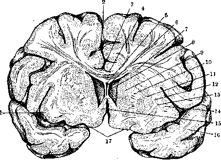
Figure 17. Frontal section of the brain through the anterior horns of the lateral ventricles: 1-insula; 2-longitudinal cerebral fissure; 3-medial longitudinal stria; 4-lateral longitudinal stria; 5-cavum septi pellucidi; 6-lamina of the septum pellucidum (left); 7-subcallosal stratum; 8-anterior horn of the lateral ventricle; 9-head of the caudate nucleus; 10-internal capsule; 11-lentiform nucleus; 12-external capsule; 13-claustrum; 14-extreme capsule; 15-parolfactory area (Broca); 16-temporal lobe (left); 17-olfactory tract. (After Rauber.)
of the hemisphere; the posterior columns, the crura of the fornix, continue into the fimbriae of the Ammon's horns. Under the fornix lies the thalamus opticus, forming the lateral wall of the third ventricle; between the thalamus opticus and the fornix is the foramen of Monro—an opening connecting the anterior horn of the lateral ventricle with the third ventricle.
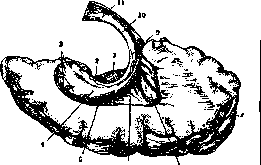
Figure 18. Inferior and posterior horns of the lateral ventricle of the brain: 1-hippocampal gyrus; 2-taenia fimbriae; 3-digitation of the hippocampus; 4-hippocampus; 5-collateral eminence; 6-collateral trigone; 7-calcar avis; 8-occipital pole; 9-fimbria of the hippocampus; 10-crus of the fornix; 11-body of the fornix. (After Rauber.)
with the paracentral gyrus, which has moved here from the outer surface of the brain. Between it and the paracentral lobule is the paracentral sulcus; posteriorly, the paracentral lobule is limited by the end of the callosomarginal sulcus, behind which is placed the precuneus—a continuation from the outer to the medial surface of the superior parietal gyrus; posteriorly from it passes the parieto-occipital fissure, and downward—the subparietal sulcus. Between the parieto-occipital fissure and the calcarine fissure lies the cuneus (wedge), belonging to the occipital lobe. Then, without sharp boundaries, the inferior surface of the brain begins. Under the calcarine fissure lies the lingual gyrus, or lingula, which narrows anteriorly and passes into the hippocampal gyrus. Under the lingual gyrus and the hippocampal gyrus passes the collateral fissure, or temporo-occipital sulcus, which separates them from the fusiform gyrus, or occipitotemporal gyrus; this gyrus goes from the occipital pole to the temporal one, fully justifying its name. Outward from it passes the already mentioned inferior temporal sulcus. On the inferior surface of the brain, anterior to the Sylvian fossa, is the frontal lobe; all three of its gyri pass here from the outer surface, which are called here the orbital gyri; the sulci passing between them bear the name orbital sulci, while the sulcus running parallel to the inner edge is the olfactory sulcus; the gyrus which is separated from the frontal ones by this sulcus is called the straight gyrus (gyrus rectus). Both the sulci and the gyri present great variations not only in the brains of different individuals but even in different hemispheres of the same brain. Such individual difference depends on the development of secondary, and mainly tertiary, sulci, on the direction, length, and depth of the permanent sulci, as well as on the arrangement of the gyri, their width, and anastomoses with neighboring gyri. Besides individual peculiarities, differences in the architecture of the brain are noted in connection with age, sex, race, mental development, and the shape of the skull. The development of sulci, which began in intrauterine life, does not end with birth but continues for some time. The width of the gyri also changes; the widest gyri are encountered in mature age; in very young and elderly people, wide gyri are usually not present. The direction of some sulci also changes in connection with the development of neighboring gyri. Racial differences boil down to the fact that in black people there is greater simplicity in structure than in white races. Some difference is also observed in the structure of the brain in women and men; this difference manifests itself in the 7th–8th month of intrauterine life. Although there is no specificity in the distribution of sulci and gyri in both sexes, some authors still find that the brain belonging to the female sex is distinguished by less deviation from the general type, greater simplicity, and regularity. It has also been noted that the brain of gifted or outstanding people is distinguished by a more complex surface architecture. Investigations undertaken in this direction have led to the conclusion that on the basis of the limbic lobe (marginal or sickle-shaped lobe); this lobe consists of two gyri: the fornicate gyrus (arcuate gyrus) and the hippocampal gyrus (hook-shaped gyrus). The fornicate gyrus, separated from the corpus callosum by means of the fissure of the corpus callosum, begins under the rostrum of the corpus callosum and ends at the splenium, gradually narrowing and passing into the hippocampal gyrus, which properly belongs to the inferior surface of the brain. The hippocampal gyrus, limited from the inside by the hippocampal fissure, ends anteriorly with a thickening—the hook (uncus), separated from the temporal pole by a small groove, the incisura temporalis of Schwalbe. The limbic lobe as a whole forms an almost closed circle, open only anteriorly and inferiorly. To the marginal lobe also belong the rudimentary marginal gyri—the dentate fascia of the Ammon's horn and the gray covering (indusium griseum) of the corpus callosum; one constitutes the continuation of the other. The dentate fascia lies on the hippocampal gyrus under the fimbria; at the anterior end, it attaches to the uncus of the hippocampal gyrus, passing into the so-called band of Giacomini; posteriorly, the dentate fascia forms a thickening, the fasciola cinerea, and, curving around the splenium, passes into the indusium griseum—a thin layer of gray matter covering the upper surface of the corpus callosum and in places forming thickenings, the longitudinal striae (such are on both sides of the seam of the corpus callosum—the internal or medial striae or the striae of Lancisi; on the lateral parts of the corpus callosum—the lateral striae, or external or tectal striae (see Fig. 19). The fornicate gyrus is limited from above by means of the callosomarginal sulcus and its branch—the subparietal sulcus. The entire remaining anterior part of the medial surface, up to the paracentral lobule, is occupied by the superior frontal gyrus.
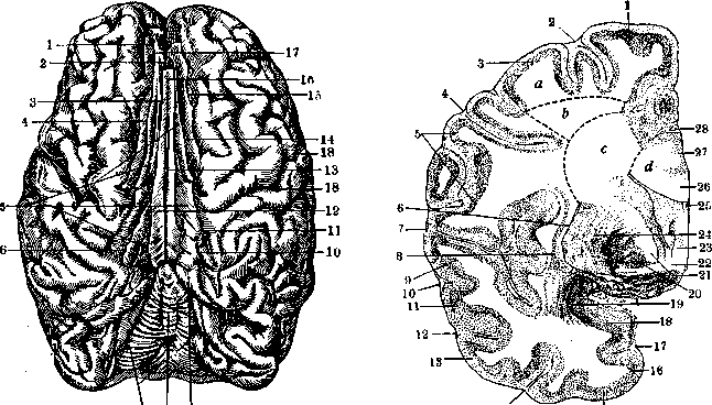
7 8 9
Figure 20. Figure 19. Superior surface of the brain (fissura sagittalis is spread apart, and the corpus callosum is visible in the depth): 1 and 5-gyr. front. sup.; 2, 6 and 9-gyr. fornicatus; 3-fiss. corporis callosi; 4, 11, 14 and 17-fiss. calloso-marginalis; 6 and 10-fiss. subparietalis; 7-cerebellar hemisphere; 8-cerebellar vermis; 12-corpus callosum; 13 and 16-striae Lancisii; 15-fiss. Rolandi. (After Dejerine.) Figure 20. White matter of the brain: a-white matter of the gyri; b-centrum semiovale; c-corona radiata; d-central white matter; 1-gyr. frontalis sup.; 2-fiss. front. sup.; 3-gyr. front. med.; 4-fiss. front. inf.; 5-gyr. centr. ant.; 6-capsula ext.; 7-fiss. Sylvii; 8-claustrum; 9-gyr. tempor. sup.; 10-fiss. tempor. sup.; 11-gyr. temp. med.; 12-fiss. temp. med.; 13-gyr. temp. inf.; 14-fiss. temp. inf.; 15-gyr. fusiformis; 16-fiss. fusif.; 17-fiss. collateralis; 18-uncus; 19-nucl. amygdalae; 20-gl. pallidus; 21-capsula int.; 22-commissura ant.; 23-fornix; 24-putamen; 25-sept. pellucidum; 26-corpus callosum; 27-cingulum; 28-fiss. calloso-margin. One cannot judge the greater or lesser giftedness of a given person based on the morphological structure of the brain surface alone, but the presence of a certain combination of signs, rarely encountered in the human mass, is necessary, such as: large brain weight, richness of third-category sulci with clearly expressed primary sulci, particular development of sulci in the region of the frontal and parietal association fields, and a predominance in the development of the left hemisphere. In addition to this, recently, in connection with the development of the doctrine of architectonics (see Architectonics of the cerebral cortex), great importance is attached in solving this question to the thickness of the cortex, even its individual layers, as well as the quantity, size of ganglion cells, etc. Besides all the conditions described above, the shape of the skull can also influence the configuration of the surface of the brain. With a dolichocephalic skull, a longitudinal arrangement of the gyri is observed; with a brachycephalic one, there is a tendency toward their transverse arrangement; in the sagittal gyri, numerous lateral bridges develop, and the position of the oblique gyri approaches the transverse. A fairly common view is that the immediate cause of the formation of gyri is the skull, which creates obstacles to growth in length and width for the walls of the cerebral hemispheres, due to which folds begin to form on the walls; other authors attribute to the skull only some influence both on the general shape of the brain and on the laying down of sulci and gyri. In turn, the growth of the skull is also under the influence of the brain (thus, in hydrocephalus, it often reaches enormous sizes). Thus, the question remains unresolved for now, but it can be said with certainty that a known connection exists between the skull and the brain. Besides this view on the formation of sulci and gyri, other views existed. The cause was sought in surface tensions caused by the uneven speed of brain growth in different directions; some saw the cause in the growth of white matter, others in the growth of gray matter. The immediate result of the development of gyri is a significant increase in the surface of the cerebral cortex without changing its constant thickness. The surface of the cortex increases gradually from lower animals to higher ones in connection with the development of intellect; in humans, the increase in surface falls mainly on the frontal lobe (41%), then on the temporal and parietal (21%), and on the occipital (17%). Both hemispheres are connected by commissures—a series of formations located on the basis cerebri [base of the brain; see separate table (cols. 503-504), fig. 2]. By basis cerebri is understood the entire middle part of the lower surface of the brain, including the brainstem part of the brain. Studying the base of the brain from front to back, one can note the following formations. In the sulc. olfactorius lies the tractus olfactorius, the anterior, expanded part of which forms the bulbus olfactorius; posteriorly, it expands into the trigonum olfactorium. Along the inner edge of the latter passes the stria olfactoria medialis, going to the gyrus fornicatus; along the outer edge—the stria olfactoria lateralis, heading to the gyrus hippocampi through the substantia perforata anterior, i.e., a gray plate covered with small holes through which vessels from the base of the brain penetrate to the subcortical formations. Along the midline is the chiasma nervorum opticorum, formed by the incomplete decussation of the optic nerves; from the chiasma outward and backward go the optic tracts—tractus optici, which then hide under the temporal lobes. Further back is located the gray tuber—tuber cinereum, extending into the infundibulum—a funnel, at the end of which is the lower brain appendage, the hypophysis; behind the gray tuber are the corpora mamillaria. The listed formations belong to the forebrain (hemispheres + diencephalon), then follows the base of the midbrain. From each hemisphere, two thick bundles head toward each other—pedunculi cerebri (cerebral peduncles), through which pass fibers connecting the hemispheres with the brainstem, cerebellum, and spinal cord; converging at the midline, both peduncles limit a triangular space covered by a perforated plate—substantia perforata posterior, through which vessels penetrate into the thickness of the midbrain and diencephalon. Behind the cerebral peduncles is located the pons Varolii (pons), which is sharply delimited from them, as well as from the underlying medulla oblongata, whereas on the sides it passes without sharp boundaries into the pedunculi cerebelli ad pontem, which go into the cerebellum. Along the midline of the pons Varolii, an indentation is observed—the sulcus basilaris—for the a. basilaris passing here. The medulla oblongata, which separates sharply from the pons Varolii, passes downward without sharp boundaries into the spinal cord; the boundary between them is considered to be the beginning of the exit of the spinal roots, as well as the decussation of the pyramids (decussatio pyramidum). Along the anterior surface of the medulla oblongata passes the fissura mediana anterior, on each side of which are visible convex formations—the pyramids of the medulla oblongata, delimited externally by the sulc. lateralis anterior; further outward are located oval formations (olivae inferiores), limited by the sulc. retro-olivaris s. lateralis posterior and the corpora restiformia, partially covered by the cerebellum. From the brainstem part of the brain emerge 10 pairs of cranial nerves. Between the cerebral peduncles emerges the III pair (n. oculomotorius); above the pons Varolii—the IV pair (n. trochlearis); this is the only nerve that emerges from the upper surface of the trunk, having formed a decussation, and then it bends around the outer surface of the midbrain and appears on the base of the brain. From the middle sections of the pons Varolii, on the border between it and the pedunculus cerebelli ad pontem, emerges the V pair (n. trigeminus) with two roots—an inner thin, motor one, and an outer thick, sensory one. Between the pons Varolii and the medulla oblongata emerge on each side four nerves: closer to the midline above the pyramid—the n. abducens (VI pair); in the cerebellopontine angle—the n. facialis (VII pair), n. intermedius, and n. acusticus (VIII pair); in the sulc. retro-olivaris appear the n. glosso-pharyngeus (IX pair), n. vagus (X pair), and n. accessorius Willisii (XI pair); to this last one approaches its spinal root, with which it connects. In the sulc. lateralis anterior emerges the n. hypoglossus (XII pair). Each of the above-described nerves emerges from the brain with several roots, which then connect into one trunk. On the base of the brain, on the sides of the pons Varolii and the medulla oblongata, the cerebellar hemispheres are visible; its middle part, the vermis, is located behind the brainstem part of the brain and is not visible from the base. The composition of the cerebral hemispheres includes the cortex, white subcortical matter, lateral ventricles, and basal ganglia. The entire surface of the brain is covered with a layer of gray matter called the cortex, cortex; the cortex also descends into all sulci, no matter how small they are, and lines them. The total surface of the cortex is on average equal to 220,000 sq. mm, of which one-third (72,000) falls on the free surface, and 2 thirds (148,000) on the sulci. The thickness of the cortex is on average 2.5-3 mm, it fluctuates in different sections of the hemisphere, reaching its greatest thickness in the region of the anterior central gyrus and the lobulus paracentralis. The basic type of cortex is the six-layered one.
Although the boundaries between the layers are not sharp, one can still see, more or less clearly, six layers when stained using the Nissl method (Brodmann). The 1st layer, the most superficial, lamina zonalis, the molecular layer with a thickness of 0.25 mm, is poor in nerve cells. The 2nd layer, lamina granularis externa, the external granular layer, is a layer of small pyramids, of the same width as the first; the cells that make up its composition are of very small size (base diameter 7 μ). The 3rd layer, lamina pyramidalis, is a layer of pyramids of medium and large size (up to 40 μ in diameter), significantly thicker; the medium pyramidal cells lie more superficially, forming the sublamina medio-pyramidalis, and the large ones lie deeper, forming the sublamina magno-pyramidalis. The 4th layer, lamina granularis interna, the internal granular layer, is of very small size, inconstant, and may be absent. The 5th layer, lamina ganglionaris (or gigantopyramidalis), is the layer of ganglionic cells or deep pyramids; it contains very large pyramidal cells. The 6th layer, lamina multiformis, the polymorphic layer, in its upper section (6a) contains cells of triangular shape, and deeper (6d) spindle-shaped ones; this layer borders on the white matter (see Vol. II, art. 347, Fig. 1). The presence of myelinated fibers in the cortex can be established by staining using the Weigert method. These fibers are of endogenous and exogenous origin; the degree of their development and their arrangement are not entirely identical in different regions, but in their main features, their grouping is as follows (Vogt): the 1st layer, lamina tangentialis, tangential fibers are located in the molecular layer, parallel to the surface of the cortex; this layer is subdivided into sublamina superficialis (1a), sublamina intermedia (1b), with two parts (pars externa and pars interna), and sublamina profunda (1c); the 2nd layer, lamina disfibrosa, is poor in myelinated fibers; the 3rd layer, lamina suprastriata, is located in the layer of pyramidal cells and is divided into three parts: sublamina superficialis (3a), or the stria of Kaes-Bechterew, s. intermedia (3b), and str. profunda (3c); the 4th layer, stria Baillargeri externa, or the external transverse stripe, corresponds to the deep layer of grains; the 5th layer is located in the region of large pyramids and is subdivided into two parts: lamina interstriata (5a) and stria Baillargeri interna (5b), or the internal transverse stripe; the 6th layer, lamina infrastriata, or the layer of intracortical association fibers of Meynert, is subdivided into lamina substriata (6a), lamina limitans interna (6b), and album gyrorum (6c). Between these belts of cohesive fibers lies a loose plexus, which is divided (Edinger) into the plexus superradialis, lying above the external stripe of Baillarger, and the plexus interradialis, situated deeper between the bundles of radial fibers. In the cortex, in the deep layers, oblique fibers are also encountered. Relationships between cellular layers and layers of myelin fibers: Layers of fibers according to Vogt: 1 - lamina tangentialis, 1a - subl. superfic., 1b - sublamina intermedia, a - pars externa, b - pars interna, 1c - subl. profunda, 2 - lamina disfibrosa, 3 - lamina suprastriata, 3a - sublamina superficialis, 6 - lamina limitans, 6 - lamina multiformis.
....
J
interna, stria Baillarger of the gyri. Perpendicular to the previous fibers, fibrae radiatae (radial fibers) run from the white matter into the cerebral cortex in radiating bundles. Local differences in the cerebral cortex consist of its width, the size and number of cells, the absence of any layer, the division of a layer into two parts, etc. The study of the morphological structure of the cortex, based on the study of local features of its structural elements, is distinguished under the name of architectonics (see Architectonics of the cerebral cortex). The cortex of the phylogenetically old olfactory region of the hemisphere and the Ammon's horn (archipallium) presents particularly many deviations from the described typical structure. In the rest of the mantle (neopallium), the structure of the following cortical regions should be noted: 1) the region of the anterior central gyrus and lobulus paracentralis are characterized by the presence of giant cells (see Betz cells) in the 5th layer, area giganto-pyramidalis; they are especially numerous in the lobulus paracentralis and in the upper parts of the anterior central gyrus; 2) the region of the fissura calcarina is distinguished by the presence of a macroscopically distinct Baillarger's stripe (Vicq d'Azyr's stripe, Gennari's stripe) in the 4th layer of the cortex; 3) the region of the frontal lobe is characterized by a reduction or almost complete absence of the 4th layer, lamina granularis interna (agranular type); 4) the region of the insula Reili presents the peculiarity that a part of the polymorphic layer of the cortex (lamina multiformis) is separated from the rest of the cortex by a layer of white matter (capsula externa) and is described under the name claustrum (according to other authors, the claustrum belongs to the basal ganglia). The cells of the cerebral cortex differ from each other in shape, size, and the character of the axial cylinders extending from them. The characteristic shape for cerebral cortex cells is pyramidal, but triangular cells and spindle-shaped cells are found in the polymorphic layer, and round ones in the granular layers. The number of ganglion cells in the cortex is approximately 14 billion, of which six billion are small. The size of the cells fluctuates within very wide limits: from 8-9 µ (cells of the granular layer) to 150 µ (Betz cells). By the character of the axial cylinders, they are divided into cells with a long axon extending beyond the limits of the cortex, and cells with a short axon, branching and ending within the gray matter of the cortex. Pyramidal cells and cells of the polymorphic layer belong to the former. Their axon usually begins from the middle of the base of the cell, goes down as part of the radial fibers, and takes part in the formation of the white matter of the brain. On its way in the cortex, especially in the 4th-5th layers, the axon gives off branches which are directed horizontally and take part in the formation of Baillarger's stripes; upon entering the white matter, it also gives off collaterals. Human pyramidal cells are distinguished by greater branching of all processes, and a larger number of axon and dendrite collaterals. The apex of the pyramidal cell is turned toward the surface of the cortex; it passes into a thick dendrite, which branches in the molecular layer; short and thin dendrites extend from the entire circumference of the cell in all directions and also branch abundantly. The axons of the cells of the polymorphic layer also go, for the most part, into the white matter. Cells with a short axon are found in abundance in all layers of the cerebral cortex; some of them are characteristic of the cortex: Cajal cells in the molecular layer with a horizontally running axon, and Martinotti cells with an ascending axon; the latter are especially numerous in the lamina granularis externa and in other deep layers of the cortex; the majority of ascending axons go to the molecular layer, in which they divide into horizontal branches, forming tangential fibers. Besides Cajal and Martinotti cells, there are several varieties of cells with short axons: 1. Small cells resembling glia, but with thin processes that branch strongly and form a dense plexus; such cells are found in all parts of the cortex. 2. Small cells with ascending short and highly branching processes penetrating into the first layer. 3. Small spindle-shaped cells provided with whole bundles of dendrites at both poles; such cells are especially numerous in the layer of large pyramids. 4. In the third and fourth layers, there are special cells whose axons, after passing a short distance up or down, divide into horizontal branches, which, with their terminal branches and collaterals, form a kind of basket around the pyramidal cells. The abundance of cells with short axons constitutes a feature of the human cortex; in the layer of pyramids, they are as numerous as the pyramids themselves. Fibers of exogenous origin enter the cortex from the white matter in an oblique direction and begin to divide in the deep layers, forming a wide and dense plexus in the middle layers of the cortex, especially in the layer of deep grains; only a small number of them reach the molecular layer; cells with a short axon (Martinotti) serve as intermediaries between them and the cells of the molecular layer. Between the nerve cells and fibers of the cortex are located the cells and fibers of the neuroglia (see). The close relationship of centripetal fibers with the cells of the upper and middle layers, especially with the granular one, shows that these layers are the main receptive layers. Pyramidal cells and cells of the polymorphic layer, with their fibers, take part in the formation of the white matter of the hemispheres, the fibers of which have different purposes, as will be seen below. The role of the remaining elements apparently reduces to the propagation of nervous excitation. Inside each hemisphere is the cavity of the lateral ventricle (ventriculus lateralis), lined with ependyma and containing a small amount of cerebrospinal fluid. The lateral ventricle communicates with all other ventricles of the brain (see Ventriculi cerebri). Each lobe of the brain corresponds to one of the parts of the lateral ventricle: the frontal lobe—the anterior horn of the lateral ventricle; the central gyri and the parietal lobe—the cella media; the occipital lobe—the posterior horn; and the temporal lobe—the inferior horn (for the figure, see Ventriculi cerebri, Vol. IV, p. 711). Between the ependyma of the lateral ventricle and the cortex covering the gyri, the entire thickness of the hemispheres consists of white matter and the basal ganglia embedded in it (see Vol. II, p. 639). The white matter of the hemispheres is formed by nerve fibers, ascending and descending, running in all directions and intertwining with each other. The fibers connect the cortex of the hemispheres with the cortex of neighboring gyri, with the gyri of the other hemisphere, and with the underlying formations. Topographically, 4 parts are distinguished in the white matter, not sharply delimited from each other (see Figure 20, p. 513-514): 1) white matter in the gyri themselves between the sulci; 2) centrum semiovale—the region of white matter extending from the sulci to the internal parts of the hemisphere; 3) corona radiata (radiating crown), a very large region extending from the previous zone to the basal ganglia and formed by fibers radiating outward, but intersected by horizontal and sagittal fibers; 4) central white matter, which includes fibers of the corpus callosum, capsulae internae, and long association fibers. (For the figure, see Vol. II, p. 413.) According to their physiological significance, the fibers of the white matter of the brain are divided into 1) association, 2) commissural, and 3) projection fibers. Association (or associative) fibers connect the cortex of the gyri of the same hemisphere. They originate from the lateral parts of the gyri and lie in the mass of white matter, the more superficially the shorter their extent. Association fibers are divided into 1) short, connecting two adjacent gyri, 2) medium, connecting gyri of the same lobe, and finally 3) long, connecting more distant gyri (see Association fibers). According to the data of many recent authors, the fibers included in the composition of long association bundles are not themselves long and do not run from beginning to end in the bundle, but gradually exit from it throughout their entire length and are replaced by new ones. Commissural (or connecting) fibers connect the gyri of opposite hemispheres, but not only symmetrical, identical places of both hemispheres, but also gyri belonging to different lobes. The commissural systems of the brain include the corpus callosum, commissura anterior, and commissura fornicis, or psalterium (see Fornix). The vast majority of commissural fibers belong to the corpus callosum, which connects all parts of the hemispheres, with the exception of the olfactory and the anterior part of the temporal lobes, which are connected by means of the commissura anterior. The continuation of the fibers of the corpus callosum in the hemispheres is called radiatio corporis callosi, and in the frontal and occipital lobes—forceps. The commissura fornicis lies between the posterior crura of the fornix and connects both Ammon's horns to each other. Projection fibers connect the cerebral cortex with underlying formations, and through them with the periphery. Two categories of projection fibers are distinguished—centripetal and centrifugal. The former carry irritation from the periphery, from the surface of the body to the centers located in the cerebral cortex, and the latter from the cerebral cortex to the periphery.
Every projection system is not continuous; on the contrary, on the way from the cerebral cortex to the periphery, it is interrupted at least once, in the gray masses or nuclei, as a result of which projection systems of the first, second, and third order are obtained. All projection systems, with the exception of the olfactory, departing from the cerebral cortex, form the corona radiata and proceed further through the internal capsule, which has five sections: the anterior limb, the genu, the posterior limb, the retrolenticular part, and the sublenticular part. Each of these parts serves for the passage of a special projection system (see Internal capsule). Projection fibers include motor conduction pathways, sensory, visual, auditory, olfactory conduction fibers, extrapyramidal systems, the frontopontine system, the temporopontine system, and fibers connecting the cortex with the thalamus, with the red nucleus, etc. [see separate table (pp. 527-528), figs. 3-6]. The brain is surrounded by three membranes: the outermost is the dura mater (or hard cerebral membrane), the middle one, lying beneath it, is the arachnoidea (or arachnoid membrane), and the innermost is the pia mater (or soft cerebral membrane). The pia mater and arachnoidea form the leptomeninx, and the hard one forms the pachimeninx. The arachnoidea (see) is separated from the dura mater by the subdural space, and from the pia mater by the subarachnoid space; cerebrospinal fluid circulates in both cavities. This division into three membranes is admitted by some authors only from a morphological point of view, with the reservation that it is actually very difficult to separate the pia mater from the arachnoidea and to find a boundary between them; embryologically they cannot be separated at all, and from a functional point of view they are a single organ performing a common function. The dura mater adheres to the bones of the skull and gives off four processes: the falx cerebri major, a large sickle-shaped process penetrating between the two hemispheres of the brain; the tentorium cerebelli, separating the upper surface of the cerebellum from the lower surface of the occipital lobes; the falx cerebelli, or falx cerebri minor, penetrating between the hemispheres of the cerebellum; and the diaphragma sellae turcicae, surrounding the pituitary gland. The arachnoidea and pia mater form one common membrane over the cerebral convolutions; at the level of the sulci, they separate: the pia mater penetrates into the sulci, and the arachnoidea passes over them, so that cavities are formed between them—the subarachnoid cisterns. In some places, these cavities reach large sizes; the most well-known of them are: the interpeduncular cistern, the cistern of the Sylvian fossa, the chiasmatic cistern, the cistern of the corpus callosum, and the cerebellomedullary cistern. The cavities are permeated by trabeculae of arachnoid tissue, and cerebrospinal fluid circulates within them. Together with the vessels, the soft membranes penetrate into the cavity of the ventricles, taking part in the formation of the tela chorioidea and the choroid plexus. Thus, the entire brain is protected by the dura mater and, as it were, by a bag with a double wall (the arachnoid bag). Both walls of this bag, together with the vessels, grow into the brain tissue during the development of the brain; the cavity between them is a continuation of the cavity of the arachnoid bag. This bag performs three functions: it is 1) the main part in the circulation system of the cerebral fluid, 2) a mechanical hydrostatic protection of the central nervous system, and also 3) an active protection of it against infection. In the thickness of the dura mater are located the cerebral, or venous, sinuses [see separate table (pp. 527-528), fig. 1]. Along the upper edge of the falx cerebri major lies the sinus falciformis major, or superior sagittal sinus; along the lower edge of the falx cerebri minor lies the inferior sagittal sinus, which connects with the straight sinus passing along the midline of the tentorium cerebelli at the place of its connection with the falx cerebri major; the straight sinus receives the great cerebral vein anteriorly and connects posteriorly with the superior sagittal sinus, forming the confluence of sinuses, which passes into the transverse sinuses lying in the transverse sulci of the occipital bone. (The lower part of the transverse sinus is called the sigmoid sinus.) The superior petrosal sinus, which runs along the petrous part of the temporal bone and receives the cavernous sinus, flows into the transverse sinus, as does the inferior petrosal sinus, which forms the basilar plexus with the sinus of the opposite side, lying on the sphenoid bone. From the confluence of sinuses, the occipital sinus descends to the foramen magnum, which forms the occipital plexus around it and communicates with the transverse sinus, which passes into the internal jugular vein; the latter flows into the superior vena cava. Blood supply of the Brain. Arteries. The cerebral hemispheres receive blood from two arteries coming from the base of the brain—the internal carotid artery and the vertebral artery. The internal carotid artery penetrates into the cranial cavity through a special canal, passes through the cavernous sinus, and upon exiting it, gives off the ophthalmic artery, which goes into the orbit, and then lies in the Sylvian fossa, where it is already known by
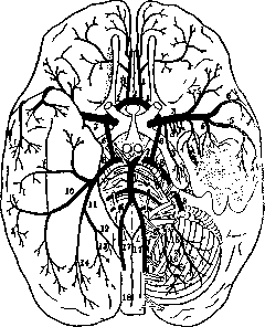
Figure 21. Arteries of the base of the brain (after Monakow). On the left side, the temporal lobe is removed; on the right side, the cerebellar hemisphere and part of the cranial and spinal nerves are removed: 1-anterior cerebral artery; 2-anterior communicating artery; 3-artery of the Sylvian fossa; 4-lenticular arteries; 5-cortical meningeal branches from the artery of the Sylvian fossa; 6-posterior communicating artery; 7-anterior choroidal artery; 8-posterior cerebral artery; 9-basilar artery; 10-temporal artery (Duret); 11-occipital artery (Duret); 12-parieto-occipital artery; 13-cuneal artery; 14-calcarine artery; 15-middle cerebellar artery; 16-inferior cerebellar artery; 17-vertebral artery; 18-anterior spinal artery.
the name of the artery of the Sylvian fossa or middle cerebral artery. Both arteries of the Sylvian fossa are connected to each other by means of the anterior communicating artery, from which the anterior cerebral artery departs. According to other data, the internal carotid artery divides into two branches—the middle cerebral artery and the anterior cerebral artery; both anterior cerebral arteries are connected to each other by means of the anterior communicating artery. The vertebral artery originates from the subclavian artery, is directed inward and backward, enters the canal,
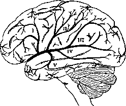
Figure 22. Schematic arrangement of the branches (I-V) of the middle cerebral artery (outer surface of the brain).
located in the transverse processes of the cervical vertebrae, leaves this canal at the level of the first cervical vertebra, enters the spinal canal, and then through the foramen magnum into the cranial cavity, where both arteries join into one common trunk—the basilar artery, which runs in the basilar sulcus of the pons Varolii to its upper end and there divides into its terminal branches—the posterior cerebral arteries; by means of the posterior communicating artery, the posterior cerebral artery and the middle cerebral artery are connected to each other. Thus, at the base of the brain, a connection of all cerebral arteries is obtained, an arterial circle, which is called the circle of Willis (circulus arteriosus Willisii, see figure 21). The anterior cerebral artery runs first along the lower surface of the cerebral hemisphere, then passes to its inner surface and serves to supply almost this entire surface, from the frontal pole to the parieto-occipital fissure; throughout its entire length, it gives off branches to the outer surface of the brain, the upper sections of which are supplied by it (see figure 23). The middle cerebral artery, or artery of the Sylvian fossa, the main cerebral artery, goes from the base of the brain along the Sylvian fissure to the outer surface and serves for its supply, with the exception of only its most extreme sections, which receive blood from the anterior cerebral artery anteriorly and superiorly, and from the posterior cerebral artery inferiorly and posteriorly (see figure 22). Having given off branches to the insula of Reil and to the claustrum, it divides into five arteries: the 1st branch supplies the frontal convolutions; the 2nd branch—the operculum and central convolutions; the 3rd—the supramarginal and angular gyri; the 4th—the posterior sections of the brain; and the 5th—the anterior sections of the temporal lobe. The posterior cerebral artery divides into temporal and occipital branches, which supply the lower surface of the brain and partly the posterior surface, posterior to the parieto-occipital fissure (cuneus, calcarine fissure) (see figure 23). Cerebral arteries are divided into two groups: cortical branches and basal, or central, branches. The cortical branches are located in the pia mater, which covers the entire surface of the cerebral cortex, penetrating into all 52(1 sulci, to their very bottom; in this membrane, the vessels form a dense network, richly supplied with sympathetic fibers; from this network, two types of arteries depart at right angles into the thickness of the brain tissue: short ones, branching in the cortex, and long ones, penetrating into the subcortical white matter; these latter enter the brain tissue at the level of the sulci. The vessels penetrating into the brain do not have innervation, and therefore the degree of supply to a particular area of the brain, as well as the pressure in the arterioles, depends on the vasoconstrictors of the meningeal network. The resistance to blood flow in the long arteries is more increased. Individual layers of the cerebral cortex differ from each other in the structural peculiarity of the capillary network: it is denser and thinner in the middle layers, and has wider loops in the superficial and deep layers.
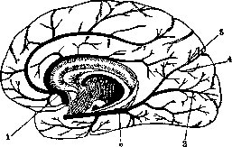
Figure 23. Schematic arrangement of the vessels
on the inner surface of the brain: 1—a. cerebri ant.; 2—a. cerebri post.; 3—a. calcarina; 4—a. cunei; 5—a. parieto-occipitalis. The rami centrales, penetrating into the brain from the base, serve to nourish the basal ganglia, the capsula interna, and the deep sections of the white matter of the brain. There is the following distribution among the cerebral arteries in the nourishment of the above-described formations. The arteria cerebri anterior gives branches to the n. opticus, chiasma nervorum opticorum, infundibulum, genu corp. callosi, septum pellucidum, fornix, caput nucl. caudati, to the anterior sections of the putamen, the anterior limb of the capsula interna, and to the substantia perforata anterior. The a. cerebri media, before its division into 5 terminal branches, gives off a large number of long and short arteries, which penetrate deep through the substantia perforata anterior and nourish the anterior half of the glob. pallidus, the genu and anterior limb of the capsula interna, and the deep sections of the white matter of the frontal lobe. One of the large arteries, the a. lenticulo-thalamica, nourishes the middle sections of the nucl. lenticularis, the capsula interna, the thalamus opticus, the white matter of the central gyri, and the insula. From the a. carotis interna, less often from the a. communicans posterior or from the a. cerebri media, the art. chorioidea anterior arises, which, after bypassing the tract. opticus and the cerebral peduncle, enters the Ammon's horn, and then the plexus chorioideus; branches arise from it that supply the posterior limb of the internal capsule, the posterior section of the nucl. lenticularis and thalami optici, the corp. Luysii, the substantia nigra, and the white matter of the parietal and temporal lobes. Long branches arise from the art. communicans posterior, nourishing the thalamus opticus, the cauda et caput nuclei caudati, the posterior limb of the capsula interna, the white matter of the brain, and terminating in the plexus chorioideus. The branches of the a. cerebri posterioris, partly through the substantia perforata posterior, and partly by bypassing the cerebral peduncle, go to the tegmentum of the cerebral peduncle, to the thalamus opticus, into the posterior limb of the capsula interna, the radiatio optica, and into the surrounding white matter of the occipital lobe; the branches penetrating into the ventricles of the brain are called aa. chorioideae posteriores, medialis et lateralis. It has been noted that the morphology of cerebral arteries in humans differs not only from that in animals, but also that there are the most diverse individual variations in the distribution of vessels in humans. These variations are observed: 1) in the arteries of the circ. arter. Willisii, in their place of origin, size, and connections: the art. cerebri posterior can be a continuation of the art. carotis interna, while the terminal branch of the art. basilaris is thread-like, and the art. communicans posterior is very wide; the art. cerebri posterior can originate from the fusion of the arter. communicans posterior with the anterior branch of the art. basilaris: an anastomosis is observed between the art. carotis interna and the art. basilaris and the art. vertebralis; the art. communicans anterior can have a different form—simple, double, reticular, in the form of a V; the art. cerebralis anterior can arise either directly from the art. communicans anterior or be a continuation of the art. carotis interna: 2) in the division and distribution of the large cerebral arteries; 3) in the distribution of the basal arteries among the various formations at the base of the brain and subcortical structures; finally 4) in the origin of branches from the art. basilaris. Recently, a study (Gindze) of cerebral arteries in prominent people was undertaken, and it was noted that morphologically they differ from the vessels of ordinary people by their length, width, and richness of branching (see figure 24). It is possible to think that, along with the weight and size of the brain and the richness of the sulci, the morphology of the vessels is of great importance for the problem of the correlation between somatic and intellectual abilities. The arteries, penetrating into the brain, divide into small arteries, diverging radially in the cortex, where they continue to divide into smaller precapillary arterioles, which pass into capillaries, from which postcapillary venules begin. According to the latest data by Pfeiffer, there may not be capillaries between arteries and veins, and then the arteries, after numerous branchings, pass directly into the branching of venous vessels. Arterioles and venules are distinguished by their thin walls and, as a result, are permeable to fluid, and in the case of pathological changes, to formed elements as well. The inner wall of the capillary in places presents thickenings, which reduces the lumen in that place and increases the resistance to the movement of blood in the capillaries (Sepp). Precapillary arterioles, due to their thin walls and pressure higher than in the tissue, transude, whereas in postcapillary venules the pressure is lower than in the tissue, and therefore absorption occurs. The capillary itself does not transude or absorb, but is an obstacle that increases the pressure difference between the transuding and absorbing sections. Being impermeable to fluid,
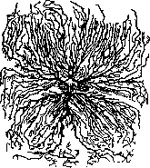
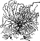
Zsr)^
Figure 24. a-arteries of a scientist's brain; b-arteries of a murderer's brain. (According to Hindze.) Capillaries have all the advantages regarding gas exchange, as they are in direct contact with the brain tissue; erythrocytes are located in them very densely, due to the transudation of fluid, and change their shape, stretching in length in the narrow and unyielding capillary. The arteries of the brain, up to their transition into capillaries, are covered with a double-walled connective tissue sheath, which serves as a diverticulum of the arachnoid sac surrounding the brain; it is called the adventitial sheath and is filled with cerebrospinal fluid, which, thus, penetrates into the brain tissue to the blind end of this sac, i.e., to the beginning of the capillaries. The blood supply of the cerebral cortex differs in that there are small anastomoses between its arterial vessels, forming a fairly extensive network. The arteries supplying the subcortical nodes do not anastomose with each other but are terminal; there are no anastomoses between the cortical arteries and the arteries of the basal nodes. The gray matter of the brain is richer in vessels than the white. Proper blood supply to the brain is an extremely important, essential condition for the proper functioning of the nervous system, for maintaining mental activity at a normal level. Increased blood supply is an essential condition for raising neuro-psychic energy. During mental work, the influx of blood to the brain increases, its vessels dilate, and intracranial pressure rises (proven by the experiments of Mosso, Gley; see Mental Work). Veins. The distribution of intracerebral venous vessels is the same as that of the arterial ones: from the network of capillaries, blood passes into a vein running together with an artery; then from the cortex of the brain through the veins of the meninges it enters the venous sinuses; the veins of the outer surface of the brain flow into the sinus falciformis major s. sagittalis and into the sinus transversus; the veins of the inner surface-into the sinus falciformis minor. The veins of the subcortical white matter, basal nodes, capsulae internae, from the cavity of the ventricles are collected into the veins of the plexus chorioidei, and then into the vena Galeni magna and into the sinus rectus. The veins of the brain do not have valves; in the cerebral cortex, they anastomose widely with each other. Chemistry of the brain. The composition of the nervous system, both central and peripheral, includes various substances; some are found in the composition of other tissues, others are found almost exclusively in nervous tissue. Nervous tissue contains proteins, various lipoids, extractive substances, and enzymes. The protein substances of the nervous system have not yet been sufficiently studied; the brain contains proteins of the nucleoprotein type (containing 0.55-0.57% phosphorus) and globulins (two globulins differing from each other in coagulation temperature). In addition, proteins have been discovered in the brain-neurostromin and neurokeratin; the latter is contained mainly in the white matter of the brain and in peripheral fibers; then there are various amino acids. Among the lipoids in the brain are: phosphatides, lecithin, cephalin, and a number of other compounds of not yet precisely determined structure containing phosphorus (myelin, leukopolin, etc.); a number of substances with undetermined structure containing sulfur, cholesterol-mainly in a free state, partly also in the form of esters; cerebrosides (cerebron, kerasin, etc.)-compounds that decompose upon hydrolysis into galactose, fatty acid (cerebronic or lignoceric), and a nitrogenous base, sphingosine. Together with lecithin, cerebrosides are part of protagon. Among the extractive substances in the nervous system, the following have been discovered: creatine, purine bases, inositol, choline, lactic acid, uric acid, and neuridine. Among the enzymes, catalase, peroxidase, lipase, amylase, protease, phosphatase, indophenol oxidase, and nuclease have been detected. Different parts of the nervous system, differing in function, also differ in chemical composition. The amount of water and proteins in different parts decreases from the cortex region to the subcortical nodes, from gray matter to white, and finally from the white matter of the brain to the peripheral nerves. Lipoids are contained most in peripheral nerves, then comes the spinal cord, and in the last place is the brain; there are more lipoids in the white matter than in the gray; the gray matter of the basal nodes is richer in lipoids and the cortex is poorer (as well as in cholesterol); a significant amount of lipoids is also detected microchemically in the walls of arterioles, venules, and capillaries of the brain. In different parts of the nervous system, there is not only a different...
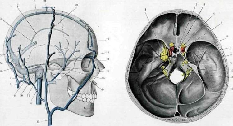
Fig. 1.
Fig. 2.
Figure 1. Venous sinuses and veins of the dura mater; 1 - sinus falciformis major s. sagittalis; 2 - sin. transversus; 3 - v. cerebri; 4 - sin. rectus; 5 - tentorium cerebelli; 6 - tentorium cerebelli; 7 - v. occipitalis; 8 - sinus sphenoidalis; 9 - sinus petrosus sup.; 10 - bulbus v. jugularis; 11 - v. auricularis post.; 12 - v. facialis comm.; 13 - v. facialis ant.; 14 - sinus petrosus inf.; 15 - v. diploica; 16 - sinus cavernosus; 17 - v. ophthalmica; 18 - v. temporalis; 19 - tentorium cerebelli. (According to Tandler.) Figure 1. Schematic section. Exit of nerves and vessels (on the left - dura mater and venous sinuses, on the right - nerves; the brain is removed; the middle cranial fossa is separated from the anterior and posterior ones): 1 - sinus cavernosus; 2 - fossa hypophyseos; 3 - tentorium cerebelli; 4 - n. opticus; 5 - a. carotis int.; 6 - n. oculomotorius; 7 - n. trochlearis; 8 - n. trigeminus; 9 - n. abducens; 10 - n. acustico-facialis; 11 - nn. glosso-pharyngeus, vagus, accessorius; 12 - a. thyreoidea. (According to Tandler.) B. M. E. To the article Brain.
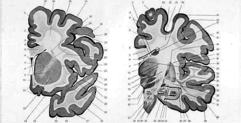
Fig. 4. Frontal section of the right hemisphere (through the anterior part of the thalamus); 1 - gyr. frontalis sup.; 2 - ventric. lateralis; 3 - ventric. tertius; 4 - septum pellucidum; 5 - nucleus caudatus; 6 - capsula int.; 7 - nucleus lentiformis; 8 - capsula ext.; 9 - gyr. temporalis inf.; 10 - v. tempor. inf.; 11 - v. tempor. inf.; 12 - subst. perfor. ant.; 13 - gyr. tempor. med.; 14 - fissura Sylvii; 15 - insula; 16 - gyr. tempor. sup.; 17 - gyr. tempor. med.; 18 - gyr. frontalis sup.; 19 - ventric. lateralis; 20 - ventric. tertius; 21 - septum pellucidum; 22 - nucleus caudatus; 23 - capsula int.; 24 - nucleus lentiformis; 25 - capsula ext.; 26 - gyr. temporalis inf.; 27 - v. tempor. inf.; 28 - v. tempor. inf.; 29 - subst. perfor. ant.; 30 - gyr. tempor. med.; 31 - fissura Sylvii; 32 - insula; 33 - gyr. tempor. sup.; 34 - gyr. tempor. med.; 35 - gyr. frontalis sup.; 36 - ventric. lateralis; 37 - ventric. tertius; 38 - septum pellucidum; 39 - nucleus caudatus; 40 - capsula int.; 41 - nucleus lentiformis; 42 - capsula ext.; 43 - gyr. temporalis inf.; 44 - v. tempor. inf.; 45 - v. tempor. inf.; 46 - subst. perfor. ant.; 47 - gyr. tempor. med.; 48 - fissura Sylvii; 49 - insula; 50 - gyr. tempor. sup.; 51 - gyr. tempor. med.
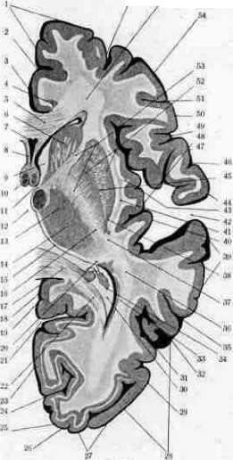
...quantity of proteins and lipoids, but the proteins and lipoids themselves do not consist of the same representatives. Mineral substances are distributed more evenly. For orientation in the chemical structure of the white and gray matter of the nervous system, see the following table (according to Winterstein). Composition of brain matter: Water........ Mineral substances ...... Extractive substances ...... Proteins....... Lipoids ...... including: Cholesterol . . . Lecithin ..... Cephalin, myelin Cerebrosides . . Sulfatides . . .
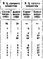
In its chemical composition, the cerebral cortex differs not only from other parts of the central nervous system, but, as is known, functionally different centers of the cerebral cortex differ among themselves in their lipoid composition; thus, it is established that along with the anat.-physiol. topography, there is also a chemical topography in the cerebral cortex. The brain of adults and children, in connection with functional peculiarities, is also not identical in its chemical composition; with age, the content of water, proteins, extractive substances, and salts relatively decreases, while the amount of cholesterol increases.
The chemical dynamics of the brain are in the stage of development; the nature of the chemical processes that occur in different parts of the nervous system during activity is still unknown, as is which processes are specific to one part and which to another, and with which chemical changes certain functional changes are associated. Brain functions are accompanied by specific chemical processes that affect both the proteins and the lipoids of the brain. With increased activity of the nervous system, the breakdown of brain proteins—aminogenesis—intensifies; with decreased activity, the phenomena of aminogenesis decrease. During starvation, proteins and lipoids decrease, while the amount of water increases, which maintains the apparent constancy of brain weight; while during prolonged starvation the spleen can lose up to 64% of its weight, the liver 54%, the kidneys 25%, the brain loses only 3%. Carbohydrate metabolism in the brain is autonomous and not closely linked to carbohydrate metabolism in other parts of the body. A constant presence of glycogen is noted in the brain, and its quantity is distinguished by stability and does not change in connection with conditions leading to general hypoglycemia; only in the presence of convulsions does the amount of glycogen fall. Under anaerobic conditions, vigorous glycolysis is observed. Along with this, both in aerobic and anaerobic conditions, there is a continuous splitting off of NH3, which increases extremely during irritation and falls during narcosis. The nerve cell during work consumes O2 and releases CO2; its respiratory quotient is less than unity. The study of enzymes in the brain has shown that they predominate mainly in the gray matter (catalase and amylase) or are present exclusively in it (glutinase, indophenol, oxidase); in connection with the change in the function of the nervous system, changes of a quantitative and qualitative nature also occur in them. Physiology of the brain. The question of the functions of the cerebral hemispheres and the localization of these functions is of exceptional interest from various points of view; besides its direct relation to the tasks of clinical diagnosis of nervous diseases, the doctrine of localization, by clarifying the pathogenesis of certain disorders, can sometimes serve to identify new disease forms; in cases accessible to surgical intervention, it will allow for orientation in the site for a brain operation; finally, the doctrine of brain functions can provide a resolution for many problems of psychology and psychiatry. The question of the functions of the brain in general is a very old question: the first hints of a doctrine of brain functions are found among ancient physicians, who were interested in the question of the place where sensations arise. Opinions diverged: some authors assigned them a place in the brain, assuming that sensations are transmitted to the brain by special channels, while others considered the blood to be the main center for sensations; the possibility was not excluded that other organs of the body could take part in psychic acts. Among other authors, Aristotle had already expressed the assumption that the brain does not have independent functions, but is an intermediary between sensations and the heart, where these sensations end. Semmering localized higher nervous functions in the ventricles of the brain, considering the fluid of the ventricles to be the center of higher feeling; under the influence of irritations coming from the organs, oscillations occur in the fluid, different for each irritation, while the oscillations do not mix with each other. A major role in the matter of isolating the functions of the brain was played by the comparative-anatomical method. As is known from the history of the development of the brain, the central nervous system in vertebrates consists of the spinal cord, the medulla oblongata, the hindbrain, the midbrain, the diencephalon, and the forebrain, i.e., the cerebral hemispheres. The development of these individual parts in different species of vertebrates is different. As one ascends to higher animals, the forebrain increases and becomes more complex in development: in fish, it is in a rudimentary state; in amphibians, noticeable development of the forebrain begins, with the midbrain still dominating; the same takes place in reptiles and birds. In mammals, the forebrain develops strongly and gains predominant importance over the diencephalon and midbrain, which lose their independence and in functional terms are subordinate to the forebrain. Proportional to the development of the cerebrum is the development of the psyche and intellect in animals: in lower animals, the cerebrum is only a receiving instance for olfactory sensations; in higher animals, higher complex processes occur in it. The question of the comparative significance in different animals of the functions of the midbrain and diencephalon on the one hand, and the forebrain on the other, is resolved by experiments with the removal of the brain; on the one hand, they reveal the loss of a known function, and on the other, they show to what extent the remaining part of the central nervous system can function correctly. Removal of the cerebral hemispheres in fish has no effect on their movements and their attitude toward their surroundings. The frog also tolerates the removal of the hemispheres very easily; after the operation, it differs little from a normal animal in its behavior: movements are preserved, it can swim and catch flies, etc.; only upon removal of the entire brain, including the thalamus opticus, are voluntary movements disturbed, becoming irregular, ataxic, the grasping of food becomes impossible, and the animal already differs sharply from the normal. In birds, removal of the brain leads to stronger disorders: the operated bird becomes as if an automaton, there are no voluntary movements; it still responds to external stimuli with movements, when thrown into the air it produces more or less correct movements with its wings and descends to the ground, it does not take food itself, it must be fed so that it does not die of hunger; the sense of equilibrium is preserved, there is no visual recognition, it does not react to the call of chicks, it is indifferent to dangers; after some time after the operation, movements improve, but initiative is lost forever. After removal of the brain in mammals by a cut through the corpora quadrigemina, tonic contraction of all voluntary musculature is observed—decerebrate rigidity (see Decerebration), due to the elimination of the regulating or inhibiting influence of the nuclei rubri on the extrapyramidal system. When only the cerebral hemispheres are removed, no major disorders in terms of movement are observed, except perhaps immediately after the operation; it is mainly psychic processes that suffer. A strong decrease in intelligence is also observed in dogs deprived of their hemispheres (see Goltz's experiments): the animals turn into automata acting only under the influence of external stimuli. Monkeys after removal of the brain survived no more than two weeks (observations by Kreidl and Karplus), remaining in a sleepy state the whole time, however, they could sit, maintain balance, look around when there was noise, as if waking up, in their paws there was aimless repetition of the same movement; in some, reflex excitability was increased; upon light touch, they raised their head, opened their eyes; upon strong irritation, a cry was often observed. Unilateral removal of the hemispheres was tolerated more easily; soon after the operation, they recovered, were able to run, climb, and reacted to external stimuli, and only with careful examination could one notice a certain defect in movements. Observations on human monsters deprived of a brain were too short-lived and insufficient, but conclusions could be drawn about the disorder of sensory, visual, and auditory perceptions, whereas reflex and vegetative functions operated more or less correctly. Thus, the higher an animal stands on the zoological ladder, the greater the development and perfection its central nervous system reaches; the dominant role in this passes to the cerebral hemispheres (cerebrum), which subordinate the activity of other parts of the central nervous system, due to which their destruction or removal strongly affects the animal: its entire behavior and reactions. The development of the cerebral hemispheres leads to the complication and perfection of relations with the outside world, due to which the volume of consciousness increases, encompassing all areas of sensory perceptions, motor acts, and psychic phenomena. The cortex of the cerebral hemispheres can be viewed as a vast projection field in which, during the process of life, engrams of perceptions received from the sense organs are deposited; thanks to this deposition, personal experience and skills are developed, and new paths for conduction are forged. Engrams are usually in a latent state, but through associations with some impulse, they become physiologically active; their durability depends on many conditions—personal exercise, the very structure of the central apparatuses. Thanks to the above, animals deprived of hemispheres react only to immediate irritation, whereas normal animals also react to previous stimuli that have already ceased to act, on the basis of accumulated previous life experience.
The hemispheres of the brain are related to movement; in the cortex there are sections corresponding to images of specific actions; within it, motor stimuli, known as volitional or voluntary, are generated. In the cortex, specific combinations are established between centripetal sensory impulses and corresponding centrifugal motor ones. Being excitable under the influence of various impulses, the cerebral cortex exerts, at the same time, an inhibitory influence on the underlying formations of the central nervous system, by which correctness and purposefulness in their actions are achieved. Localization of functions in the cerebral cortex. The first attempts at localizing cerebral functions in separate sections of the cortex of the hemispheres belong to the phrenologists (Gall). From the very beginning, the phrenologists went down a false path and, instead of simple centers, such as centers of movement, sensitivity, etc., began to search for centers of abilities, inclinations, affections, etc., while trying to find indications of internal qualities of the mental makeup based on the external features of the skull. Such attempts at a biased solution to a most complex psycho-physiological problem, of course, had no success. The first scientific attempts to clarify the question of localization in the cerebral cortex were undertaken by Flourens. Working according to the experimental method of destroying individual parts of the brain, Flourens came to the conclusion that the cortex in its entirety is an integral and unified organ of 'intellect and will,' and that the removal of any part of it leads to a general weakening of its functions—separate centers for different functions do not exist in the cortex. Until 1870, Flourens's doctrine dominated among physiologists, although among clinicians it had already aroused some doubts. Broca precisely established some facts proving a definite localization of cortical centers, namely, he proved the dependence of speech disorders on the destruction of the third frontal gyrus in the left hemisphere. The doctrine of Flourens was dealt a particularly heavy blow by the experimental works of Hitzig, Fritsch, and Ferrier, who pointed out the presence of centers in the cerebral cortex, the irritation of which causes contraction in individual muscle groups. This discovery constituted an entire epoch in the physiology of the brain and entailed a huge number of studies. It was then verified on the human brain during operations (Horsley, Bergmann, Krause, Bekhterev, and others). Another method of studying the cerebral cortex led to approximately the same results, namely, the method of destroying its specific sections. Especially important in this field were the studies of Munk, which led him to a doctrine of strict localization, which received wide distribution, especially among clinicians. Goltz and his students opposed this doctrine in a series of experimental studies; Goltz noted the possibility of broad substitution of cortical functions by subcortical nodes, drew attention to the general phenomena of depression following the operation, and pointed out the necessity of distinguishing functions that have truly permanently dropped out from those that are only temporarily depressed. Later, Monakow dwelt on this question, proving that phenomena of depression can manifest themselves in the form of loss of function even in formations spatially located far from the site of the injury (see Diaschisis) and that, thus, not all functions lost after an operation must be localized at the site of destruction. Finally, a whole series of authors (Anton and others) pointed to the existence of vicarious substitution of functions—i.e., other areas of the cerebral cortex can take upon themselves the functions of destroyed parts. Flechsig approached the doctrine of localization from the point of view of myelination, based on the assumption that all fibers having the same function are myelinated at the same time and that the maturation of fibers proceeds according to their function—the higher the function of a given area, the later its fibers are covered with myelin. On the basis of the myelination method, Flechsig identified four projection centers and three association centers. The projection centers are located around the primary sulci: 1) the center of all body sensations, where sensory fibers end and motor ones begin, located near the Rolandic sulcus; 2) the olfactory center—in the gyrus hippocampi; 3) the visual center in the occipital lobe near the fissura calcarina; 4) the auditory center in the gyrus temporalis superior. These centers occupy one-third of the entire surface of the brain, while the other two-thirds are occupied by association centers intended for higher mental functions: 1) the posterior—in the occipital and parietal lobes, 2) the middle—in the insula Reili, and 3) the anterior—in the frontal lobes. Between the projection and association centers are intermediate zones. Projection fibers are myelinated and begin to function first, whereas the beginning of myelination of the association centers relates only to the first years of life, and the end—to the twentieth year. As the internal organization becomes more complex, the child's intellect develops. Thus, association fibers and centers are, in Flechsig's opinion, the substrate of human experience, knowledge, speech, and other higher mental processes in the human brain. The modern doctrine of myelo-cytoarchitectonics speaks in favor of the doctrine of separate cortical centers as carriers of various specific functions, and it becomes obvious that the localization of centers by gyri must give way to a more subtle localization by architectonic fields—areae; physiological data on the localization of individual functions in the cortex generally correspond to the division of the cortex into architectonic regions and fields, but the question of whether any brain function exactly coincides with the limits of one area or another has not yet been fully resolved. In any case, it is very interesting that the areal maps, one compiled by Vogt based on experimental data, and the other by Foerster based on the irritation of the cortex in humans during operations, coincide to a significant degree. Despite the huge number of studies, much remains controversial and obscure in the question of the localization of functions in the cortex of the cerebral hemispheres. While some authors (Foerster, Dejerine, Vogt, and others) strive for strict localization of certain brain functions (movement, various types of sensitivity) in specific anatomical centers, others (Goltz, Monakow, Goldstein, and others) reject the view of brain activity as a simple sum of individual functions and relate negatively to attempts to localize these functions in separate, sharply limited sections of the cortex. They believe that these functions are the result of the activity of the entire brain and that the processes occurring within it are so complex that they cannot be attached to any one of its sections. Sherrington denies the localization of individual functions in the cerebral cortex: in his opinion, antagonistic movements can arise sequentially in the same place depending on the stimulation of receptors. With simultaneous stimulation, such antagonistic movements inhibit each other, while homogeneous ones sum the strength of the effect. Even the very concepts of centers do not agree. Some authors understand by centers areas in the cerebral cortex that are strictly limited anatomically or by the commonality of architectonic structure or by sulci and gyri; in these areas are located cells that give rise to projection centrifugal (motor) fibers or are the site of termination of centripetal (sensory) fibers. The doctrine of the localization of a particular function in the corresponding areae has become somewhat more complex in connection with the doctrine of laminar, or layered, structure of the cortex (see Architectonics of the cerebral cortex). According to this doctrine, the cortex is divided into two sections: the outer section, including the four upper layers, performs a receptor-associative function; in it, centripetal fibers end and associative ones begin, connecting different areas of the cortex; the inner section includes the fifth and sixth layers of the cortex, which have a projection-commissural function; in them, commissural and centrifugal fibers begin, connecting the cortex with underlying formations. Thus, according to the data of the laminar doctrine, one and the same area carries out very complex functions, being the site of origin or termination of diverse fibers. From the brief review provided, it is evident how different the existing views on the localization of functions in the cerebral cortex are. At the present time, data on focal localization can be considered accurately established only for primitive functions. The motor area is located in the cortex of the brain hemispheres on the outer, and partly on the inner surface, along the Rolandic sulcus, mainly anterior to it, in the gyrus centralis anterior and lobulus paracentralis (see separate table). Irritation of specific places or their destruction has shown that the uppermost section is occupied by centers for the movement of the lower extremities; highest of all are located the centers for the movement of the big toe, slightly lower—for the ankle joint, then the knee joint, and even lower the center for the movement of the thigh.
The centers for movement of the upper limb are located in the middle part of the gyrus centralis anterior; of these, the center for movement of the shoulder is the uppermost, followed by the center for the forearm, the hand, and the fingers; the centers for the muscles of the face, tongue, mouth, and larynx are located in the lower part of the same gyrus; in the posterior part of the gyrus frontalis superior is the center for movement of the trunk, and in the gyrus frontalis medius is the center for movement of the eyes and head. Data regarding the localization of motor cortical centers in humans have been obtained mainly by observing cases of brain injury or its pathological changes, whereby a complete coincidence with the results of artificial stimulation was obtained. Upon destruction of these areas, there occurs a loss of movements of the opposite half of the body. These centers were considered centers of volitional, or voluntary, movements. Monakow introduces some corrections. A volitional motor act consists of 4 components: 1) the idea of a goal evokes an idea of the movement by which the goal can be achieved; 2) the general idea of the movement is transformed into a separate motor act in the form of a sequential series of coordinated motor impulses; 3) these impulses excite the aforementioned motor centers, and 4) these excitations pass into the pyramidal tract. The first of the four components cannot be focally localized; it originates from the entire cortex; motor ideas do not disappear upon the destruction of even a large area of the cortex; the second moment has a localization, but of a very general kind, in an area of the cortex wider than the central gyri; the third moment is expressed by the excitation of individual motor centers in the vicinity of the Rolandic fissure; thus, only the output of motor impulses from the cortex is subject to localization. The centers of sensitivity are located in the same area, only posterior to the Rolandic fissure. According to the data of some authors (Munk, Luciani), the sensory-motor centers have a common localization; other authors sharply distinguish the centers of movement and sensitivity from each other, and finally, there exists a third opinion, according to which the centers of movement are functionally separated from the centers of sensitivity, but at the same time their anatomical proximity and even coincidence are admitted. The distribution of sensory centers in the posterior central gyrus corresponds to the distribution of motor ones in the anterior central gyrus, i.e., the uppermost sections of this gyrus are occupied by the center of sensitivity for the lower limb, then for the upper, and in the lowest part are the centers of sensitivity for the face. The posterior central gyrus undoubtedly has a relation to tactile and muscular sense, but apparently the cortical area of sensitivity is not limited only to it, but extends to the anterior and to the parietal (for details, see Sensitivity). Sensitivity, like movement, is a complex phenomenon; in it, conscious excitations and unconscious ones are distinguished; some of them are localized in specific centers, others are the result of the action of the entire cerebral cortex. Monakow also distinguishes several stages here: 1) cortical diffuse sensitivity, in which excitations are already felt, but summarily, without differentiation into quality and place; 2) then this differentiation occurs, and the excitations are localized, i.e., connected with a specific part of the body; this stage corresponds to localization in the physiological sense of the word in specific centers of the cortex; 3) psychic processing of sensations and perceptions of objects of the external world; since other sensations also take part here, it is considered as a complex aggregate activity of various parts of the cortex. The center of visual perceptions is located on the inner surface
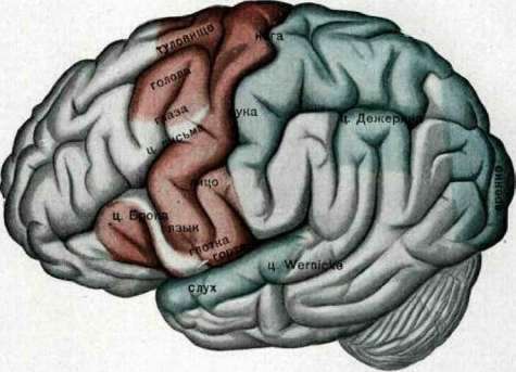
Fig. 1.
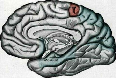
The visual center is located in the occipital lobe near the fissura calcarina (cuneus, gyr. lingualis). As is known, the cortex of this region is characterized by the presence of a special stripe—the stria of Gennari (see Stria of Gennari). Some authors allow for the extension of the visual center to the outer surface (see Visual pathways, centers). Fibers from the peripheral parts of the retina go to these centers after their interruption in the subcortical visual centers; due to the incomplete decussation of the optic nerve, each hemisphere is connected to half of the retina of both eyes, and therefore destruction of the occipital lobe causes not complete blindness in one eye, but the loss of the corresponding halves in both eyes, i.e., hemianopsia (see). As for the localization of central vision, which is conducted by the macular bundle, the question of its localization has not yet been finally resolved; it is localized in both the anterior and posterior sections of the floor of the fissura calcarina; some assume a more extensive termination, encompassing the cortex of the entire cuneus. Color perception is also localized by some authors in the region of the fissura calcarina, but the possibility is admitted that other elements of the cortex serve it (see Visual pathways, centers). The center of hearing is located on the outer surface of the temporal lobe, mainly in the gyr. temporalis superior and in the anterior part of the gyri transversi Heschli; damage to these gyri leads to a decrease in hearing in both ears, mainly in the opposite one. It is possible that individual sections of the temporal lobe have a relationship to different types of auditory sensitivity, to tones of different pitch (see Hearing). The olfactory center is localized in the uncus gyri hippocampi, partially extending to the hippocampus. Since each tractus olfactorius is connected to the olfactory centers of both hemispheres, damage to one hemisphere leads only to a decrease in the sense of smell on both sides. The center of taste has not yet been precisely established; there are two opinions: according to one opinion, this center is located near the olfactory center in the gyr. hippocampi, according to the other—in the lower sections of the gyri centralis posterioris, in the neighborhood of the sensory-motor centers of the mouth and tongue; clinical data confirm the second assumption rather (see Taste). Among other parts of the brain to which specific functions were attributed, it is necessary to note those whose damage leads to a disorder of speech—aphasia (motor and sensory). The center of motor aphasia was discovered by Broca, bears his name, and corresponds to the posterior sections of the gyri frontalis inferioris (pars opercularis, pars triangularis) of the left hemisphere, fields 44 and 45 (Brodmann) and FCBm (Economo). The center, damage to which leads to sensory aphasia in the form of word deafness (Wernicke's center), is also located in the left hemisphere in the posterior sections of the gyri temporalis superioris and gyri transversi Heschli, fields 41 and 42 (Brodmann). Economo localizes the perception of sounds in general in field TC, the understanding of words in TB, the comprehension of words in TA1, and musical hearing in TA2. The second sensory center, damage to which leads to word blindness, alexia—the Dejerine center—is located in the gyr. angularis of the left hemisphere, corresponds to field 39 (Brodmann) and PG (Economo). Monakow speaks out against the localization of special centers related to speech in the indicated places: the speech disorder observed during damage to these sections, in his opinion, depends on diaschisis (see) and on the disruption of association pathways between different sections of the cerebral cortex (see Aphasia and Architectonics of the cerebral cortex—areal maps). The part of the brain whose damage causes apraxia has not yet been finally established; it is placed in the corpus callosum, in the gyr. marginalis—field 40 (Brodmann), PE (Economo), and in the frontal lobe of the left hemisphere, but there are also objections against the localization of apraxia, since it too must be considered as a disruption of association, of the coordination of movements, into which the general preliminary idea of a motor act transitions, and therefore it cannot be precisely localized. There are attempts to localize complex functions of the brain—judgment, reasoning, consciousness, abstract concepts—in association centers, mainly in the anterior, i.e., in the frontal lobe; these attempts are based on the structure of these centers—late myelination, lack of connection with the periphery, the relatively large size of the frontal lobe, increasing in proportion to mental development; extirpation of the frontal lobe in animals, its disease in humans causes dementia; in idiots and microcephals, its underdevelopment is observed. There are objections against all these justifications. Monakow, without denying the important role of the frontal lobes in psychic life and admitting that their damage leads to significant psychic defects, nevertheless denies the localization of any definite function in them. The teaching of I. P. Pavlov on conditioned reflexes (see) opened a new era in the physiology of the cerebrum. According to this teaching, the entire nervous system consists of a collection of analyzers (optical, acoustic, etc.). Lower degrees of analysis are characteristic of the lower sections of the nervous system; the organism is capable of them even without the hemispheres of the brain, but the highest, most subtle analysis is achieved only with the help of the hemispheres of the brain. This analysis of external agents of the environment occurs with the help of conditioned reflexes. 300 years ago, Descartes established the concept of "reflex" as the basic act of the nervous system—this or that activity is a lawful response to one or another external agent, and this connection of the active organ with the given agent, as cause with effect, is established with the help of a specific nervous pathway. In the 18th, 19th, and 20th centuries, physiologists used the idea of the reflex in detail, at first only in the lower sections of the central nervous system, but gradually rose higher and higher through its sections; the first step toward applying the concept of the reflex to the hemispheres of the brain was taken by I. M. Sechenov (1863), representing the activity of the hemispheres as reflex activity. This idea found further development in the teaching of Pavlov and his school on conditioned reflexes. The basic nervous reactions of both animals and humans are innate in the form of a reflex, and all other nervous activity of the organism is built upon the foundation of these reflexes. These innate reflexes serve as elements of constant adaptation, of balancing with the environment. Although they ensure the existence of the organism, they do not do so to a sufficient degree. Life requires more detailed special relationships of the animal with the surrounding world, which is achieved by the gradual development of a series of new reflexes, called by Pavlov conditioned, since their appearance and work depend on a very large number of conditions; they are also called acquired or individual. They are developed in the cerebral hemispheres, the removal of which leads to the disappearance of already developed reflexes and the cessation of the development of new ones. An infinite mass of natural phenomena constantly conditions the formation of positive and negative conditioned reflexes through the cerebral hemispheres and thereby determines the entire activity of the animal. For each reflex, there is a point of application in the cortex in the form of cells; consequently, one unit of the cortex is connected with one activity of the organism, another with another; one causes activity, another inhibits it. Thanks to this, the cortex represents a grandiose mosaic, a grandiose analytical board, on which, however, there always remain places for the formation of new "signal" conditioned stimuli; the occupied places are constantly undergoing changes in connection with the different activity of the organism. The complication of the connection of the animal organism with the surrounding world, the more precise adaptation to external circumstances in connection with the development of conditioned reflexes, proceeds in parallel and inseparably with the analytical activity of the cerebral cortex. Thus, the basic activity of the cortex, in Pavlov's opinion, along with the development of conditioned reflexes, is analytical activity—the analyzing of the stimuli of the external world; furthermore, connections are formed in the cortex between the perceiving cortical points of conditioned and unconditioned stimuli; in other words, excitations received by different sections of the cerebral cortex simultaneously or at short intervals are associated with each other, and subsequently, the presence of one excitation also calls forth others even without any external stimuli. The received external stimuli are synthesized by the cortex into one whole, and a differentiated conditioned reflex is formed for such a complex stimulus. Two excitations that have arisen simultaneously in a specific area of the cerebral cortex inhibit each other; if two nervous excitations follow one after another, then one paves the way for the other, and the second of them reaches a greater strength than it would have without the preceding excitation.
Along with inhibition, disinhibition also occurs in the nervous system; an extinguishing conditioned reflex can be revived by some new stimulus. Excitation, upon reaching the cerebral hemispheres, does not immediately localize in any one brain center, but irradiates over the entire surface of the hemisphere and only gradually begins to concentrate at one specific point. Thus, in Pavlov's opinion, the cortex of the Brain represents a collection of the endings of cerebral analyzers; the activity of the cortex is analyzer-synthetic; the boundaries of individual cortical synthesis-analyzers are not sharp and merge at the periphery, mutually diffusing and imperceptibly passing into one another. Pavlov rejects the existence in the cortex of special associative zones, or sharply delimited psychomotor and psychosensory centers. Only a perfectly balanced nervous system reacts in a constant manner to specific stimuli; when its equilibrium is disturbed by the presence of a center possessing the greatest excitability, previous stimuli evoke different reactions. This center acquires the significance of a dominant, governing factor in the work of other centers: it accumulates excitation from various sources within itself, but inhibits the ability of other centers to respond to impulses that have a direct relation to them. The existence of such a focus of increased excitability was established simultaneously by I. P. Pavlov and Ukhtomsky. Pavlov called this center the center of optimal excitability, and Ukhtomsky called it the dominant (see). Thanks to this, the aggregate work of the centers is mobile, and this mobility is determined by the place of origin of the governing focus of excitation.
E. Kononova. Pathology of the brain. General concepts. The brain participates in pathological processes in various ways. During acute infections (typhus, croupous pneumonia, scarlet fever, etc.), the brain reacts to the general disease with disturbances in thermoregulation, the function of consciousness, work capacity, general well-being, the appearance of headache, seizures, etc. The same is observed during external intoxications (alcohol, opium, hashish, etc.) and internal ones (uremia, diabetes, etc.). General reactions of the brain also manifest under the influence of general impacts of a mechanical (general contusion, fall from a height), thermal (sunstroke), or psychic (severe fright, shock) nature, to a large extent depending on individual characteristics, constitution, predisposition, age (in children, for example, a general convulsive seizure can be likened to a chill in an adult), sex, cultural level, etc. Besides general reactions to the disorder of the functions of the entire organism, in the pathology of the brain, there are observed morbid processes that proceed as if only within it itself. These processes can in some cases have a coarser organic character and be local, focal, occupying a limited space in the brain (tumor, hemorrhage, etc.) or diffuse, affecting more or less the entire substance of the brain (hemorrhagic encephalitis, arteriosclerosis, etc.), sometimes spreading predominantly only to the cerebral cortex (progressive paralysis, endarteritis of the small arteries of the cortex, etc.). In other cases, diseases of the brain have a functional character, representing this or that transient disorder of circulation (anemia, hot flashes, migraine, edema), nutrition, or arising on the basis of exhaustion (neurasthenia, cerebrasthenia, etc.). Regarding mental disorders, see Psychoses, Soul, mentally ill; regarding functional disorders, see Neuroses. Below, we refer predominantly only to organic diseases of the brain. Historical data. Although as early as the 6th century B.C., Alcmaeon of Croton was the first to begin performing dissections of animals, engaged in various anatomical-physiological studies, and taught that the brain is the central organ of all mental activity, and the first experiments with brain dissection belong to Anaxagoras, nevertheless, the pathology of the brain in the form in which it is depicted in modern neuropathology is predominantly a creation of the second half of the 19th century. The Hippocratics (4th century B.C.) already had an excellent description of apoplexy of the brain; there was also a description of hysterical phenomena, although the explanation was given by them depending on the supposed wandering of the uterus. Galen (2nd century A.D.), besides developing anatomical data (vena magna Galeni), performed very subtle experiments on the nervous system. In the period of the 13th–19th centuries, one encounters the works of remarkable researchers (Italians Rolando of Parma and Constanzo Varolio, Englishman Thomas Willis, Dutchman Franciscus de le Boë Sylvius, Germans Reil, Blumenbach, Frenchman Magendie, Englishman Monro), who left their names in the pathology of the brain in connection with a whole series of anatomical designations of its parts. Before the 19th century, printed works on encephalopathology certainly appeared, but they all had a somewhat accidental, amateurish character or were included as undeveloped or very little developed chapters in general treatises on medicine. However, this time left the mid-19th century, when modern encephalopathology began to be created, a number of monographs (as can be seen, for example, in relation to epilepsy) that represented and represent great value. The doctrine of diseases of the brain in its modern form was created by the works of a whole series of individuals belonging to different countries. Among them, in the first place, one must place French scientists [with predecessors, contemporaries, and followers—Charcot, Broca (doctrine of aphasia), Bayle (description of progressive paralysis), Bravais (cortical focal epilepsy), Bouchard, Ballet, Brissaud, Pitres, Féré, Raymond, Dejerine, Pierre Marie, Babinski, and others]. The role of German researchers in the creation of encephalopathology is enormous, starting with Franz Gall with his phrenology. Here, in the first place, one must place the Wernicke school (aphasia, etc.) with his students: Hermann Liepmann (apraxia), Bonhoeffer, Otfried Foerster, Kleist, Goldstein. Besides them, the pathology of the brain was created by such classics of neurology as Hitzig, Fritsch, Westphal, Erb, Oppenheim, Strümpell, Flechsig, and by such histopathologists as Nissl, Alzheimer, Spielmeyer, O. and C. Vogt. To this group of strictly German encephalopathologists, one must add the Viennese school, tracing its origin to Meynert and Nothnagel—Anton, Obersteiner, Frankl-Hochwart, Wagner-Jauregg, Economo. The role of English scientists in the development of questions of the pathology of the brain is very great. Starting from Sydenham, Parkinson, Bell, Huntington, Little, the creation of the pathology of the brain in England continued with the works of Jackson, Horsley, Ferrier, Gowers, Wilson. Among the Swiss, as having great significance for encephalopathology, one should name the Russian-born C. von Monakow and the surgeon Kocher; among the Italians—Bianchi and Mingazzini; among the North Americans—Beard, Sachs, Cushing; among the Swedes—Henschen. Among Russian researchers, one must name Bekhterev, Darkshevich, A. Ya. Kozhevnikov, Korsakov, Muratov, Rossolimo, and others. It is important to note that the progress in the field of encephalopathology was enormously influenced by successes in other fields of medicine, physics, chemistry, and natural science in general. The progress of bacteriology is reflected in the research and treatment of tetanus (Kitasato), meningitis, syphilis of the brain, and in the establishment of the concept and role of filterable agents in encephalopathology. The application of Quincke's lumbar puncture creates a new era and leads, among other things, to the study of the etiology of meningitis (Weichselbaum—meningococcus, Netter—pneumococcus, Fraenkel, Benda—streptococcus). The discovery of the ophthalmoscope by Helmholtz makes it possible to study the pathology of intracranial relations. The discovery of X-rays by Röntgen is reflected in the diagnostics of diseases of the brain and skull, establishing changes in the bones, the presence of foreign bodies, and in recent times, by means of new methods of introducing air and some other substances into the cavities of the skull and brain, makes it possible to conclude about changes in intracranial pressure, about the state of the brain cavities, about existing adhesions, etc. (encephalography). The development of a new methodology for examining the ear labyrinth, such as rotation according to S. von Stein, galvanic examination by Babinski, caloric testing according to Bárány, further improves the diagnostics and understanding of processes in encephalopathology. The creation of serology and the doctrine of immunity, and then questions of biochemistry, colloid chemistry, and physical chemistry, in turn, are reflected in the deepening of the development of the pathology of the brain and provide an opportunity for a new illumination of the processes related to this. Etiology of diseases of the brain. The causes of diseases of the brain are very diverse. Infections are most frequently observed. Among them, the main role is played by: syphilis, tuberculosis, malaria, various acute general infections, such as typhus, erysipelas, sepsis, scarlet fever, measles, anthrax, influenza, epidemic cerebrospinal meningitis, epidemic encephalitis, diseases with filterable agents (rabies, smallpox, herpes and zona; the latter two give a group of diseases successfully named by Levaditi "neurotropic ectodermoses"). The pathways of penetration of infectious agents, besides the blood, are also the direct transition of infection from the nasal cavity and nasopharyngeal region through the openings of the ethmoid bone into the cranial cavity or from the region of the inner ear and surrounding bones during ear diseases to the meninges and especially easily into the temporal lobe of the brain, etc. One must especially name the group of diseases of the brain in connection with the introduction into it of animal parasites, such as cysticerci, echinococcus, protozoan infections. Traumatic etiology plays a large role in the origin of diseases of the brain: concussion of the brain, fractures of the base of the skull, gunshot wounds, air contusions, injuries during childbirth, etc. In some cases, trauma is the directly acting cause, producing a general or local disturbance; in other cases, trauma turns out to be an etiological factor influencing after a more or less distant time (sometimes even several years). It is often possible to trace the role of trauma in the occurrence of brain abscess, traumatic inflammatory reaction, the development of a brain tumor, progressive paralysis, epilepsy. Among exogenous pathogenic factors, one must also name poisonings in connection with industrial poisons (lead, carbon disulfide, carbon monoxide, manganese, etc.), intoxications with alcohol (see Alcoholism), opium, hashish, and other narcotic poisons (morphine, cocaine, etc.). Mass poor nutrition in connection with famine (ergotism, lathyrism) must also be considered as an etiological factor in encephalopathology.
Important etiological factors also include: intrauterine disease of the fetus, leading to various anomalies of its development—agenesis, porencephaly, deformities, and hereditary-familial predisposition, which affects the development and activity of various systems of the nervous apparatus, causing their premature degeneration, wear, and abiotrophy not only in members of one family, but also in a whole series of generations. Such degenerative changes of a systemic nature lie at the basis of a whole series of diseases, such as, for example: Wilson's disease, Westphal-Strümpell pseudosclerosis, Parkinson's paralysis agitans, Huntington's chorea, bilateral athetosis, certain forms of idiocy, etc. Less studied are diseases of the brain in connection with metabolic disorders, internal secretion, and blood diseases. Finally, a special group of diseases is represented by changes developing in connection with old age (involutional diseases of the brain).
Pathological anatomy and general pathology of brain disease. During an autopsy of the brain, macroscopically, attention is paid from the surface to the degree of its blood supply, changes in the vessels (sclerosis, gaping, arteriosclerotic plaques), edema, the state of the meninges (turbidity, pus, hemorrhages), the total amount of cerebrospinal fluid, and the presence of encapsulated accumulations of it. On a cross-section, one can see the degree of dilation of the brain ventricles (hydrocephalus), their contents, the thickness (thinning, atrophy) of the cortical layer of gray matter, the presence of hemorrhage (large and small foci), tumors, abscesses, areas of inflammation, softening, cysts, porencephaly, traumatic injury, cysticercosis, etc.
Developmental defects include asymmetries in brain development, underdevelopment of individual parts, e.g., the frontal lobes, anencephaly, microcephaly, macrocephaly, cerebral hernia, etc.
Extremely important is the state of the bones of the skull, the correspondence of the cranial cavity to the cranial contents, the thinning and thickening of the cranial vault, premature or late closure of the cranial sutures, the prominence of 'digital impressions' on the inner side of the skull, the state of the sella turcica, the presence of any process in the bones of the skull, periostitis, hyperostosis, etc. (see Skull).
Before opening the dura mater, attention is paid to the degree of its tension, tightness, existing adhesions to the cranial vault, the development of Pacchionian granulations, the state of the sinuses (thrombophlebitis), the presence of hemorrhages (hemorrhagic pachymeningitis), extradural abscesses, etc.
Regarding inflammation of the meninges (acute, chronic, serous, purulent)—see Meningitis.
Regarding hemorrhage into the brain—see Apoplexy.
Regarding inflammation (acute, epidemic, hemorrhagic) of the brain—see Encephalitis.
Brain edema can be inflammatory, congestive, traumatic, or toxic, and can be related to general hydrocephalus (see Hydrocephalus).
Brain abscesses usually have a more or less well-defined pyogenic membrane or pus-forming capsule, sometimes isolating them very well from the surrounding tissue. Often one can see not one, but several abscesses located in the vicinity. Abscesses are located both deep in the brain and close to its surface, sometimes breaking through outward into its meninges or, conversely, inward, into the cavity of, for example, a lateral ventricle.
The blood vessels of the brain can show very sharp atheromatous changes in elderly people. Aneurysms are encountered. Especially characteristic are small, miliary aneurysms of the cerebral vessels. In young people, thickening of the vessels and the development of thrombotic processes usually depend on a syphilitic process in the vessels; however, one cannot deny the existence of early arteriosclerosis of the brain (in general, a rare phenomenon).
As a result of thrombosis, embolism, etc., a focus of brain softening is observed; in this case, it can be white or red due to the subsequent infiltration of the focus with blood elements of the infarct type. During life, a focus of brain softening turns into a yellow-colored plaque, and eventually into a cicatricial formation.
As a result of the resorption of a hemorrhage, a traumatic focus, or softening, a cyst can form and remain in the brain. Sometimes the cyst is filled with blood-colored contents.
A large focus of softening in the brain, after its partial resorption, can lead to the formation of a large scar, sometimes running through the entire thickness of the brain from its surface to the lateral ventricle (porencephaly).
Special mention should be made of thrombotic processes in the cerebral sinuses (thrombophlebitis). They develop in connection with general or local (in the vicinity, in the bones of the skull) causes, most often of infectious origin, especially frequently in ear processes.
How often tumors are encountered among diseases of the brain can be judged by the number of observations belonging to individual authors: Allen Starr cites data on 600 cases, Collier-Thue-Stern have 630 cases at their disposal, and Cushing has (1922) 1,344 cases of undoubted brain tumors. According to the consolidated data of the pathological-anatomical departments of the hospitals of the city of Moscow for 1923-27, out of 37,899 autopsies, 225 brain tumors were observed (mainly gliomas), which constitutes 8.9% of all tumors.
Brain tumors are diverse in their structure. There is not yet one established classification, therefore data from different authors turn out to be different. Interesting is the statistics of Allen Starr (North American United States), in which the structure of the tumor and the age of the patients are taken into account: Tuberculomas......
152 in children
37 », 34 », 30 », 10 », 2 », 139 cases, 123 », 137 », 65 », 33 », 362 cases, 16 », 11.4%, Neuromas (auditory nerve) . 77 », » Granulomatous tumors ..., 13 », Metastatic tumors ..., 34 », 3.9% Choroid plexus tumors
8 » 0.9% Others............. ._____^6___»___13% 868 cases. Roussy and Cornil propose the following classification of brain tumors: 1. Brain tumors proper. They are formed from neuroglia, from the epithelium of the ependyma or choroid plexus, from nerve cells, from cranial nerves. Gliomas, formed from elements of neuroglia, have several subspecies: a) astrocytomas, or fibrillary gliomas, b) cellular, or non-fibrillary gliomas (round-cell, spindle-cell, polymorphic, with amoeboid cells), c) glioblastomas, or spongioblastomas. In gliomas (see), hemorrhages, the formation of false cysts, and colloid degeneration can be observed. Tumors originating from the ependyma and choroid plexus are called neuroepitheliogliomas, ependymogliomas, ependymomas, neuroepitheliomas. Tumors from actual nerve elements of an embryonic character include neuroblastomas, neurocytomas, and undifferentiated neuromas. Closely related to these are peculiar brain diseases in the form of limited foci of brain tissue proliferation; they are designated as neurogliocytomas, focal neuroglioblastomatosis, Bourneville-Brissaud tuberous sclerosis, Pellizzi's diffuse histo-atypia of the cerebral cortex, and diffuse neuroglioblastomatosis (Westphal-Strümpell pseudosclerosis of the brain; see Ganglioneuroma). These diseases constitute a transition from actual brain tumors to congenital malformations. The last group of actual brain tumors is formed by neoplasms developing at the expense of cranial nerve tissue, especially the auditory nerve. This usually includes tumors lying in the cerebellopontine angle. By their structure, they are fibromas, fibrosarcomas, neurinomas (peripheral gliomas); they can be unilateral, bilateral, or multiple (neurofibrosarcomatosis, neurolipomatosis, von Recklinghausen's neurofibromatosis). 2. Tumors of the meninges (meningiomas), originating from the membranes and invading the brain substance, are the tumors most successfully enucleated during surgery, since they usually do not grow into the brain tissue, but only press into it. Oberling and Masson consider meningiomas to be tumors formed from meningoblasts. 3. Tumors of the pituitary gland and pineal gland. In most cases, these are adenomas, cystic tumors, ependymogliomas, teratomas (see Pituitary gland). 4. Miscellaneous tumors: osteomas, cholesteatomas, lipomas, enchondromas, sarcomas, or formations that proceed clinically as tumors—teratomas, serous cysts, parasitic cysts, etc. Among brain parasites, the echinococcus and cysticercus are especially interesting. Sometimes the parasite's bladder floats in the brain cavity, occupying, for example, its fourth ventricle. 5. Metastatic tumors are most often cancerous. Primary cancer of the brain itself usually does not occur. Metastases in the brain are often given by hypernephroma and bronchial cancer. Comparative data on the frequency of tumor involvement in different parts of the brain are very interesting. According to Allen Starr's data: Location of tumor in the brain Total number of cases Adults Children Cerebellum......... White matter of the brain (centrum semiovale)..... Large gray nodes of the base of the brain and ventricles Multiple tumors . Pons varolii...... Corpora quadrigemina and peduncle 148 141 60 55 127 45 27 17 14 9 4 2 21 96 27 33 38 21 8 5 6 Base of the brain (skull). Fourth ventricle . . Medulla oblongata . . . Colle, Toue, and Stern give the following figures on almost the same numerical material: Location of tumor in the brain Number of cases 129 112 6 5 4 3 1 Besides tumors, various kinds of deviations in development and brain deformities are encountered in the brain (see above, and also Anencephaly, Agenesis of the central nervous system, Macrogyria, Microgyria, and other deformities). The brain, in relation to its diseases, must be considered not only as an organ consisting of a known (in particular, nervous) tissue, like muscles, the liver, some gland, or bone, but as an organ having a complex structure and being in close relations with the formations and tissues surrounding it. The brain has a complex surface (sulci and gyri) and a cavity (ventricles) complex in its shape, located inside it, containing not only fluid but also a gland peculiar in its structure (choroid plexus). The entire organ is surrounded by fluid, as if floating in a sheath of membranes, and in some places, whole reservoirs (cisterns) of fluid form between the brain and the membranes. Over the soft membranes is stretched a resilient dura mater with venous sinuses, into which Pacchionian granulations penetrate, and resilient plates stretched between individual parts of the brain (falx cerebri, tentorium cerebelli). All these formations are enclosed in a dense cranial box with a whole series of openings through which the brain is connected by means of cranial nerves with the organs surrounding it, etc. If one adds to this the entire complex circulatory system of the brain, it becomes understandable why a local injury to the brain (e.g., hemorrhage) is accompanied not only by local, focal symptoms but also by general cerebral and reflex phenomena, as a result of which a general impact on the skull (e.g., trauma, heat irritation, contusion) can lead to very complex disorders of brain functions. Therefore, a concussion of the entire organism, atmospheric fluctuations, etc., can lead to a peculiar disturbance of the functions of this very organ, which is complex in its structure and in its interrelations with other tissues. The brain pulsates; it is very sensitive to changes in its blood supply and to fluctuations in blood pressure. Cerebrospinal fluid, being secreted in increased quantity, requires a correspondingly increased outflow, not to mention the need for its free passage through the connecting openings in the brain. Retention of fluid inside the ventricles stretches the latter and leads to phenomena of hydrocephalus. Venous outflow is also very complex and requires its proper functioning. As a result of the resulting disturbances in blood and lymph circulation, intracranial pressure increases, which leads to a series of further phenomena (change in vision, stasis in the optic nerve, retina of the eye, etc.). The brain in its cranial box, in the case of an external mechanical impact (trauma) or an internal one (sudden hemorrhage), is subjected to a blow or shock, as a result of which the entire complex system experiences a concussion, which suddenly gives a series of general cerebral phenomena of varying degrees depending on the strength of the blow or shock. This is how general cerebral symptoms develop. They also arise as a result of a slowly increasing rise in intracranial pressure, for example, in the case of tumor growth. General cerebral symptoms can also appear as a consequence of an infectious or toxic disease of the brain. In addition to general phenomena, in the case of one or another local injury to the brain, focal, regional, or local symptoms arise. Besides general cerebral and focal phenomena, disorders of brain functions of a secondary, reflex character may appear due to the interruption of connecting pathways between individual parts of the brain or temporary disturbance or disconnection of brain mechanisms. This is how symptoms of disintegration or splitting of functions arise—diaschisis (diaschisis; a term introduced by Monakow, denoting the disconnection of a complex function into its constituent elements due to the exclusion or inhibition of the guiding main connecting system). Individual symptoms, whether they are focal or manifestations of the splitting of functions, can in turn be the result of irritation (excitation), inhibition, or paralysis (loss) of local nerve mechanisms. In addition to the just-named disorders of brain mechanisms, it is also conceivable that a disturbance of function with the character of excitation arises due to the cessation of the arrival of impulses to a given brain mechanism from damaged parts of the brain (“isolation phenomena” of Munk, “dynamosis” of Davidenkov); such pathogenesis can also be understood as a particular type of diaschisis. Of particular importance in the emergence of certain disorders are the so-called secondary degenerations of associative nerve fibers between individual parts of the brain, which are a consequence of the death of the nerve cells themselves or damage to nerve fibers by a pathological focal process. Secondary degenerations lead to anatomical and functional loss of connections between various parts of the central nervous system, and subsequently to the disorder of the functions of the parts themselves. Secondary degenerations develop 1–2 months or more after the injury has occurred (see Wallerian degeneration). The development of symptoms on the basis of secondary degenerations and subsequent degenerative phenomena in the brain begins to manifest itself after a more or less prolonged period from the onset of the local disease of the cerebral cortex, white matter, or subcortical centers. Nerve cells and nerve fibers in a brain disease can turn out to be destroyed, and then their restoration no longer takes place. For the most part, damage to cells and fibers occurs in the form of compression or partial damage, so that with the regression of the pathological focal or diffuse process, the noble elements of the brain recover, and their function is restored.
In certain diseases of the brain, such as general paralysis, senile dementia, certain types of idiocy, etc., the destruction of nerve cells of the cerebral cortex and subcortical ganglia, as well as fibers, proceeds in a more or less continuous manner, in a diffuse way, as if undergoing the widespread action of some poison. In other cases, the action of the harmful agent is more selective, systemic, affecting only certain layers of the cerebral cortex, predominantly one type of nerve cell, while sparing others, and so on. Just as there can be various specifically acting nerve poisons in a selective sense, so too do different elements of brain tissue and the nervous system apparently possess different special receptors for poisons (Khoroshko). Both experimental and pathological-anatomical data lead to this conclusion. Cellular poisons of the neurotoxin type illustrate a number of interesting and important facts here. Oskar Vogt introduced a new term for this kind of phenomenon, "pathoclisis," denoting a change of a uniform character in one direction or another of a known unified system of nerve elements; this designation partly corresponds to the former concept of "systemic disease." To a certain extent, Gowers' "abiotrophy" (see) is close to the same type of nomenclature, denoting the premature wear and tear of known tissue elements, in particular of some system of nerve pathways and nerve cells. Edinger emphasized one more point in the disease and degeneration of individual nerve elements, namely, in dependence on increased function, introducing the concept of "diseases of exhaustion" (Aufbrauchkrankheit), and confirmed again the expressed systemic nature of the disease, indicated even by old authors for some brain diseases. These questions lead to the concept of hereditary-constitutional, familial, and degenerative diseases of the brain. Various types and forms of chromatolysis, vacuolization, pyknosis, disintegration of fibrils, shrinkage, and neuronophagia are characteristic expressions of the disease and death of nerve cells; secondary degenerations are the most typical picture of nerve fiber suffering. Besides nerve cells and fibers, neuroglia, blood vessels, and connective tissue elements take part in the pathological processes of the brain. The reaction of neuroglia to pathological moments is most peculiar, since neuroglia, in various pathological processes, performs very complex functions that are still far from fully studied. The protoplasmic elements of neuroglia, fibrous glia, and microglia each in turn give a special reaction. It is far from always possible to distinguish between the reaction of histiogenic and hematogenic elements. The most typical reactions of neuroglia are the pictures of neuronophagia, marginal gliosis, perivascular gliosis, and gliomatosis. The vascular reaction in the brain can be of a diverse nature: exudative, proliferative, degenerative; in some cases, there is a picture of what seems to be the new formation or emptying and even disappearance of vessels, in others, a picture of endarteritis, mesarteritis, perivasculitis, etc. Besides more destructive changes, such functional disorders as hyperemia, brain edema—local and general—and the like can arise from the vessels. In addition to changes in nerve elements, neuroglia, and vessels, other pathological phenomena are noted in brain tissue, serving as manifestations of various kinds of processes and playing a certain role in the pathogenesis of disorders: various granules characterized by their special staining, amyloid bodies, senile plaques, and abnormal metabolic products (calcium, iron, glycogen, pigments, cholesterol, fats). All these formations accumulate mainly in the interstitial substance, in the perivascular and adventitial spaces, etc. A special question is the relationship of the brain to infection in general. Infection can penetrate the brain in the following ways: 1) through the lymph spaces around vessels and nerves, 2) through the blood, 3) through peripheral nerves by way of absorption or the ascent of the infectious agent along the nerve trunks themselves. Infection penetrating the brain through lymph fissures causes inflammatory phenomena on the part of the meninges, which consist of endothelium and other connective tissue elements. This is most typically expressed in the case of meningitis (see Meningitis). In the area where the olfactory filaments and auditory nerves exit the cranial cavity, the fissures between the cavity of the meninges and the environment external to the brain are the widest; due to this, it is apparently comparatively easier for infection to penetrate the brain here. With hematogenous penetration of infection into the brain, a vascular reaction and a proper glious reaction of the nerve tissue (gliosis, microglia, satellite cells) arise. The entry of infection along nerve trunks (axial cylinders)—rabies, tetanus, encephalitis—apparently causes the weakest reaction. Regarding the penetration of infection into brain tissue, one can also speak of the permeability or impermeability of nerve cells to an infectious or toxic agent [lipoid membrane (Overton), or the boundary layer of cells], as well as the blood-brain barrier (see Barrier function). The blood-brain barrier is not something homogeneous or unified (epithelium of the choroid plexus, endothelium of the meninges and vessels, histiocytes, certain glial elements), but is in essence a concept of the working hypothesis type, denoting the performance of a function, which is currently also attributed to the reticulo-endothelial tissue. The function of the blood-brain barrier or the reticulo-endothelial apparatus extends not only to infectious agents but also to other chemical poisons, narcotics, etc. Narcotics weaken the strength of the brain's reaction to infection. Symptomatology (phenomenology) of brain diseases. The manifestations of brain diseases are very diverse, expressing themselves as a disorder of all functions of the nervous system: movement, sensitivity, reflexes, vegetative innervation, and psychic activity. These disorders can be of a dual nature, being phenomena of either irritation or deficiency: convulsions and paralysis, pain and anesthesia, increase and disappearance of reflexes, psychic depression and excitation. To the phenomenology of brain diseases, one must attribute various kinds of asymbolic disorders (aphasias, apraxias, agnosias, etc.), psychomotor and psychic phenomena of deficiency (memory, memorization, etc.), disorders of consciousness, and trophic, vasomotor, and other vegetative symptoms. Certain symptoms or groups of them are formed as a result of general cerebral disorders, being a consequence of damage to the brain as a whole, while others turn out to be predominantly local symptoms and symptom complexes. A. General cerebral symptoms. a) Unconscious state. Consciousness may be absent at the beginning of a whole series of brain diseases [hemorrhage (apoplexy), meningitis, encephalitis, uremia, diabetic coma, concussion of the brain, poisoning, sunstroke, etc.]. But an unconscious state can also enter into the picture of other brain diseases, such as syphilis, tumor, general paralysis, arteriosclerosis, etc. The unconscious state has special significance in a general convulsive seizure, most often epileptiform. When examining a patient, one should determine whether there are focal symptoms (hemiplegia, monoplegia, oculomotor—deviation of the eyes to the side, disorders on the part of the cranial nerves, etc.), and further, determine the state of the pupils, pupillary reaction, urination, and swallowing. It is always necessary to perform a urine test. b) General convulsive seizures, in particular epileptiform seizures. These refer specifically to general seizures, and not to the so-called Jacksonian ones, which develop in the order of the arrangement of the cortical motor centers and represent more of a symptom complex of a focal nature. c) Symptom complex of irritation of the meninges (meningism): headache, vomiting, slowed pulse, tension of the neck muscles, position usually on the side with legs drawn up to the abdomen, retracted abdomen, Kernig's sign. d) Symptom complex of increased intracranial pressure: headaches, dizziness, bouts of vomiting, staggering gait, slowed pulse, general psychic retardation and depression, weakening of vision; in the fundus of the eye—hyperemia, choked disc, and subsequently even atrophy of the optic nerve; with a lumbar puncture—increased pressure (fluid comes out in a stream or fountain). The named general cerebral symptoms are observed in apoplexy, inflammatory and intoxication diseases of the brain, tumors, hydrocephalus, pseudotumor cerebri, etc. In addition to the listed symptom complexes, general symptoms of changes in the blood supply of the brain must also be attributed to general cerebral phenomena. e) Symptom complex of hyperemia of the brain—red face, dizziness, sensation of pulsation in the head, headaches, tense pulse, increased blood pressure. f) Symptom complex of anemia of the brain—headaches, especially in connection with fatigue, dizziness of a fainting nature, weakness, fatigability, noise in the ears, pallor of the face, weak pulse.
General cerebral symptoms characterize the general state of the entire brain, such as shock (stroke, apoplexy), coma, irritation or depression of the cerebral cortex, increased intracranial pressure, etc. Disease of individual parts of the brain is signaled by individual local (focal) symptoms. In view of the existing decussation in relation to motor and other cerebral pathways, in case of disorders in one half of the body, one must look for the pathological process in the opposite hemisphere. In disorders of a cerebellar nature, the side of the clinical phenomena corresponds to the side of the cerebellar lesion. Aphasic and apractic disorders in right-handed people are usually observed with left-sided localization of the pathological process; in left-handed people, the opposite is true. Brain
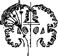
Figure 1. Course of motor fibers from the cortex to the internal capsule inclusive. Black rectangles indicate the various localization of lesions.
B. Local symptoms are also called focal (hence topical diagnosis). The study of local symptoms in a patient is the basis for a conclusion about a topical diagnosis. 1. The most studied area in terms of symptomatology is the region of the central gyri of the Brain. Thanks to this, other symptoms can be considered as localizing anterior to the central gyri—in the direction of the frontal lobe, or posterior to them—in the direction of the occipital lobe, or inferior to the central gyri—in the temporal lobe. The region of the central gyri, in the case of its disease, manifests itself by motor deficit disorders (pareses, paralyses; see Figure 1) or irritation (convulsions, seizures). Anterior to the Rolandic fissure are located parts of the brain having a primary relationship to the act of movement, while posterior to it—to the act of perception, i.e., sensitivity. A lesion of the central gyri is expressed by disorders of the leg if it involves approximately their upper third, by disorders of the arm in case of disease of their middle part, and by impairment of the functions of the face and tongue in case of a lesion of the lower section. Here, in the majority of cases involving a lesion of the anterior central gyrus, convulsions, pareses of a monoplegic nature, and disorders of individual muscle groups and even isolated movements of any one muscle can be observed. Typical symptoms of irritation in this area should be considered attacks of cortical convulsions, Jacksonian type cortical epilepsy (epilepsia corticalis; see Epilepsy). In the case of disease of the posterior central gyrus and areas posterior to it, there are disorders of sensitivity, anesthesia, hyperesthesia, pain in the corresponding limbs or parts thereof or the whole body. Depending on the lesion in this area, one can observe the sensory aura of an epileptiform seizure, epilepsia corticalis sensoria, etc. The most typical for a lesion of the cortical sensory zones should be considered the loss and disorder of deep sensitivity, kinesthetic sensations (the so-called "muscle sense"), and astereognosis. Of particularly great importance for a topical diagnosis is the order of development of a Jacksonian epilepsy seizure, namely—whether the seizure develops in the order of the arrangement of the cortical motor centers. When observing a patient, it is always extremely important to clarify how the seizure developed, where the convulsions began, how they spread to other muscle groups, etc. From the central gyri, the motor pathways for voluntary movements go downward through the corona radiata to the internal capsule
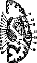
-thalamus opticus.
and further into the brainstem. The closer a lesion is to the cerebral cortex, the greater the chance that a focal disorder of the same size will produce a more isolated (monoplegic) lesion. This depends on the fact that nerve pathways in the internal capsule lie more closely together, while in the direction of the cerebral cortex they diverge fan-like. Conversely, capsular lesions usually produce not mono-, but hemiplegic symptoms. Signs of damage to the motor pathway for voluntary movements: pathological reflexes of Babinski, Rossolimo, Mendel-Bechterew, Oppenheim, etc., increased tendon reflexes, weakening of cutaneous reflexes, clonus. In the posterior limb of the internal capsule, nerve pathways are arranged in a specific way, which is also reflected in the symptomatology of this region (see Capsula interna; see Figure 2). Figure 2. Horizontal section of the right cerebral hemisphere (schematic). Distribution in the internal capsule of motor fibers going to various organs (1-12): 1-for the eyes; 2-for the head; 3-for the mouth; 4-for the tongue; 5-for the shoulder; 6-for the elbow; 7-for the wrist joint; 8-for the fingers; 9-for the trunk; 10-for the hip joint; 11-for the knee; 12-for the toes. 13-fiss. parieto-occipit.; 14-nucleus caudatus; 15-nucleus lenticul.; 16-claustrum; 17-sensory fibers; 18-fiss. Sylvii; 19-radiatio optica; 20- If, in hemiplegia, there is a sensory disorder on the same side of the body, this means that there is damage to the posterior part of the posterior limb of the internal capsule or the posterior parts of the thalamus (see Thalamus opticus). Regarding movement disorders in diseases of the brainstem, see Alternating syndromes. When the motor neuron in the brain is damaged, the paralyzed limb appears to be in a state of contracture: in the arm, a flexion contracture with adduction to the trunk is usually observed; in the leg, with the knee extended, the foot is flexed and turned inward (the picture of spastic hemiplegia). In the case of symmetrical damage to the central gyri in their upper part, near the midline, a symptom complex of spastic weakness of both legs (paraplegia spastica inferior cerebralis) is observed. This is most often observed in cases of trauma and wounds to the skull along the midline. In congenital Little's disease, which develops in cases of difficult labor with a narrow pelvis, symmetrical spastic pareses and paralyses of the upper and especially the lower extremities are also observed. In the latter case, the picture of sharp contracture of the adductor muscles of the thighs is especially characteristic, so that the legs cross over one another. If the disease (trauma, wound, tumor) involves the region of the central gyri along the midline over a larger extent, then symptoms of damage from the upper and middle parts of the central gyri may appear, i.e., from the legs and arms in the form of tetraplegia, or quadriplegia, triplegia, depending on which areas are involved by the pathological process. Similar types of motor disorders are observed in the case of bilateral lesions of the motor pathways in the white matter of the brain, for example, as a result of two strokes. With symmetrical bilateral damage to the pyramidal tract in the brain, there are disorders of speech, swallowing, and facial musculature (pseudobulbar disorders). These disorders of a bulbar nature do not depend on damage to the nuclei of the cranial nerves located in the medulla oblongata, but only on bilateral damage to the central pathways of voluntary innervation going to these nuclei, which is why they are called pseudobulbar. Since the brainstem apparatuses for swallowing, speech, and the musculature of the upper part of the face receive innervation from both hemispheres of the brain, these disorders are usually not observed in unilateral focal disease of the brain in the region of the pyramidal tract (in simple hemiplegia). In cerebral damage to the motor pathway in the brain, there may sometimes exist on the paralyzed limbs of the patient, in addition to diffuse muscle wasting, quite significant muscle atrophies without a reaction of degeneration. It is interesting that these atrophies in some cases of cortical lesions have the character of isolated wasting of individual muscles or atrophies of a selective nature, as if of radicular or spinal origin, but without degenerative phenomena in them. Usually, in hemiplegias or monoplegias of cerebral origin, one can also note other vegetative disorders on the part of the affected limb: changes in blood circulation, cyanosis, lowering of temperature, edema, changes in sweating, trophic disorders on the skin, osteoporosis of the limbs on radiographs. Sensory disorders of an encephalopathological nature are expressed most often, especially with foci within the internal capsule, by a picture of hemianesthesia or hemihypesthesia, whereby sensitivity is usually more severely affected in the distal parts of the limbs than in the proximal ones. Sometimes hemianesthesia is profound throughout the entire half of the body. Sensitivity suffers particularly sharply in cases of damage to the thalamus (predominantly pain and temperature sensitivity). When the focus is localized in the latter, pains of cerebral origin may be observed. These pains are sometimes extremely severe, involving the entire limb or half of the body or any one segment of a limb. 2. Anterior to the anterior central gyrus are located the frontal lobes of the brain, namely, the posterior parts of the three frontal gyri. Regarding questions of topical diagnosis, it is important to know that when a pathological focus is localized in the posterior part of the 1st frontal gyrus, a disorder of movements on the part of the trunk may be observed. The posterior part of the 2nd frontal gyrus, when diseased, causes a disorder of conjugate movements of the eye muscles and head movements (pareses, convulsions, turning of the head and eyes to the side, increased blinking, etc.). When localized in the same area or slightly anterior to it, a disorder of writing (kinetic or motor agraphia) is sometimes also observed. Finally, the posterior part of the 3rd frontal gyrus, when diseased, signals with speech disorders; in this case, the classic symptom complex is Broca's motor (kinetic) aphasia in left-sided lesions in a right-handed person (see Aphasia). More anterior (frontal and prefrontal) parts of the brain, when diseased, produce a whole range of symptoms that are not yet amenable to strict localization. These symptoms concern both the physical and mental state of the patients. Somatic disorders include: frontal ataxia, changes in the manner and peculiarities of performing movements (similar to phenomena of catatonia, stereotypy), and disorders of tendon, cutaneous, and pupillary reflexes in the form of a difference between them on one side and the other, or the loss of some of them. The following symptoms relate to the psychomotor sphere: disturbances in the functions of speech, action (ideational apraxia, or apraxia of intent), behavior, and writing, predominantly in terms of their activity, voluntariness, initiative, and initiation. Symptoms of a mental order are expressed especially by disturbances of active attention, clarity of consciousness, memory, observation, volitional functions (unmotivated, inappropriate actions), and mood (inappropriate cheerfulness, forced smile, tendency to make jokes)—in general, by a disturbance of the functions of activity, inhibition, regulation, self-awareness, and self-control. The symptoms of the frontal lobes just listed are observed in focal diseases of them, but these symptoms play a major role, bearing a more softened or generalized character, also in general diseases of the cerebral cortex, as is the case in psychoses, e.g., in progressive paralysis, dementia praecox (schizophrenia), and senile forms. At the present time, when knowledge of the focal symptomatology of the frontal lobes of the brain has improved, one can discern or anticipate the involvement of the frontal lobes in the pathological process in the picture of one or another mental illness. Such frontal symptoms should be considered especially the disorder of behavior, the weakening and general change of active attention, initiative (Khoroshko), and what is called volitional functions in the language of psychology. As symptoms of the frontal lobes of the brain, but from the side of its base, there also occur disorders of smell (anosmia) as a consequence of pressure, e.g., from a tumor, on the olfactory tract and bulb. 3. Posterior to the region of the central gyri are located the parietal and occipital lobes. The symptomatology of the parietal lobes is expressed predominantly by disorders of deep sensitivity, astereognosis, tactile agnosia (epilepsia sensoria corticalis); these symptoms are observed in damage to those parts of the parietal lobes which are adjacent to the posterior central gyrus and occupy the more superior parts of the parietal lobes. The lower part of the parietal lobe is composed mainly of the gyrus supramarginalis and gyrus angularis, which, when diseased, produce asymbolic disorders.
A very characteristic symptom of a lesion of the gyrus supramarginalis on the left side is kinetic (motor) apraxia, and for the gyrus angularis, which lies at the junction of the parietal, occipital, and temporal lobes of the brain, the most important phenomenon in disease is a disorder of reading (alexia, optic agnosia, and the symptom complex of psychic blindness). The latter symptom complex is observed more often with bilateral localization, but sometimes also with only left-sided lesions. Moving further posteriorly, toward the occipital pole of the brain, one finds on the side of the cerebral cortex, namely in the region of the cuneus (cuneus, area striata), i.e., on the inner side of the hemisphere, a place whose lesion is expressed by a very characteristic sign—a disorder of vision in the form of so-called cortical blindness. In this case, with a lesion of the left hemisphere, a loss of function of the left halves of the retinas of both eyes is observed, i.e., right-sided hemianopsia; with a right-sided disease—left-sided hemianopsia, i.e., in both cases, it is crossed. With bilateral lesions of both inner parts of the occipital lobes in the region of the cunei (bullet wound, tumor, softening, etc.), complete cortical blindness may develop (see Hemianopsia).
4. Below the central gyri lies the temporal lobe of the brain, the symptomatology of whose diseases is expressed by Wernicke's sensory aphasia—word deafness (with a lesion of the posterior part of the 1st temporal gyrus on the left), psychic deafness (with extensive lesions), and complete cortical deafness (with bilateral lesions of the 1st temporal gyrus). With disease of the anterior end of the temporal lobe, a disorder of taste (ageusia) is noted. With a lesion of the posterior part of the left inferior temporal gyrus, amnestic aphasia may be observed. Sometimes with disease of the temporal lobe, disorders of smell are found (anosmia, olfactory hallucinations) as a result of a lesion on the inner side of the uncus gyri hippocampi. As a supplement to the description of the cortical symptomatology of the Brain, see Asymbolia, Aphasia, Apraxia, Agnosia, Agraphia, Alexia, Amusia, Anosmia, Psychic deafness, Psychic blindness, Agrammatism, Hemiplegia, Contractures.
5. Symptom complexes of the base of the brain have the following localizations [see separate table (cols. 527-528), fig. 2]: a) Anterior cranial fossa. Here, painful phenomena are characterized by disorders of smell (I pair of cranial nerves), symptoms from the frontal lobes (see above), and a disorder of vision (pressure on the optic chiasm). In the case of, for example, a large tumor, the pressure may extend to the orbit and cause protrusion of one eyeball. b) The middle cranial fossa, upon its disease, gives symptoms from the chiasm, pituitary gland, oculomotor nerves, and the Gasserian ganglion. This symptomatology reduces to bitemporal or binasal hemianopsia (depending on how the chiasm is compressed: from its inner or outer sides), to symptoms from the pituitary gland (acromegaly, changes in the sella turcica on an X-ray, etc.), to paralysis of the eye muscles, and to pain and anesthesia in the region of the trigeminal nerve. In the middle cranial fossa also lie the temporal lobes of the brain, and therefore their symptomatology is of definite interest here. c) Posterior cranial fossa; the symptomatology of this region is very complex. On one hand, a large mass of cranial nerves lies and passes here (oculomotor, facial, trigeminal, auditory, glossopharyngeal, vagus, hypoglossal), the brainstem, and the cerebellum; all these organs are covered from above by the tentorium cerebelli, and this entire region represents, as it were, a special part of the cranial cavity, somewhat closed off from the brain, but the closure here is only relative, since the relations with neighboring parts are variable, and the spread of the pathological process, either directly or through pressure (specifically on the occipital and temporal lobes of the brain), can take place. The most important localization of the process relating here is localization in the cerebellopontine angle (see). Most often, the growth of a tumor originating from the auditory nerve is observed here; in this case, hearing is impaired, and the adjacent facial nerve and the corresponding hemisphere of the cerebellum are involved in the process (“hemiasynergy,” “adiadochokinesis,” etc.). In addition, see additionally—Alternating syndromes, Cerebellum.
6. Besides the already described symptoms in the field of encephalopathology, relating predominantly to the phenomenology of diseases of the cortex of the Brain, individual signs and complexes of them are also observed depending on lesions of the white matter, subcortical nodes, and also the diencephalon. Lesions of the projection systems of the Brain are best known in relation to the pyramidal tract, the signs of the disease of which were already described in connection with the lesion of the central gyri; in addition, damage to the visual pathways (radiatio optica, fasciculus Gratiolet) is of great importance, especially in the space from the posterior part of the internal capsule to the cortex (cuneus) of the occipital lobe (hemianopsia). Furthermore, the frontopontine system, passing through the corona radiata of the frontal lobes and going to the cerebellum, can result in ataxia as a result of its lesion. Disorders of hearing are observed only with bilateral diseases of the pathways from the internal capsule to the temporal lobes of the brain. The corpus callosum also relates to the white matter, among the symptoms of the disease of which general dementia and apraxia of the left hand are best known (the latter is observed with a lesion of the pathways going from the gyrus supramarginalis sinister to the right anterior central gyrus).
7. Large subcortical nodes can give a very diverse symptomatology; most characteristic in it are motor disorders of so-called extrapyramidal origin: dystonias, chorea, athetosis, spasms, paralysis agitans (parkinsonism), rigidity, involuntary movements, and constant hyperkinesis of various kinds.—See also—Amiostatic symptom complex, Athetosis, Wilson's disease, Torsion spasm, Epilepsy, Chorea, etc. All these diseases, disorders, and symptom complexes are now linked to a number of formations, such as the corpus caudatum, thalamus opticus, nucleus lenticularis and its parts: putamen and globus pallidus, locus niger, etc. However, besides motor disorders, the complex system of subcortical formations and the diencephalon also gives a whole range of vegetative disorders, namely from the side of blood circulation, lymph circulation, thermoregulation, mineral metabolism, carbohydrate metabolism, water metabolism, etc. (see Vegetative nervous system, pathology). It is also necessary to mention disorders of hearing and vision linked to the internal and external geniculate bodies, as well as the quadrigeminal plate; in connection with these formations, disease of the pineal gland (premature sexual development and adipositas epiphysaria) is also of great interest. As symptoms from the brainstem, namely the gray matter around the Sylvian aqueduct and the III cerebral ventricle, it is necessary to name increased drowsiness and somnolence, as is observed in lethargic encephalitis or tumors. The ventricles of the Brain, being stretched by accumulated fluid or involved in a pathological process, e.g., by a tumor, can give a number of symptoms, the recognition of which is far from easy. Since the pathogenesis of these symptoms must be attributed to pressure from inside the ventricles, especially the lateral ones, in hydrocephalus, and since the direction of this pressure can vary greatly depending on a whole range of circumstances, the uncertainty and variability of the symptoms become understandable. Here are observed: paretic-spastic phenomena up to bilateral hemiplegia, disorder of coordination, ataxia, epileptic seizures, sometimes of the Jacksonian type, paralysis of cranial nerves due to their compression by the entire mass of the brain, and endocrine-vegetative disorders (e.g., pathological obesity, virilism, hirsutism, etc.) as a result of pressure on the region of the III ventricle, pituitary gland, pineal gland, etc., and psychic symptoms.
General diagnostics of diseases of the Brain. Examination of a patient with a disease of the Brain is required to be performed systematically in relation to movement, sensation, reflexes, functions of the sense organs, and vegetative and psychic functions. It is very important to have a sequential and thorough study of the state of all cranial nerves. In the future, knowledge of certain symptom complexes relating to the pathology of the Brain is very helpful. Special examinations of the fundus of the eye, the auditory apparatus, the nasal cavity and larynx, X-ray diagnostics of the skull, including encephalography, and examination of the cerebrospinal fluid are of enormous importance, not to mention the examination of urine, blood, and internal organs. When establishing a diagnosis, the question is first decided whether one is dealing with an organic or functional disease. One must remember the ever-present possibility of the existence of functional layers in an organic disease.
Diagnosis of brain diseases is complex, as in addition to a symptomatic diagnosis (hemiplegia, monoplegia, hemianopsia, dystonia, athetosis, etc.), it is required to establish the site of the lesion (topical diagnosis), the etiological (syphilis, tuberculosis, arteriosclerosis, etc.), and the nosological (tumor, progressive paralysis, etc.) diagnosis. The course of the disease—acute, subacute, or chronic, remissions, or steady progression of the disease—is of great importance for the diagnosis. It must be kept in mind that in some cases (especially with tumors, hydrocephalus), compression of individual parts of the brain may occur, e.g., of the cranial nerves (most often the nervus abducens), due to their being pressed by the mass of the brain against the base of the skull or due to counter-pressure by the mass of one hemisphere, increased in size, against the other hemisphere or the cerebellum. In this way, one can sometimes explain the appearance of certain symptoms that do not fit into the strict framework of the intended topical diagnosis. Besides diseases localized in any one place in the brain, there may be diseases with multiple localization (syphilis, multiple sclerosis, cysticercosis, encephalitis). Special pathology of brain diseases. 1. Circulatory disorders in the brain are observed in various forms. Symptom complexes of cerebral anemia and hyperemia were mentioned above. a) Cerebral anemia in an acute form is observed with a large loss of blood by the organism (operation, injury, internal bleeding), with the outflow of blood to other organs (collapse, etc.). Chronic cerebral anemia occurs with anemia, states of weakness, and intoxications. Treatment is causal; as symptomatic therapy—stimulants (camphor, valerian, caffeine), physiological saline. b) Cerebral hyperemia can be active (due to intense mental work, dilation of arteries under the influence of intoxication, etc.), congestive (due to difficulties in venous outflow), and constitutional (as a result of metabolic peculiarities, general plethora, etc.). Treatment is causal; as symptomatic agents, the following are used: laxatives, bloodletting (leeches, venesection), counter-irritants (cupping, cantharides, mustard plasters), hot foot and hand baths, diet. c) Hemorrhage (see separate table, fig. 3)—one of the most frequent diseases of the brain. The disease develops suddenly, in a stroke-like manner ("stroke") with loss of consciousness. The preceding threatening state is expressed by hypertension, habitus apoplecticus, and general phenomena of arteriosclerosis. The age of the patients
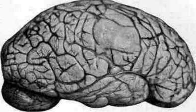
I'HI- I

'*T[t $
Brain [hemorrhages] usually [occur] above 40-50 years of age, but hemorrhages also occur at a young age. (For more details on brain hemorrhage, see Apoplexy.) d) Thrombosis of the cerebral arteries. Thrombosis of the cerebral arteries usually occurs at the same age as in cases of hemorrhage. The main pathological process is arteriosclerosis. Thrombosis usually develops without loss of consciousness, gradually, with the presence of a non-tense, but on the contrary, weak pulse and cardiac insufficiency. The area of the middle cerebral artery is most frequently affected. Paralysis does not develop immediately in this case. Sometimes one can observe how, with the timely administration of agents that increase cardiac activity, the phenomena of paresis disappear, only to reappear upon weakening of the heart. If the clinical picture of brain thrombosis develops in a young subject and there are no signs of heart defect or any other definite causes (e.g., encephalitis, migraine), then in this case, the thrombosis is most often of syphilitic origin. In these cases, it is extremely important to begin specific treatment as early as possible, since the success of therapy and the outcome of the disease are usually inversely proportional to the time of initiation of treatment. Thrombosis on the basis of syphilitic vascular lesions of the brain is too frequent a phenomenon in general not to apply specific therapy in doubtful cases. Fatigue, infections, weakening of the organism, pregnancy and the postpartum period, and the state of deep sleep contribute to the development of thrombosis. The outcome of thrombosis depends on the location and degree of the lesion and the earliest possible improvement of circulatory conditions. Treatment: rest, raising cardiac tone, reducing blood viscosity (cardiac, iodine preparations, not to mention specific therapy), diet. The patient must remain in bed for at least three weeks. Treatment of residual phenomena is symptomatic; for spastic paralysis—massage, heat, passive and active gymnastics, of course in a cautious form. The use of electrotherapy in relation to the affected limb in spastic phenomena is contraindicated as it intensifies contractures and increases reflex excitability. It is very important from the very beginning of the disease to take care that contractures do not develop (correct positioning of the limb, splints, passive movements). Great importance should be attached to active gymnastics, individualized in each separate case. Instilling in the patient faith in success regarding the necessary active movements and their correct selection is by no means an easy task. In aphasic, apractic, agraphic, alexic, and other asymbolic disorders, treatment with exercises, in particular according to the method of initial education, is undoubtedly expedient. Unfortunately, success here does not always correspond to the efforts of the physician and the patient; at an older age, the chances of success in training decrease. Treatment of arteriosclerosis according to general rules is mandatory, since in the case of the development of a cerebral stroke, whether on the basis of hemorrhage or thrombosis, there is always a fear of the development of a new stroke. In some cases, strokes are multiple. e) Arteriosclerosis is the most common disease of the brain, being a particular case of arteriosclerosis (atheromatosis) in the organism. Separate pathological-anatomical forms of brain arteriosclerosis: lesion predominantly of the large vessels of the brain, arteriosclerosis of its small vessels, obliterating endarteritis. On the basis of arteriosclerosis, hemorrhage, thrombosis, and softening of the brain usually develop; among other clinical forms, one must mention: initial arteriosclerosis of the brain, late epilepsy, arteriosclerotic dementia, etc. For more details, see Arteriosclerotic psychoses. f) By softening of the brain, or encephalomalacia (ramollitio cerebri, sive encephalomalacia), one must understand the formation of necrotic areas in the brain due to an obliterating process in an artery, i.e., the formation of an infarct (see separate table, figs. 4 and 5). Softening of the brain is also observed in thrombosis, in embolism of a cerebral vessel, in leukemia, tumor metastases, and in primary tumors (e.g., gliomas), in poisonings (CO, mercury, manganese, botulism, etc.). Most often, the causal factor lies in heart disease (manifested by cardiac weakness and capable of being a source of embolism, thrombosis), atheromatosis of vessels, syphilis. Softening of the brain can be observed not only in the elderly, but at all ages. Especially interesting in clinical and practical terms are encephalomalacias in connection with poisonings by industrial poisons (brain toxicosis and encephalopathies). In softenings, just as in thrombosis and embolism, syndromes of lesion of the basin of one of the cerebral arteries are observed: anterior, middle, or posterior (see above the diagrams of blood circulation in the brain). The area supplied by the middle cerebral artery is most frequently affected, less often by the other two: a) The syndrome of the middle cerebral artery is expressed by hemiplegia or monoplegia, mild sensory disturbances, homonymous hemianopsia, sensory aphasia, motor aphasia, anarthria, apraxia, etc. b) The syndrome of the anterior cerebral artery gives the following symptoms: mental disorders (see above the symptomatology of the frontal lobes of the brain) and sometimes weakness of the leg (lobulus paracentralis). c) The syndrome of the posterior cerebral artery can be divided into two: the syndrome of its anterior and the syndrome of its posterior branches. Lesion of the posterior branches is expressed by homonymous hemianopsia, psychic blindness, alexia. Softening in the anterior branches of the posterior cerebral artery gives the syndrome of the optic thalamus (hemianesthesia, hemiplegia, hemianopsia) and the region of the red nucleus (cerebellar symptoms). It must be kept in mind that not the entire area of a known basin may be involved in the softening, and then the clinical picture also turns out to be narrower, underdeveloped. Since in softening a large area of the brain is usually involved in the pathological process and a necrotic lesion takes place, treatment and prognosis in such cases appear more or less dismal. Treatment is, if possible, causal and predominantly symptomatic. According to data from the pathological-anatomical departments of Moscow hospitals for 1923–27, softening of the brain (red and white) as a cause of death was found 719 times, which constitutes 1.9% of all autopsies and 28% of all nervous diseases. g) Embolism of cerebral vessels constitutes one of the most frequently encountered causes of softenings. Most often, embolism occurs in mitral stenosis and septic endocarditis. The material for the embolus usually consists of thrombotic deposits on the valves. The embolus may come from an aneurysm of the aorta, carotid artery, or vertebral artery. Its source can be fragments of tissue during a suppurative process, pulmonary tuberculosis (transfer of tubercle bacilli to the brain), traumatic injury (fat embolism, e.g., in bone fractures; air embolism, e.g., in whooping cough). Embolism of a larger vessel usually develops in a stroke-like manner with loss of consciousness. Fat embolism of the vessels of the medulla oblongata can lead to sudden death. The diagnosis of embolism is based on establishing the presence of a source of embolism. Treatment of brain embolism: rest, if possible, causal therapy, symptomatic therapy; iodine is also used as an agent of influence (as a means of promoting the resorption of the focus), but apparently without special results. Treatment of the consequences of embolism is carried out according to the same methods as in other cases of brain disorders. h) Aneurysms of cerebral arteries are observed in the form of small, miliary aneurysms, which are sometimes a source of hemorrhages, and in the form of larger aneurysms of such vessels as the vertebral artery, internal carotid, artery of the Sylvian fissure, the arterial Circle of Willis, etc. Usually, brain aneurysms proceed covertly, sometimes having a course similar to brain tumors; in the majority of cases, they are a finding at autopsy. In some cases, patients feel special noises in the head, and sometimes this noise can be heard by a physician from the side of the skull; noises can be observed in sepsis lenta. Regarding therapy, one must first of all think about syphilis. i) Thrombophlebitis, thrombosis of the venous sinuses. In weak, emaciated children after severe infections or diseases, in cachectic patients with tuberculosis, cancer, etc., marantic thrombophlebitis of the cerebral sinuses can develop. In other cases, thrombosis of the sinuses develops due to the local spread of infection from surrounding parts to the sinus. From the face, pharynx, oral cavity, nose, and accessory cavities, the process can pass through the vena ophthalmica or plexus pterygoideus to the sinus cavernosus. Most often, the complication of thrombophlebitis is observed in ear suppurations - the spread of infection to the sinus sigmoideus and sinus transversus, from where it further proceeds into the vena jugularis.
Finally, from the surface of the skull through the emissaria parietale et occipitale, the inflammatory process can spread to the sinus longitudinalis superior. As sources for thrombosis of the cerebral venous sinuses, there can be boils, erysipelas, infected head wounds, anthrax (pustula maligna), dental infection, and suppurative processes of the jaw, eye, orbit, nose, ear, etc. Sometimes sinus thrombosis develops sequentially after thrombosis of the small veins of the brain itself. The clinical picture consists of general symptoms of exhaustion or infection, phenomena of increased intracranial pressure, and local disorders. In addition to local painful phenomena, redness, and edema, symptoms from the nervous system are also observed. In thrombosis of the transverse and sigmoid sinuses, signs of damage to the glossopharyngeal nerve (taste disorders, paresis of the soft palate), the vagus nerve (anesthesia of the pharynx, disorders of the larynx, swallowing, pulse), the accessory nerve of Willis (m. sterno-cleido-mastoideus, m. cucullaris), and the hypoglossal nerve (symptom complex of the foramen lacerum posterius) are noted. In thrombosis of the superior longitudinal sinus, signs of venous stasis of the entire surface of the cerebral cortex are noted, up to epileptiform seizures and paralysis, and in addition, swelling of the skin on the surface of the head (vertex), recurrent nosebleeds, and swelling of the veins on the scalp (caput medusae). Sinus thrombosis differs from meningitis in that the cerebrospinal fluid in thrombosis appears without special changes. In view of the great importance of the factor of exhaustion and infection in the origin of sinus thrombosis, preventive measures must be of very great importance here in the sense of special attention to children exhausted by disease and to the most thorough possible treatment of infectious diseases on the head. Timely surgical intervention, and in particular ligation of the internal jugular vein, are in certain cases life-saving measures. Otherwise: rest, treatment of infection, and exhaustion according to standard rules. 2. Syphilis, multiple sclerosis, and hysteria (see) are diseases that often cause the greatest difficulties for differential diagnosis and clinical analysis. 3. Inflammatory-infectious processes in the brain (see Meningitis, Encephalitis) produce a whole gamut of diseases that are different in their characteristics, both in etiological and clinical respects. One of the most important moments in diagnostic terms is an increase in temperature, although hyperthermia is also possible depending on local damage to thermoregulatory centers. 4. A brain abscess can be considered a severe consequence of the penetration of infection into the brain. Most often, abscesses develop in the temporal lobe and cerebellum through the spread of infection from the ear. Abscesses of the frontal lobes of the brain are observed more often in inflammatory processes in the paranasal sinuses and bronchiectasis. Metastatic abscesses are observed in septicemia, endocarditis, osteomyelitis, empyema, etc. Disease of the bones of the skull (osteomyelitis) can be a source of the development of a brain abscess in the vicinity. In cases of skull trauma and traumatic brain injuries, an abscess sometimes develops as a complication. In some cases, it is not possible to isolate any infectious pathogen from the contents of the abscess or find one in the body. In such cases, one can think of an idiopathic sterile abscess, arising as a result, perhaps, of general brain trauma or after an initial necrotic process of another kind in the brain tissue. A brain abscess can sometimes develop over the course of several days, and sometimes several years. According to Moscow data for 1923–1927, a brain abscess was encountered 70 times in 37,899 autopsies. In addition to focal symptoms, an abscess causes an increase in temperature (not always significant), a picture of increased intracranial pressure (see above), congestive phenomena in the eye, and leukocytosis in the blood; if the abscess is located close to the meninges, an inflammatory-purulent reaction is noted in the cerebrospinal fluid. The general picture of the progression of phenomena and their character are similar to a brain tumor. Trial anti-syphilitic mercury treatment can sometimes give temporary improvement of phenomena and thereby confuse the diagnosis. Prophylactically, thorough treatment of skull wounds, ear diseases, and generally purulent-inflammatory processes in the body is very important. When an abscess is recognized, treatment is surgical. The results of the latter vary among different authors, but in general, they are far from hopeless (from 37% to 60% recoveries). Unpleasant outcomes of an abscess: rupture of pus into the meninges or into the cerebral ventricles; the latter complication quickly leads to death with a picture of collapse. When operating on a brain abscess, it is necessary to take into account that often, in the vicinity of the opened abscess, there may be additional encapsulated abscesses. 5. Tumors of the Brain. The pathological anatomy of brain tumors and the symptom complex of increased intracranial pressure were described above. Local symptoms arising with one or another localization of a tumor correspond to the data that form the basis of the phenomenology of Brain diseases in connection with questions of topical diagnosis. Brain tumors can have an extremely typical course and picture, starting gradually with indefinite headaches and malaise, then passing to classic symptoms: congestive phenomena in the fundus of the eye, choked disc, bouts of vomiting, slowing of the pulse, in the presence of certain local phenomena (paresis, paralysis, convulsions, seizures, hemianopsia, hearing impairment, speech disorder, etc.) and general stupor, inhibition, staggering when walking, and ending with atrophy of the optic nerves, increasing paralysis, loss of memory, intellect, interests, the onset of drowsiness, general fading of life, and finally death. The general scheme of such a course of a brain tumor is expressed by a line of gradual worsening of phenomena, increasing symptoms, and progression of the disease in accordance with the growth of the tumor itself and the gradual compression of the surrounding parts of the brain and the pressing of more distant parts and the entire mass of the brain against the bones of the skull. In the initial stage of tumor development, general cerebral symptoms arise, apparently due to changes in blood circulation, disturbances in the secretion of cerebrospinal fluid and circulation, stasis, irritation of the meninges, intoxication by the products of the tumor itself, and the general reaction of the brain to the foreign growing body that has appeared in it. Subsequently, general cerebral symptoms, and in particular mental disorders, are likely derivatives not only of the influence of the tumor on the general state of the brain but also of the compression of its frontal lobes, since the dementia observed in this case often bears the picture of mental defects encountered in diseases of the anterior parts of the brain, whereas the tumor may be located in another part. Sometimes in the course of a brain tumor, one notes, as it were, stroke-like worsenings of the disease, stages in its progressive course. They either depend on hemorrhages into the substance of the tumor or can be considered as certain crises in the relationship between the tumor, on the one hand, and the brain and the entire organism, on the other. In these crises, the advantage usually remains on the side of the tissue of the foreign progressive growth, but at times, clinically, one can observe a kind of calming of the process, a temporary stop, even some improvement, for example, under the influence of mercury therapy. In addition to the more or less typical course of a brain tumor, the latter often has a very masked development, proceeds secretly or under the guise of another disease: syphilis, arteriosclerosis, progressive paralysis, encephalitis, even functional disorder. Headaches, vomiting, choked discs, slowed pulse, and other classic symptoms for a tumor may not occur for a long time or at all until death. The patient dies with one diagnosis, and at autopsy, a brain tumor is found. Tumors of the anterior parts of the brain and tumors located deep in the brain produce general cerebral phenomena and, in particular, changes in the fundus of the eye relatively later than tumors located at the base, in the posterior cranial fossa, near the meninges of the brain. Tumors of the frontal lobes, the right temporal and parietal lobes can sometimes exist for a very long time without local symptoms. If a tumor originates from the meninges and has a capsule, then with slow growth, it can very gradually compress the brain substance, giving the latter the opportunity, by adapting, to maintain its functions up to a certain moment. This explains such facts when focal phenomena develop suddenly, making it possible to make a topical diagnosis, while on the operating or anatomical table, an extremely large tumor is found, the location of which could not be determined until the indicated last moment of the development of local symptoms. The symptom complex of increased intracranial pressure is observed not only with a tumor but also with other diseases, such as, for example, brain abscess, syphilis, hydrocephalus, meningitis, encephalitis, brain parasites, aneurysm, meningitis serosa cystica circumscripta, etc. It is not always possible to differentiate.
The following data can serve as reference points for diagnosis: an abscess develops faster than a tumor, causes a febrile state and leukocytosis, although not always, and there is a source for pyogenic infection. For syphilis, serological reactions and anamnesis are significant, but often the question must be decided by prescribing specific treatment (the latter, in doubtful cases, should be prescribed for no more than 2–3 weeks so as not to delay the question of surgical treatment). An acute and even stormy course in meningitis and encephalitis, and corresponding data from the cerebrospinal fluid (lumbar puncture is not recommended when a tumor is suspected to be localized in the posterior cranial fossa) can be a guiding thread for the diagnosis. Chronic hydrocephalus, chronic meningitis, meningitis serosa cystica circumscripta, and pseudotumor cerebri sometimes present great difficulties for differentiation if there are no such striking indications as a large hydrocephalic head or sharply pronounced meningeal phenomena. In such cases where diagnosis is difficult, encephalography and the study of pneumo-radiographic images obtained in different positions of the patient's head are indicated. However, even these additional data do not always have decisive significance. It is also possible to use the Neisser-Pollack brain puncture (a type of brain biopsy). When there is difficulty in diagnosis, one has to leave the decision of the question to time, wait, or apply trial surgical intervention. The described difficulties are understandable if one takes into account that usually with brain tumors there are accompanying phenomena in the form of both chronic meningitis and hydrocephalus. In the case of brain parasites (cysticercus, echinococcus), facts such as finding cysticerci in the subcutaneous tissue or echinococcus in the liver, eosinophilia, and a positive result of the Weinberg reaction can be significant. Besides the general diagnosis of a brain tumor, it is necessary to determine its location and its nature. The development of congestive phenomena in the eye to a greater extent on one side, unilateral pain upon percussion of the zygomatic arch (Bekhterev's symptom) and the skull, unilateral localization of pain, and predominant decrease in the conjunctival and corneal reflex on one side, in addition to focal symptoms, can serve as data for resolving the question of the side of the disease. The most reliable are symptoms from the central gyri (convulsions, seizures, paresis, disorder of kinesthetic sensations). Mental symptoms are more often observed with frontal localization. Sometimes the patient's tendency to witticism is particularly demonstrative (however, this sign does not have absolute significance). Particularly pronounced drowsiness along with nutritional disturbance and other vegetative symptoms is an indication of the probability of localization in the region of the base of the brain, near the third ventricle. The question of tumor localization is decided on the basis of the most thorough and comprehensive (do not forget the necessity of repetition) examination of the patient and the evaluation of these data from the point of view of topical understanding. It must be admitted that the question of topical diagnosis is far from always resolved successfully. A classic example of a supposedly gross diagnostic error must be considered the fact when, instead of a frontal lobe tumor, it turns out to be a cerebellar tumor or vice versa; meanwhile, now, after establishing the existing connections between the frontal lobe of one side and the cerebellar hemisphere of the other, and also after taking into account the possibility of the development of symptoms due to counter-pressure and pressing against the bones of the skull, such a, in reality, regular connection of cerebral pathological phenomena should not seem surprising and, on the contrary, should sometimes lead to practical conclusions, for example, to shifting the field of operation in the opposite direction during surgical intervention in case of negative data. It is very important to clarify the sequence of the appearance of individual symptoms and to establish the earliest symptoms in time, on which data one should sometimes build a hypothesis about the topical diagnosis.
The nature of the tumor during life and before the operation has to be established rather conjecturally. First of all, one should think about syphilis, and in a practical order, specific treatment has to be carried out as a necessary and consistent measure in a methodical respect. In children, tubercles are most often observed, in adults-gliomas. Besides gliomas in adults (and in children), sarcomas (from the membranes and bones) are encountered relatively often. If in an adult the tumor is localized on the surface of the brain, then in the majority of cases these are meningiomas. Tumors localized deep in the brain, in the cerebellum, or in the brainstem belong more often to gliomas [see separate table (p. 559-560), figs. 1 and 2]. If a large development of vessels on one side or naevus pigmentosus is noted on the surface of the head, then this can sometimes serve as an indication that the tumor belongs to angiomas (see Angioma). Tumors of the cerebellopontine angle (most often of the acoustic nerve) belong to neuromas or peripheral gliomas; in these cases, the presence of signs of general neurofibromatosis is sometimes noted in patients (see Cerebellopontine angle). Cancers of the brain are usually only metastatic, especially cancer of the bronchi; among metastases in the brain, hypernephroma is often encountered. On a radiograph, one can sometimes see accumulations of lime in the tumor (psammomas); deposition of lime can also occur in tubercles, cysticerci, and teratomas. The state of the sella turcica is of great importance, although even here an enlarged sella turcica does not always indicate a tumor, but is also observed with internal hydrocephalus. A radiograph (in profile) gives an indication of increased intracranial pressure (enlarged sella turcica, pronounced digital impressions, thinning of the bones). Destruction and deformation of the sella turcica speak for a tumor of the appendage (acromegaly, adipositas hypophysaria). Encephalography can provide very interesting and valuable data in a diagnostic respect for brain tumors in those cases where the recognition of the nature of the process and its location presents difficulties for an experienced neuropathologist. Treatment of brain tumors to this day remains predominantly surgical. If a radical operation cannot be performed or the localization of the tumor is unknown, then a decompressive trepanation is performed. Deep radiotherapy in cases of tumors of the infundibulo-hypophyseal region, where surgical intervention is associated with very great and severe trauma, should be preferred to surgery, especially since it often yields certain positive results. In the case of gliomas located deep in the brain, which usually do not possess a capsule, radiotherapy is also more indicated and is preferred to surgical intervention. Regarding tumors of the choroid plexus - see Chorioideus plexus. 6. Among the rarer diseases of the brain, one must also name its parasites. As such, cysticercus and echinococcus are observed. The clinical picture is similar to a brain tumor (see there regarding diagnosis). However, cysticercus of the brain is often not recognized clinically, as it can proceed very diversely, for example, under the guise of epilepsy, multiple sclerosis, psychosis, etc. Cysticercus is often multiple, scattering throughout the entire brain—both in its substance and in the membranes. Sometimes cysticercus floats in the ventricles of the brain and can develop in the fourth ventricle. In isolated cases, it was possible to remove cysticercus from the fourth ventricle of the brain by surgical means. Echinococcus can reach the size of a fist in the brain. Treatment, if possible, is surgical. 7. Traumatic diseases of the brain from the point of view of encephalopathology are of great interest in that the place of damage to the brain is often visible to the naked eye in them, as, for example, in cases of bullet wounds of the skull, thanks to which the topical diagnosis does not present difficulties. It must be remembered that with skull injuries, it is still not always possible, despite the obviousness of the bullet track or damage to a certain part of the brain under the cranial defect, to speak with complete certainty about the destruction exclusively of this place; it is necessary to admit the possibility of a remote result of the trauma in connection with the development of the action of forces during the inflicted external injury in different directions (hydraulic pressure, tangential wound, penetrating injury, introduction of shell and bone fragments into the substance of the brain, contrecoup, hemorrhage, etc.—see Skull, general doctrine of traumatic injuries of the skull). The traumatic etiology of brain diseases is...
S70 is read from injuries during the act of childbirth due to a narrow pelvis and other causes. Congenital traumatic diseases are most often expressed by the picture of diplegia spastica inferior, tetraparesis spastica, forming various forms of the manifestation of Little's disease. In addition to local, simple, and complex brain injuries, general traumas are observed, such as concussion of the brain, contusion of the brain. Phenomena that developed directly as a result of damage to the brain substance are called the pathogenic effect of the trauma. Furthermore, one speaks of the pathotropic effect of brain trauma, meaning by this the influence of a previously sustained trauma on the subsequent development of hemorrhage into the brain, its arteriosclerosis, abscess, tumor, epilepsy, progressive paralysis, multiple sclerosis, paralysis agitans, meningitis, etc. Along with more severe traumatic diseases of the brain, accompanied by gross violations of its integrity, lighter functional disorders of brain activity are observed in the form of traumatic neurosis or psychoneurosis, in which the nature of the pathological-anatomical changes in the brain is unknown. Transitional forms between the former and the latter are such general disorders as concussion of the brain, contusion of it. Organic brain injuries can often be complicated by psychic trauma. Traumatic psychoneurosis often remains as a consequence of a previously sustained concussion of the brain or contusion. When the skull and brain are wounded, an unconscious state usually develops immediately due to brain shock. After the smoothing out of the general phenomena of shock, if there is no complication due to an infection introduced during the wounding, local symptoms of the damage appear particularly clearly in accordance with its location, described above in the symptomatology of the brain. The most severe picture is observed at the beginning of the injury, if the latter proceeds without complications, and then it gradually smooths out and is reduced in the end to a cicatricial change remaining in the brain in its substance. In some cases, after a certain time has passed since the previous trauma, so-called late traumatic apoplexy may occur, apparently due to damage to a vessel that does not manifest itself immediately, sufficient for the development of a hemorrhage. It is very important to radiograph the damaged skull, since cracks in the skull and fragments inside it can be revealed more accurately and definitely only on the X-ray. Complications of wounds and injuries of the skull can be encephalitis, meningoencephalitis, abscess. In case of suspicion of a complication of the injury by infection, surgical intervention is necessary. If there is no complication of an infectious nature, traumatic aseptic encephalitis can be observed as a complication as a reaction of the tissue to decay products, and there may be a reaction of a cytotoxic (neurotoxic) nature. The course of these complications for the most part proceeds favorably in the sense of the absence of a fatal outcome, but it can lead to progressive traumatic dementia. In traumatic diseases of the brain without complication by infection, an increased protein content is often noted in the cerebrospinal fluid (hyperalbuminosis greater than 3-6‰), the amount of urea, sugar may be increased, and pressure may be elevated; the number of formed elements is usually not increased. Similar changes during a lumbar puncture are also observed in general concussion of the brain and contusion by air due to the explosion of a shell; in some cases, an admixture of effused blood is also noted. In general concussion of the brain, besides general cerebral symptoms, local ones are also observed, including often paralysis of the cranial nerves (oculomotorius, abducens, facialis, acusticus, etc.) due to a fracture of the base of the skull (discharge of blood from the nose, ears). General symptoms of concussion of the brain are: unconscious state, vomiting, slowed pulse, headache, dizziness, amnesia, psychic inhibition, confusion, uncertainty, apathy, etc. In concussion of the brain, small hemorrhages, vascular disorders, changes in nerve cells, local edema are found, but the essence of the matter apparently consists in a general shock of the brain enclosed in the cranial box and surrounded by fluid. In air contusion, there is also a blow, since the air in such cases strikes like a solid object. In air contusion (see), due to the flight or explosion of a shell, there is present now a compression, now a rarefaction of the air, consequently now an increase, now a decrease in pressure, as a result of which the occurring hemorrhages and necroses of the brain substance can be considered similar to the phenomena developing in caisson disease, namely due to the release of gases from the blood and subsequent gas embolism. Different degrees of concussion or contusion of the brain are distinguished. In severe cases, the absence of pupillary reaction, urinary incontinence, and an increase in pulse and respiration are noted. Regarding traumatic epilepsy, see Epilepsy. Traumatic diseases of the brain require complete rest in the acute period. Proper rest, especially for the head of the patient, must be taken care of in case of transporting him. Surgical intervention is undertaken only in case of suspicion of infection or if it is necessary to clean the wound, remove fragments. Otherwise, the therapy is expectant. A lumbar puncture is indicated for reducing intracranial pressure. In the case of concussion of the brain, rest should be as prolonged as possible in accordance with the severity of the case. Getting out of bed is permitted only with a normal pulse and the disappearance of all general cerebral physical symptoms. In the case of residual phenomena after brain trauma, such as hemiplegia, etc., treatment is conducted according to general rules. Patients suffering from phenomena of commotional or traumatic neurosis are inclined to fall into a hypochondriacal state; therefore, their treatment must be conducted from the angle of psychotherapy, and the physician must necessarily ensure that the patient does not lose his ability to work and does not fall into the position of a psycho-traumatized person wandering through medical institutions. In the case of injuries at an older age, due to the increased possibility of the development of arteriosclerosis, one should also think about appropriate therapy. In the case of cicatricial changes remaining after cranial injuries, local ion-galvanization with iodine can often be successfully used to soften the scar. In the case of the localization of scars near the motor zone, one must keep in mind the possibility of the development of convulsive seizures during ion-galvanization. In such cases, combined therapy with the prescription of anticonvulsants (luminal, bromides) internally sometimes helps. V. Khoroshko. Surgery of the brain. Brain operations fall into two large groups: the first includes surgical interventions that are undertaken for diagnostic purposes. These surgical interventions usually precede radical surgical interventions. The second group includes therapeutic operations—palliative, radical, and plastic. First of all, the first group includes various kinds of punctures of both the cranial cavity and the brain and its ventricles. The methodology of the latter was developed particularly carefully by Neisser and Pollack; these authors gave an exact description of diagnostic punctures based on the doctrine of cranio-cerebral topography. On the basis of data from cranio-cerebral topography regarding the location of various sulci of the brain and the arrangement of one or another of its sections, a puncture is performed under appropriate indications. The technical execution of the puncture, according to the authors, should be reduced to the minimum possible trauma to both the soft coverings of the skull and the bone box and, of course, be performed with special care in relation to the vessels of the brain, both venous and arterial. For this purpose, the above-named authors proposed special instrumentation developed by them. Punctures as a diagnostic technique were initially met with great sympathy from both neuropathologists and surgeons. It seemed that with specific, cautious, and essentially sparing manipulations, one could significantly refine topical diagnostics without harm to the patient; however, recently (and even earlier), this intervention has been subjected to strict criticism, since one can never have a guarantee of the accuracy of the data of cranio-cerebral topography. Secondly, the very nature of the pathological process may be such that the most cautious puncture causes substantial and irreparable harm in the form of hemorrhage into both healthy brain substance and one or another of its tumors, and in the case of infectious processes, one can also pave the way with a puncture to healthy cavities and sections, which can occur in the case of an abscess or limited meningitis. Recently, Heymann subjected the significance of diagnostic brain punctures and their danger to critical analysis. He points out that during brain punctures, besides the danger in the sense of hemorrhages, brain centers that are very important for the nervous regulation of all body functions can be wounded with every prick. Pincus (1915) collected a large amount of material published in the literature about unpleasant complications with this methodology.
It has indeed turned out that although in the majority of cases the danger of hemorrhage is not so great, nevertheless cases of injury to vessels with subsequent fatal outcome are known. Every surgeon engaged in brain surgery has at his disposal observations of either very large hemorrhages after punctures or even fatal hemorrhages; punctures on the exposed brain are especially instructive in this regard. In these cases, fields of the cerebral cortex devoid of visible large vessels on its surface are usually chosen for punctures. And yet, quite often, a rather persistent hemorrhage occurs from the puncture channel. It is known that an extracerebral hematoma or a hematoma of the cerebral cortex, despite injury to meningeal, cortical, or subcortical vessels, arises rarely if the cranial cavity remains closed. If, however, the skull and the dura mater are opened, then hemorrhage begins from the damaged vessels. This circumstance, of course, speaks very much in justification of the wide diagnostic application of brain punctures, but it cannot be elevated to a general rule. In cases described by Pincus, there are examples of the opposite nature. F. Krause saw after a puncture a brain that was entirely covered with a hematoma, which entailed the death of the patient. Heymann also saw similar cases. The difference in hemorrhages with an unopened and an opened cranial cavity is very peculiar. Proof of the difference in hemorrhages from damaged brain vessels with an open and closed skull can be served by the following observation by Heymann: a puncture was performed on one patient for diagnostic purposes, which yielded very valuable materials for diagnosis and proceeded without any severe complications; however, upon opening this patient's skull for the purpose of performing a radical operation, strong arterial hemorrhage occurred from the puncture hole, which had not been present before; thus, the very quietly occurring vessel injury was revealed only when the skull was opened. The second circumstance that can take place during brain punctures is injury to important nerve centers (understanding by nerve centers not only ganglion cells or nerve pathways, but also specific areas of one or another important vital center); these injuries can sometimes be caused by the puncture of areas of the brain located far from important vital centers. If, with an opened skull, one punctures a brain with a tumor, one can often observe a rapid enlargement of the brain, as if a rapidly occurring edema of it, which leads to the prolapse of the brain before the eyes of the operator. Simultaneously with this, uncontrollable venous hemorrhage occurs from countless visible and invisible vessels; to this is added paralysis of the respiratory center. If this paralysis does not pass in a short time, then a fatal outcome is the rule. Many surgeons claim that this fact is observed equally both in those cases where the puncture needle passed far from or near the IV ventricle, and in the so-called silent areas of the cerebral cortex. It must, however, be noted that different categories of patients relate to these punctures differently, and this circumstance must be taken into account. Punctures undertaken on the mentally ill, on epileptics, on syphilitics, can proceed completely differently than in patients with a brain tumor, especially in those cases where there is strongly increased intracranial pressure. The first category of patients has a different blood circulation in the brain and different intracranial pressure. With brain tumors, on the basis of surgical experience, one can with great probability assume the presence of dilated vessels, mainly veins, with high blood pressure in them. Every surgeon knows how strong the hemorrhage can be from the emissary venous vessels of the skull. But as a result of diagnostic punctures, one can observe not only hemorrhage but also brain edema. One can assume brain edema, in the absence of signs of hemorrhage, on the basis of the deterioration of the general condition of patients with a brain tumor. Besides these general phenomena from the brain after diagnostic punctures, one can also point to a very important hemorrhage into the tumor itself. A puncture into the tumor can provide much diagnostic data regarding the nature of the tumor, but hemorrhage into the tumor in some cases costs the examined patients their lives. Perhaps this depends partly on the diseased state of the vessels themselves or on the aforementioned increased intracranial pressure. Dangerous hemorrhages are observed not only when the vessels of the tumor are damaged, but also when the vessels of the areas of the brain bordering the tumor are damaged by the puncture needle. Whether a change in the vessels or a change in the nerve tissue itself, which can affect blood clotting, takes place here is difficult to say. For topical neurological diagnosis, a trial puncture, conducted on the basis of neurological data in the areas of the suspected tumor, can clarify the diagnosis regarding the nature of the tumor, but it is hardly possible to find a tumor with a diagnostic puncture, the location of which is not exactly known. If one pays attention to different areas of the brain, then many punctures that it is desirable to undertake to determine the location of the tumor will have to be abandoned: from puncture of the cerebellum, puncture in the area of speech centers, the pituitary gland, and the infundibulum (although recently a lot of attention is being devoted to punctures in the area of the pituitary gland). Punctures undertaken to detect one or another inflammatory process of the brain carry with them the same dangers as with brain tumors: and with inflammatory processes, just as with brain tumors, the peculiarity of blood vessels to give strong hemorrhage is observed both in the area captured by the inflammatory process and in the bordering areas. Furthermore, to this is added the aforementioned danger of laying a path for pus into healthy areas of the brain and into the subarachnoid space through the puncture channel. It would seem that in the presence of pus, an operation following immediately after the puncture would eliminate this danger thanks to the drainage of the purulent cavity, but one must reckon with the fact that diagnostic brain puncture does not always, even in the presence of pus or a tumor, yield positive results: it is not always possible to suck into the needle both particles of the tumor and elements of pus. These negative results naturally force one to repeat the puncture and, with a persistent desire to obtain pus or a particle of the tumor, to plunge the needle deeper or give it another direction. It is necessary to point out the negative sides of brain punctures because they are undertaken mainly in particularly severe conditions of patients. If one poses the question about the percentage ratio of failures and complications during brain punctures compared with harmlessly proceeding punctures, then one must admit the percentage is not high; however, one should not forget that a diagnostic procedure should not be more dangerous than the disease itself; therefore, without denying in principle the significance of brain punctures and applying them in a significant number of patients, one must also reckon with their danger. To the question of whether one should abandon punctures altogether and whether one can do without them, it is necessary to answer quite definitely: according to the state of modern knowledge, in some cases, a puncture is inevitable, and in a certain number of cases, it can help to sort out the topical diagnosis of a tumor or abscess, especially in relation to silent areas of the brain.
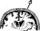
Along with brain puncture, puncture of the subarachnoid spaces and brain ventricles, which has as its goal to obtain cerebrospinal fluid for diagnostic purposes, plays a significant role in the surgery and diagnosis of diseases of the central nervous system. These manipulations are much more innocent than brain punctures, especially when cerebrospinal fluid is obtained from the cranial region by means of a suboccipital puncture, although even with the latter intervention, severe complications have been described, up to and including a fatal outcome. The technique of this puncture is as follows: puncture of the lateral ventricles from the anterior (see Figure 1), posterior, or inferior horn is performed through the brain substance, and everything that has been said about the dangers of damaging brain tissue during punctures remains in force for these operations as well. As much as these punctures are easy with dilated brain ventricles, they are difficult with normal sizes of the ventricles; in the latter case, one has to make several movements with the needle to find the cavity of a normal ventricle. To punctures of the ventricles with, Figure 1. Puncture of the anterior horn of the lateral ventricle. (According to Bier-Braun-Kümmell.)
Diagnostic operations, known as ventriculography (see), also fall under the category of procedures aimed at obtaining cerebrospinal fluid from them. Sometimes this intervention is performed not for the purpose of introducing air, but either for therapeutic purposes—to introduce serum into the ventricular cavity—or for diagnostic purposes—to introduce dyes or salt solutions, the presence of which is subsequently detected in the cerebrospinal fluid or in the urine [dyes (Dandy) or sodium iodide solutions (Förster)]. The second group of operations. This includes operations of the emergency surgery type: a) operations for intracranial hemorrhages, b) operations for skull injuries and infectious processes and their immediate and long-term consequences (traumatic epilepsy), c) plastic-type operations (for congenital cerebral hernias and hydrocephalus), and d) operations for brain tumors. Surgical intervention for intracranial hemorrhages (arterial and venous). The most common hemorrhage in the cranial cavity is an extradural hemorrhage from the middle meningeal artery, usually occurring during skull fractures in the temporal region. It very often leads to phenomena of brain compression with all the resulting consequences. A classic sign of this hemorrhage, as well as of intracranial hemorrhages in general, is the well-known "symptom of the lucid interval" (observed in 60-70%). To combat hemorrhage from the middle meningeal artery, one reaches the bone of the temporal region through the soft tissues and resects it in the places corresponding to the passage of the artery (see Figure 2). To determine the projection of the passage of the middle meningeal artery, the classic Krönlein scheme is used (see Figure 3). Finding the artery does not present much difficulty. The blood clot (see Figure 4) is removed, and the bleeding site is usually easy to find. The artery is dissected from the thickness of the dura mater or the bone canal and
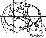
Brain is ligated with a double ligature. The wound in closed skull fractures is closed tightly; in open and contaminated fractures, a drain is inserted. Hemorrhages inside the dura mater (intradural), according to Küttner, most often occur in significant volume from the cerebral veins draining into the sinuses, or from the latter during injuries. In closed fractures, diagnosis is very difficult, and among surgeons, the question of intervention for subdural hematomas is decided differently. And this is understandable: in addition to hemorrhages from the sinuses, there can be hemorrhages during traumatic injuries of arterial trunks or after brain operations or punctures; there are also parenchymal
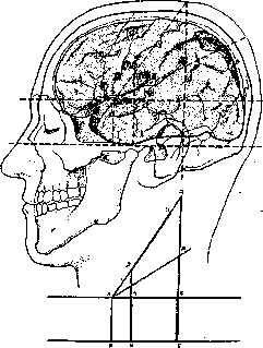
Figure 3. Krönlein scheme: VG—German horizontal line, passing from the lower edge of the orbit to the upper edge of the external auditory meatus; AB—upper horizontal line—from the upper edge of the orbit—parallel to the previous one; AV—anterior vertical line from the middle of the zygomatic arch perpendicular to the VG line; ZI—middle vertical line from the articular head of the mandible; L-G—posterior vertical line from the posterior edge of the mastoid process to the intersection with the sagittal line (L) from the base of the nose to the external occipital protuberance; AL—line connecting the point (A) of intersection of the anterior vertical line with the upper horizontal line with point L—position of the central sulcus (Rolandi); AM—line bisecting the angle—position of the lateral sulcus (Sylvii); Roman numerals—centers of the corresponding cranial nerves. Centers: 1—writing; 2—movements of the head and eyes; 3 and 4—speech; 5—taste; 6—eye muscles; 7—reading; 8—muscle sense; 9—parieto-occipital sulcus. (According to Bier-Braun-Kümmell.)
hemorrhages; in these cases, a difficult situation is created for the surgeon: in wounds, resp. injuries of significant arterial branches, intervention is usually delayed and useless; parenchymal hemorrhage is difficult to diagnose; postoperative hematomas are the easiest both for diagnosis and for intervention. Usually, after aseptic brain operations, surgeons close the cranial cavity without drainage, trying to restore the coverings of the brain in all their layers, starting from the dura mater and ending with the skin. If interrupted sutures are applied to the dura mater, then subdural hemorrhage does not threaten with a catastrophic increase in intracranial pressure—the cerebrospinal fluid and partly blood can exit through the edges of the wound under the skin of the skull, and through them to the outside; with careful continuous sutures, the hematoma can sometimes, on the contrary, cause very formidable phenomena, and then the surgeon is faced with a dilemma: either open the wound or perform, perhaps several times, lumbar punctures. Bergmann advised, in relation to subdural hemorrhages, as a rule, to take a wait-and-see approach. Measures are much clearer in open injuries of the skull and brain causing intradural hemorrhages, and in closed injuries of the skull bones with clear localization in the region of the sinuses of the dura mater. Hemorrhage from the sinuses sometimes proceeds with mild symptoms from the central nervous system, sometimes, on the contrary, with very severe ones, which depends on where the main mass of blood rushes: either only into the subdural space (resp. subarachnoid) or pours out partly, and sometimes in a larger mass, to the outside. In view of the fact that hemorrhage from the sinuses can be life-threatening, a whole series of techniques has been proposed to combat it. In the first place (both in simplicity and in the history of its origin) is simple tamponade of the sinus lumen. Tamponade is performed either with gauze or catgut threads or pieces of tissue (both free and pedicled). Methods of suturing the sinus wound have been proposed. Sutures are applied either directly to the edges of the gaping wound or, according to Reversdrop, to healthy areas of the dura mater. Methods of plastic closure of sinus wounds have also been proposed by cutting out a cover for the wound from the entire thickness of the dura mater (Chugaev) or from its superficial layer (Raug, Burdenko). During the Imperialist War, Retterer proposed placing a wooden plate over the site of the sinus wound, which was pushed between the bone and the site of the sinus wound and was intended to act as a compressor. This idea was taken from observations of sinus wounds that were compressed by displaced bone fragments. Repeatedly during operations undertaken to remove skull bone fragments, increased hemorrhage from the sinuses was observed, which occurred immediately upon opening the cranial wound and upon lifting the depressed fragments. Regarding each of the listed methods of combating hemorrhage from the sinuses, much can be said for and against; the last of the methods is the least expedient, since the removal of the wooden plate subsequently is very difficult, and leaving it as a foreign body, incapable of resorption, is highly undesirable. As for operations for hemorrhages into the substance of the brain on the basis of trauma or arising spontaneously due to vascular diseases, these operations have not received widespread use. Operations for infections and skull injuries. This includes operations aimed at treating purulent processes that cover predominantly or only the meninges (purulent meningitis) or also affect the substance of the brain itself (encephalitis and abscesses). Treatment of purulent
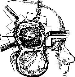
Figure 4. Trepanation for hemorrhage after rupture of the middle meningeal artery: after turning the osteoplastic flap down, the middle meningeal artery is ligated at the site of the rupture (1), and the blood clot (2) is removed. (According to Bier-Braun-Kümmell.)
of meningitis represents a rather modest chapter of brain surgery. This applies entirely to cases of general meningitis. In cases of localized meningitis, surgical intervention has somewhat greater success. Here, the main attention in a certain number of cases should be directed toward prophylactic operations, as can be the case with skull injuries or in the presence of a purulent focus in the accessory cavities of the skull or in one or another area of the skin or bones of the skull. The treatment of every infectious focus must be approached from the point of view of the possibility of its complication by purulent inflammation of the meninges, and the more that is done in this regard concerning these purulent foci, the less the danger of purulent inflammation of the meninges. In those cases where it is necessary to surgically treat diffuse purulent meningitis, surgical measures are reduced to interventions that are either simple or very complex. The multiplicity and variety of the proposed methods testify to the difficulty of combating this severe disease and, on the other hand, to the impossibility of recognizing one method or another as the most rational. The simplest measure is repeated punctures, both simple and with the use of irrigation with a physiological saline solution. Along with this, various authors have proposed using punctures for the introduction into the spinal canal of various medicinal substances, such as electrargol, collargol, vucin, optochin, mercury cyanide, and various sera (meningococcal, streptococcal, pneumococcal, and others). Along with spinal punctures performed in the lumbar region, ventricular punctures and suboccipital punctures have been proposed. These punctures can be used for the introduction of one fluid or another for the purpose of irrigation or for disinfection. More complex measures are trepanations and drainage of the cranial cavity.
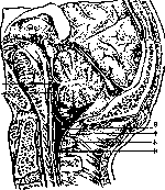
Figure 5. Sagittal section through the occipital region: 1-ventric. IV; 2-velum medullare post.; 3-cisterna cerebello-medullaris seu magna; 4-membrana atlanto-occipitalis post.; 5-tuberc. post. atlantis; 6-dura spinalis. (According to Bier-Braun-Kümmell.) as well as drainage of the spinal canal by means of laminectomy of the vertebrae. Drainage, in one place or another, has as its task the diversion of infected cerebrospinal fluid to the outside. Drainage is sometimes used for irrigating the spinal cavity with fluids. Dandy proposes performing sequentially: 1) repeated lumbar punctures, 2) drainage of the cisterna magna (see figures 5 and 6), and 3) irrigation of the subarachnoid space. Eagleton considers prolonged irrigation necessary in this process. In addition to these measures, various authors have proposed drug treatment by means of intravenous administration of various medicinal substances (the most popular of which are urotropin, trypaflavine, electrargol, and various therapeutic sera). There are isolated proposals regarding blood transfusions from patients who have recovered from streptococcal sinus thrombosis and purulent otitis. In cases of developed severe symptoms of increased intracranial pressure, decompressive trepanations with ventricular drainage have been proposed. Each of the proposed methods of treatment has premises that are very weakly substantiated from a biological point of view. The literature on the subject also provides a considerable amount of critical material. Regarding all the listed
Figure 6. Quadrangular window in the membrana atlanto-occipitalis: 1-opening; 2-membrana atlanto-occipitalis. (According to Bier-Braun-Kümmell.)
To these methods of treatment, one must add a whole series of proposals regarding the intravenous administration of medicinal substances into the vascular system of the brain—both into the veins and into the carotid artery. These proposals are connected with the doctrine of the blood-brain barrier. The vessels of the brain—arterial or venous—are chosen for the direct administration of medicinal substances under the assumption that medicinal substances introduced into peripheral veins may suffer damage in the quality and quantity of their therapeutic effect on their way. To this end, some authors propose injecting Pregl's solution into the confluens sinuum and hope by this means to produce direct disinfection of the brain (Anton and Werker), which physiologically appears poorly substantiated. Knauer injected neosalvarsan into the common carotid artery; Lang injected vuzin. All the proposed methods concern isolated cases and have not yet received any significant dissemination, although 25 years ago Gussenbauer stated the position that the therapy of purulent meningitis should be surgical. At the present time, for these diseases, the usual method of treatment is mainly punctures and lavages with physiological saline using therapeutic sera, which are introduced under the skin, or into a vein, or directly into the cerebrospinal cavity. The treatment of brain abscesses is conducted according to the principle of the surgical treatment of abscesses in general: as soon as the diagnosis of an abscess is made, its location is established, and accessibility to it is justified, it is subject to removal. In the case of brain abscesses, therapeutic measures vary depending on whether there is an abscess that arose by extension (abscess in inflammation of the middle ear, abscess in osteomyelitis of the bones of the skull) or an abscess that arose via a metastatic route. In the first case, the primary focus very often indicates the path of surgical intervention; in the second case, diagnostics appear difficult, and surgical intervention is less certain. In the methodology for treating a discovered abscess, along with the old and proven method of wide opening, a method recently proposed by Lemaitre and others has appeared, which boils down to treating the abscess by drainage without wide opening of the abscess cavity, and to treating the abscess with punctures. The Lemaitre method was recently debated at the congress of otolaryngologists in Amsterdam and received wide recognition. As for the therapeutic success of treating brain abscesses, it must be said that the percentage of favorable outcomes has recently risen to 56. The therapeutic success of treating a brain abscess depends not only on the method of surgical intervention but also on the nature of the abscess: the most favorable results are obtained with encapsulated abscesses; diffuse abscesses give worse results. Regarding the first forms of abscess, mortality is calculated at 50% to 60%. In cases of encapsulated abscesses, mortality according to the latest data falls to 25 percent. Regarding infectious processes of the brain, prophylaxis, as nowhere else, has decisive significance both in the sense of preventing the disease and in the sense of the outcome during surgical intervention. This determines the necessity of surgical intervention in fresh traumatic injuries, especially regarding open fractures of the skull resulting from a blow, from a fall, from contusions, from wounds by blunt, piercing, cutting, chopping instruments, and firearms. At the present time, it is accepted as a rule to treat every open fracture by means of surgical intervention. It has the goal of removing the infection and thereby preventing its spread. Operative intervention consists of removing damaged infected parts, opening the bone wound down to the dura mater, controlling it, and, in case of need, opening and controlling the brain substance. At the same time, blood clots, grossly damaged parts of brain substance, as well as foreign bodies (fragments of skull bones, parts of skin and hair, parts of headgear, remnants of wounding projectiles) are removed. The question of what to do with a fresh wound in such cases is decided in two directions: before the World War, it was considered a basic rule to treat the wound by the open method, with one or another type of tamponade, whereby the edges of the skin wounds were partially pulled together over the tampon with sutures. Tampons were usually removed partially or completely after a few days. During the Imperialist War, Barany, at the siege of Przemyśl, first applied the method of closed treatment of fresh gunshot wounds. The starting point of his method was the doctrine of Fischer that bacteria in the first 12 hours after introduction can be removed from the infectious focus almost completely. Among Russian authors, the Barany method was applied by Britnev, Burdenko, Golyanitsky, and others. The method consisted of complete closure of the wound, however, it was modified by the majority of surgeons who used it in the direction that small drains were placed in the corners of the wounds. This method has the goal of preventing necrosis of brain tissue due to pressure from tampons and is a preventive measure against a severe complication of brain wounds—brain prolapse. Observations by various authors have provided many confirmations of the expediency of this method, but it can be applied only under certain strict indications: early operation, perfect cessation of bleeding, absence of too great contamination of the wound, and finally, an environment allowing for strict control over the condition and course of the wound. The latter is important in that sometimes a tight suture has to be partially opened. Military field surgery after the Imperialist War must consider early intervention and a broad indication for the surgical method of treatment of all open wounds of the skull and brain as a firmly established rule. Peacetime practice likewise recognizes this principle as mandatory in the treatment of traumatic injuries of the skull. Under all circumstances, however (be it conditions of peacetime or wartime), it is necessary to wait for the disappearance of the phenomena of traumatic shock, which usually lasts several hours. The number of authors who consider it possible to conduct conservative treatment (Tillman) is becoming smaller and smaller, and at the present time, questions are no longer being disputed about conservative or active treatment, but about when, and under what environmental conditions, early intervention should be performed. According to the data of various authors, the percentage of mortality after operations grows progressively depending on the timing of intervention after trauma: for operations in the first 24 hours, the mortality percentage = 20; for late operations (over 24-48 hours), the mortality percentage = 50-70-100. Defenders of the conservative method cite in support of their arguments the statistics of old authors, as well as the fact that a certain percentage of the wounded are able to cope with trauma and infection even without an operation. One cannot help but agree with the authors of this trend that it is necessary to more strictly develop indications for operative intervention, but the number of late abscesses in operated and non-operated patients still does not speak in favor of conservative treatment. Direct observations in hospitals, where conservative treatment was conducted by some doctors by virtue of one or another consideration (including theoretical ones), were infinitely sad and dismal (Burdenko). Military-sanitary statistics from wars of the pre-antiseptic and pre-aseptic period also shed little light on questions of conservative treatment. Furthermore, when evaluating the figures cited in reports of various wars (from 20% to 32% mortality), one must remember that these are final, summary figures collected in the rear. It is known from the indication of a whole series of authors that 2/3 of those wounded in the skull perish in the zone of dressing stations and divisional hospitals. Thus, relatively light material reaches the rear. In old reports, wounds of the head and wounds of the skull were not always precisely distinguished. True, even with primary and early operations, it is not always possible to protect the wounded from late meningitis and abscesses. Injuries of the skull and brain, even though subjected to early intervention, often lead to complications—encephalitis, meningoencephalitis, abscess; besides these infectious complications, a complication no less severe may occur, such as serous meningitis and aseptic encephalitis. Both these forms sometimes proceed clinically very severely and can lead to a fatal outcome. Here, apparently, phenomena of a cytotoxic nature take place as a consequence of brain trauma, concussions, contusions, hemorrhages, sometimes distant from the site of the wound. Besides direct infection in skull trauma, cases of infectious damage to the meninges and the brain itself are observed via secondary infection, which is observed in closed fractures of the skull with cracks at the base of the skull and simultaneous damage to the accessory air-bearing cavities of the skull. From here, infection, especially in cases of ruptures of the dura mater, can easily penetrate into the cranial cavity and cause infectious processes in the form of localized or diffuse meningitis or abscesses.
These forms of infection usually run a very severe course, and only recently have indications for their surgical treatment been developed, particularly by French authors. In cases of trauma—closed, uncomplicated fractures of the skull—surgical intervention is also sometimes indicated, which aims to restore the integrity of the bony vault. The operation in these cases is performed according to the condition of the traumatized areas of the skull and consists either of elevating the depressed parts of the bone, removing them, freeing the depressed areas, or restoring normal relationships. In doing so, it is necessary to verify the absence of hemorrhage under the dura mater, which can be judged by the degree of amplitude of the brain's pulsation or its complete absence. If a subdural hematoma is suspected, opening the dura mater and removing the blood clot is required, because although it is known that many patients who have received such injuries recover safely despite the breach of the cranial vault, there is no possibility of guaranteeing a favorable outcome for this type of injury—a certain percentage of so-called traumatic epilepsy owes its origin to residual phenomena after these injuries: the formation of depressions in the cerebral cortex, serous cysts, and localized serous meningitis. Intervention in open and closed wounds of the skull and brain, in addition to preliminary control of infection and the possibility of severe complications of the type listed above, has the task of preventing the development of one of the serious complications of skull injuries—epilepsy, which may arise in connection with the trauma—either shortly after the injury or after several months or years—and proceed either in the form of Jacksonian epilepsy or in the form of generalized epilepsy. Traumatic epilepsy is observed, according to world literature in the post-war period, in approximately 12% to 22% of cases, and sometimes independently of the site of the skull wound in terms of proximity to the motor centers. This complication is one of many long-term consequences of wounds to the skull and brain, but among others, it occupies a significant place. The long-term results of trepanations after gunshot wounds are, in general, far from brilliant. In the post-war period, the question of the fate of trepanned patients was studied in detail, and French authors developed a special term, 'syndrome des trepanes,' characterizing both mental disorders and functional damage to the central nervous system. Weitzel provides a review of 340 cases, of which a more or less pronounced syndrome of the trepanned was noted in 230 cases; of these, 46 cases of severe brain amnesia and 7 cases of disorders of mental activity without hope for improvement. Out of 340 cases, 13 remained with an unremoved foreign body, 45 developed local and general epilepsy, 116 had defects of the motor zone, and 30 had phenomena of visual impairment. Burdenko's data, covering 178 cases out of 1,054 cases trepanned in the front-line zone in 1914–16, are as follows: out of 178 cases (according to social security agency data), mental defects—32 cases, epilepsy—32 cases (local and general), defects of the motor zone—94 cases, visual impairments—19, hearing impairments—2. Burdenko's material proved to be more severe, as it was taken from the category of registered invalids. Of all these complications, epilepsy is usually the object of surgical intervention. The question of the treatment of traumatic epilepsy, and mainly Jacksonian, arouses little disagreement; the majority of both neuropathologists and surgeons consider surgical intervention indicated for these forms of epilepsy after unsuccessful attempts at medicinal, dietary, and hygienic treatment. Only a few authors consider surgical intervention for these forms of epilepsy to be poorly indicated. In most cases of traumatic epilepsy, surgery reveals more or less sharp morphological changes in the form of adhesions, scars, cysts, and degeneration of a larger or smaller section of the cerebral cortex. Recently, with the help of the encephalography method, it has been possible to establish the presence of ventricular dilation on the side of the injury. (For the surgical treatment of generalized epilepsy, see Epilepsy.)
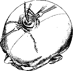
Plastic-type operations. Plastic operations in the region of the brain and the ventricular system mainly include operations for hydrocephalus—internal and external. Their task is to produce a series of plastic adjustments which would restore the proper outflow of cerebrospinal fluid from the ventricles into the subarachnoid space and from there into connective tissue reservoirs or cavities or into the vascular system. Depending on one or another theoretical conception of the origin of hydrocephalus—closed and communicating—a whole series of operations was proposed, which were the logical consequence of theoretical premises. After the doctrine of the production of cerebrospinal fluid by the choroid plexuses of the ventricles was accepted, Dandy conducted experiments with the artificial closure of the foramen of Monro and the aqueduct of Sylvius. These experiments established with certainty the fact of the formation of unilateral or bilateral accumulation of fluid in the right or left lateral ventricle upon closure of one or two foramina of Monro, the accumulation of fluid in the lateral ventricles and the third ventricle upon closure of the aqueduct of Sylvius, and the absence of fluid accumulation upon closure of the named openings in cases where the removal of the choroid plexuses was performed. These experiments were repeated by various researchers, who confirmed Dandy's data. From the point of view of these experiments, Dandy himself put forward the hypothesis that in cases of communicating hydrocephalus, one must look for Figure 7. Site of the soft tissue incision and bone removal during the Anton-Bramann operation. (According to Bier-Braun-Kümmell.)
an explanation for the accumulation of fluid in two factors: either the choroid plexuses produce too much cerebrospinal fluid, or the absorption apparatus is unable to remove the normally produced fluid. In view of this, a number of the following operations were proposed: for closure of the aqueduct of Sylvius, Dandy proposed its drainage; for closure of the foramen of Monro or the Sylvian aqueduct, Anton-Bramann proposed an operation on the corpus callosum (see figures 7–9); for closure of the foramen of Magendie, Anton-Schmieden proposed an operation to open the foramen of Magendie. In addition to restoring closed passages, Dandy proposed an operation with the task of reducing the secretion of cerebrospinal fluid by removing the choroid plexuses, in the hope of influencing the cause of the formation of hydrocephalus with this operation. With the aim of creating a better outflow from the ventricular system, Payr proposed an operation on the great vein of Galen, which is the collector for the venous system of the ventricles. By means of these operations, it is indeed possible to create a free outflow of fluid from the ventricular system for a more or less long period. In communicating hydrocephalus, when one can expect insufficiency of the functions of the apparatus that normally absorbs cerebrospinal fluid, a whole series of operations has been proposed by various authors, which have the goal of either strengthening the insufficiently functioning absorption apparatus or to some extent creating it anew. The task of these operations consists of creating constant drainage from the cerebrospinal canal either into loose connective tissue space, or into the venous system, or into the urogenital system, or into the peritoneal cavity. Operations of the first kind consist of opening the dura mater and creating connections between this artificial opening and connective tissue reservoirs, such as, for example, the connective tissue of the orbital cavity (Hildebrandt and Zaaijer operations). Connection with the venous system was proposed by Payr by means of connecting the ventricular cavity with the longitudinal sinus (free implantation): a segment of the saphenous vein was sutured into the sinus, and the free end was lowered into the ventricular cavity. McClure connected the cavity of the posterior cranial fossa with the internal jugular vein by means of the implantation of a saphenous vein taken from the patient's father. Recently, Foerster proposed the following method: a vein of the scalp, which is always more or less dilated in hydrocephalus, is dissected out and then sutured into the opening of the dura mater; recently, an operation has also been proposed which attempts to solve the question of constant drainage of the cerebrospinal space by means of suturing the ureter into the dura mater at the level of the lumbar vertebrae. This operation requires extirpation of the kidney. Many hopes are placed on this operation both for its relative ease of performance and for the guarantee of the absence of adhesions and cicatricial secondary changes in the ureter as a drainage channel. This consideration is of great value in view of the fact that with other methods, for example, when diverting fluid into connective tissue spaces, degeneration of the cellular tissue occurs, which consequently ceases to play the role of an absorption apparatus (Rozanov), and with the implantation of veins, their thrombosis may occur. Regarding operations for connecting the cerebrospinal cavity with
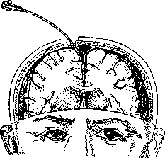
Figure 8. Schematic representation of the puncture
of the corpus callosum with a catheter according to Anton-Bramann in the frontal plane. (According to Bier-Braun-Kümmell.) Regarding the urinary system, the following must be said: however tempting this operation may be in its simplicity, it is necessary to take into account some of its negative aspects. This operation is based on the assumption that the opening of the ureter into the bladder is constantly closed and that the urogenital system is completely healthy both at the present moment and for the future. Regarding the first assumption, it cannot be said with certainty that in a given subject the opening of the ureter is closed in a perfect manner. Facts are known about urine entering from the bladder into the ureteral stump during nephrectomy. Furthermore, the urogenital system may
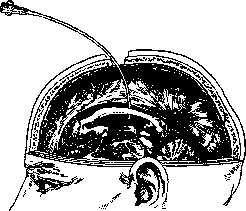
Figure 9. Schematic representation of the puncture
of the corpus callosum (Balkenstich) in the sagittal plane. (According to Bier-Braun-Kümmell.) easily become infected, which creates a great danger of infection for the cerebrospinal cavity. Recently (Burdenko), an operation has been proposed for connecting the cerebrospinal cavity with the abdominal cavity by means of implanting the omentum into an incision of the dura mater at the level of the lumbar vertebrae. Regarding the latter operation, not enough facts have yet been accumulated to express one judgment or another. The second cycle of plastic operations, aimed at restoring the normal relations of the brain and skull in so-called hernias, congenital and acquired after trauma, as well as operations for defects of the skull and meninges, belongs to the field of plastic surgery. Operations for brain tumors. Surgical intervention for brain tumors is currently considered by the majority of neuropathologists to be a rational method of treatment in a certain number of cases. Regarding views on the surgical treatment of brain tumors, three stages can be established: the stage of optimism, pessimism, and compromise, or reconciliation. These three stages, which brain surgery has passed through, cannot at the present time be considered to have settled into completely rigid forms. Some authors set the indication for surgical intervention unusually broadly, others express themselves with restraint, remaining dissatisfied with the final results of the surgical intervention; in the end, one has to admit the validity of the old medical truth about the necessity of individualization. Surgical intervention for brain tumors falls into two groups: the first group includes palliative operations, the second—radical operations. Palliative operations. Palliative operations include operations,
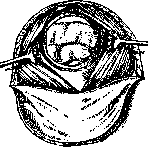
aimed at lowering intracranial pressure; Horsley proposes performing them in the early symptoms of neuritis optica. These operations are used: 1) in those cases where the tumor cannot be accurately localized, 2) when the tumor is inaccessible to surgical intervention due to its topographical position, 3) when the tumor is too large, and 4) in cases of exhaustion of the patient or in synchronous diseases that are a contraindication for radical surgical intervention. Decompression operations have as their task, as their very name indicates, to create such conditions under which intracranial pressure can be lowered. They are performed according to various methods. 1. The Cushing method consists of removing a part of the bony coverings of the skull in the region of the right temporal bone (see Figure 10).
The details of this operation are as follows: a skin incision is made, slightly below the upper linea temporalis, of a horseshoe shape, with the base at the bottom. The skin flap is dissected down to the temporal fascia and
thrown down. Figure 10. Decompression trepanation. (According to Cushing.) The temporal fascia lying at the bottom of the wound, covering the temporal muscle, is cut together with the latter by a longitudinal incision from top to bottom along the course of the muscle fibers. This incision begins from the highest point of the oval skin incision and reaches the zygomatic bone. The incision penetrates to the periosteum. After this, the periosteum is detached with a raspatory on both sides, i.e., forward and backward. The exposed bone is removed approximately over an area of 5x6 cm. Most surgeons remove the bone according to Cushing together with the periosteum. The dura mater, lying at the lower edge of the trepanation opening, carries on itself the branches of the middle meningeal artery, which is ligated below with two ligatures. The dura mater is opened and removed entirely, corresponding to the bone defect. Some authors do not open the dura mater during this (Lecene and other French authors). Goldberg has recently proposed removing not the entire dura mater, but only its upper layer. Some authors open the dura mater in a cruciate manner and turn its flaps onto the edges of the bone wound. The listed modifications of the basic Cushing operation have as their goal to protect from too rough trauma the brain, which is under increased intracranial pressure, which immediately upon opening the dura mater bulges through the wound surface and can be severely traumatized against the sharp edges of the bone, which can give undesirable consequences in the form of damage to those or
Figure 11. Valve according to Krause-Sänger. The bone flap is reduced. The incised dura mater is turned back.
of other areas of the cerebral cortex and cause hemorrhage. This circumstance lies at the basis of Cushing's proposal to perform trepanation on the right side and submuscularly. With right-sided trepanation, the opening lies over silent areas of the cortex. According to the data of cranio-cerebral topography, the bone defect will correspond to the third frontal gyrus, the first and second temporal sulci, or the lower edge of the Sylvian fossa. However, the lower part of the central gyrus may also be located here. 2. Along with the decompression according to Cushing, another type of decompression operation was proposed by F. Krause, the essence of which consists in the formation of a skin-bone flap lying freely on the cerebral surface. To ensure the freedom of the bone flap, the surgical opening of the skull is expanded parallel to its edges by 1/2-1 cm (see Figure 11). The dura mater is treated differently: either it is removed entirely (Kocher, Lamy-Longue) or it is folded back onto the bone edges after a cruciate incision (Krause-Sanger). These flaps are usually cut out on the parietal bones in the region of the Rolandic sulcus—in cases of epilepsy (Kocher), primary or secondary, accompanying tumors—or over the site of the suspected tumor. In some cases, decompression trepanation is essentially a diagnostic trepanation. According to Krause, it is performed as follows: an ordinary skin-bone flap is cut out, which is folded downwards. The dura mater is opened in such a way that the base of its flap lies directly opposite the base of the skin-bone flap, thanks to which it is folded back to the upper line of the skin-bone incision. If, upon examination of the skin-bone area of the brain, the tumor is not found or proves to be unresectable, then a significant portion of the skull bone lying below, as well as portions of the bone of the lateral edges, are removed with bone forceps. After this, the dura mater is placed back in its former position, and the skin-bone flap is placed over it. Along with these methods, it has been proposed for certain cases to perform a decompression operation in the region of the cerebellum with removal of the arch of the atlas—sometimes with opening of the dura mater, sometimes without it. The significance of palliative operations is rated very low by many authors: firstly, because they provide only temporary relief, and secondly, they are far from as safe as it might seem at first glance. It is known from statistical data that for some authors these operations yielded a 50% mortality rate (Lecene), but the point of these operations is that thanks to these relatively simple interventions, it is possible to relieve patients, at least temporarily, from the severe symptoms of their primary ailment—progressive blindness and unbearable headache. The duration of these beneficial moments is, of course, different, depending on how fast the tumor grows and how far the visual impairment has progressed. In neglected cases, these operations do not bring any benefit. For the sake of completeness, one must also mention the palliative operation proposed by Schüller: to decompress the sella turcica and simultaneously perform a puncture of the third ventricle. This operation is extremely difficult in its technique and dangerous in terms of infection. Radical operations. The French surgeon de Martel characterizes this group of operations uniquely and with exceptional expression in such propositions: brain tumors are generally inoperable. To perform operations, it is necessary 1) that a neuropathologist can localize them based on the symptomatology of the tumors and 2) that they are limited, visible, and favorable for enucleation. In surgical textbooks, therefore, there are sections dealing with unlocalized tumors, localized tumors, and tumors that truly correspond to topical diagnosis. For unlocalized tumors, in the majority of cases, the above-mentioned palliative operations are used. If, with the application of all modern methods of auxiliary examinations (radiography, encephalography, lipiodol, introduction of contrast solutions into the internal carotid artery, punctures, and biopsies), the percentage of accurately established topical diagnosis has risen to 76, then in relation to the requirement of the second point, the percentage at the present time is very low. By their topography, brain tumors, depending on the surgical technique, fall into three groups: 1) supratentorial (cerebral) tumors, 2) subtentorial (cerebellar) tumors, and 3) pituitary tumors (see Acromegaly). The general principles of the operative technique for removing intracranial tumors at the present time cannot be considered fully developed and generally accepted. The question of anesthesia is discussed with great passion. Some surgeons speak out exclusively for local anesthesia (de Martel, Foerster), others for general (Cushing), considering the former "inhumane" and frivolous (Frasier). The question of hemostasis is still being developed with exceptional thoroughness, and Cushing sacrifices much time during the operation, scrupulously stopping the slightest bleeding. The question of an electric motor or manual force for cutting instruments also does not have a definite solution. Usually, all trepanations undertaken for the removal of supratentorial brain tumors are done osteoplastically, and only after orientation and at the end of the operation is the question of removing the bone flap decided; for subtentorial tumors, the majority of authors perform a resection of parts of the occipital bone (see Figures 12 and 13). The question of great importance regarding a one-stage or two-stage intervention does not leave the pages of the periodical press, but apparently, the question is incorrectly posed in its essence and cannot be set forth in a regulatory form. The decisive moment is the state of the patient's blood pressure; sometimes with brain tumors, bleeding from the scalp alone is so strong,
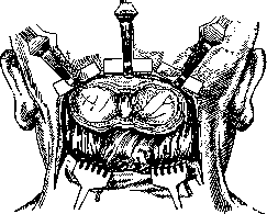
Fig. 12. The U-shaped skin-muscle-periosteal flap is turned down. The bone in the region of both cerebellar hemispheres has been removed.
that it is necessary, before trepanning the skull bones, especially during operations on the posterior cranial fossa, to take measures to maintain blood pressure. Trepanation and resection of bones with the sometimes inevitable damage to emissary veins also create dangerous moments, and sometimes stopping the operation becomes inevitable. Many surgeons perform constant blood pressure monitoring during the operation and are guided by it.
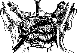
Figure 13. The dura mater has been incised after preliminary ligation of the occipital sinus and turned down in the form of a flap. With a spatula, the right cerebellar hemisphere is pressed down, and the tumor appears in the depth. (According to Bier-Braun-Kümmell.)
in their manipulations of the manometer readings. Some cases, on the contrary, are more advantageous to operate on in one stage, even with some risk. In supratentorial operations, the question is not so acute. After careful hemostasis in the one-stage method, one usually proceeds to open the dura mater with preliminary ligation of the arterial branches running through its thickness. Sometimes, already upon inspection of the dura mater, parts of the tumor and the infiltration of its various sections by the tumor are visible. Usually, the dura mater is opened parallel to the edges of the osteoplastic flap, but with an inverted base: the osteoplastic flap has its base downward (toward the base of the skull), while the flap of the dura mater has its base toward the cranial vault. Orientation regarding the state of the pia mater and arachnoid, the gyri, their outlines, and the vascularization provides much for judging the location of the tumor. The very act of removing the tumor varies in difficulty depending on the nature of the tumor. The most satisfying to the second requirement of the de Martel formula are endotheliomas (meningiomas, in Cushing's terminology), neurinomas (tumor acusticus), parasitic cysts, cholesteatomas, teratomas, and some forms of tuberculous granulomas (solitary tubercles). Tumors of the glioma type, on the contrary, are not very favorable for complete removal. All types of these tumors—fibrillary gliomas (astrocytomas), cellular gliomas, glioblastomas with hemorrhages, with cysts, or with colloid degenerations, usually even macroscopically demarcated—do not have capsules and in this respect do not differ from diffuse gliomas. Therefore, surgical intervention for them is considered inexpedient by many surgeons. Dandy considers it possible to perform intervention for these tumors by the resection type; he excises tumors according to the principle of tumor removal in other parts of the body—within the limits of healthy brain tissue, i.e., he performs an extensive resection, sometimes capturing the entire frontal, temporal, or occipital part of the hemisphere, completely or partially, without regard for subsequent defects. However, the majority are very reserved regarding such interventions. Much more expedient is the Cushing method for some forms of cystic gliomas, mainly cerebellar gliomas. In the case of irremovable tumors, the cyst is opened, its contents and a part of the tumor protruding into the cyst cavity are removed, and then fixation is performed with Zenker's fluid or a formalin solution. The results of such treatment were in some cases very favorable. Technically, tumors of a vascular nature must be recognized as very difficult to operate on, because coping with bleeding in the area of brain tissue is, on the one hand, not easy, and on the other hand, stopping the bleeding can cost very dearly—ligation of arteries appears to be no less dangerous than the bleeding itself. The listed tumors are the most frequent objects of surgical intervention, as can be seen from Cushing's statistics, covering 1,146 cases: gliomas—492, meningiomas—141, neurinomas—100, congenital tumors—67, granulomatous tumors—37 cases. The accessibility of tumors for surgical intervention can be judged to some extent by the statistics of Allen Starr and Collier, Tooth, and Stern [see above—pathology of the Brain (art. 547)]. The data of these authors speak of a comparatively small number of tumors which, due to topographical conditions, can be an object of intervention. If one also takes into account their histological structure, size, and relationship to vessels, it will not be surprising that in their time Oppenheim and Bruns considered an extremely insignificant number of brain tumors to be subject to operation: from 2–9% (Oppenheim) to 14% (Bruns). Recently, cases of intervention on the gl. pinealis (Dandy) and the region of the corp. quadrigem. (Foerster) have been published, but these operations will in all respects remain exceptions for a long time. When the question is raised about the results of operations for brain tumors, it is usually impossible to give a satisfactory answer. To provide general statistics on mortality, recovery, improvement, deterioration, and remaining unchanged means to leave the question unanswered; the general question must be broken down into more detailed questions, but unfortunately, at the present time, it is not possible to provide exhaustive material, and the answers can for now only be of an indicative nature; the most favorable figures are given by Cushing—12% immediate mortality after operations undertaken for brain tumors. In explanation of these comparatively high losses, even for a surgeon such as Cushing, it is necessary to point to the exceptional severity of the postoperative course in patients with brain tumors and the extraordinary tendency toward complications in the postoperative period. These factors alone must be the subject of exceptional attention on the part of the operating surgeon, and here, as nowhere else, the principle of traumatic prophylaxis during the operation and the principle of conscious, iron discipline in postoperative care must be especially strictly implemented. Subtentorial tumors yield a higher percentage of mortality than supratentorial ones: thus, for supratentorial gliomas, Cushing calculates the immediate mortality at 10%, for cerebellar gliomas—from 25% to 30%, and for neurinomas of the n. acusticus—at 12%. Of course, as in other areas, the outcome of surgical intervention depends on a combination of many factors and individual characteristics, both on the part of the patient and the operator. The figures are taken from Cushing as an operator who is exceptionally authoritative in this field, and who, moreover, has his own statistics (relating to 1909). Cushing's mortality for subtentorial interventions is 70% (out of 34 operations—24 deaths). As for the long-term results of surgical intervention, they must also be evaluated according to the type of tumor, the method of operation, and the observation periods, and here one must limit oneself only to the illustrative data of the same author: thus, for supratentorial gliomas cured after a three-year interval, Cushing gives 40%. No less valuable an illustration is the assessment of long-term results for tuberculomas—despite all the ease of technical execution and the excellent immediate result, the long-term results are exceptionally bleak; recurrences and dissemination of the process are common phenomena in these operations. The cited figures of Cushing are so brilliant that German authors, even on the pages of textbooks, discuss the question of the enormous difference in the data of German authors. To understand this, it is sufficient to cite the results of Eiselsberg (1913). In 162 cases of tumors, the fatal outcome immediately after the operation was calculated at 33%; in 33 cases of cerebellar tumors, the tumor was not found in 12 cases, of which 7 cases died immediately after the operation; out of 17 cases of cerebellar tumors, the tumor was removed in 15 cases—of these, 10 died during the operation; of the 5 remaining alive, only 2 patients lived longer than 3 years. Subsequently, Marburg and Ranzi published Eiselsberg's material, covering 318 cases with a mortality of 40%. The data of Krause and Küttner are almost identical to the figures of Eiselsberg: 45% mortality in 109 cases for Krause and 45% in 100 cases for Küttner. Even more severe consequences were observed by German authors during interventions in the posterior cranial fossa, where mortality reached 60–75%. Thus, surgical thought is undergoing great trials in resolving the problem of the rational treatment of brain tumors, and the results of the persistent, decades-long work of Cushing give the right to hope for a bright future for this difficult field of surgery.
N. Burdenko. Social significance of brain diseases. The social significance of brain diseases has a whole range of aspects; insofar as, etiologically, brain diseases are connected with many social diseases and calamities, tasks of a preventive nature of a social order arise here; insofar as brain diseases develop as a result of its wear and tear, the preventive struggle against overwork, atherosclerosis of vessels, and premature aging is of enormous importance; insofar as the treatment of brain diseases requires great effort and difficulties, the allocation of funds for this purpose must be more significant; finally, insofar as patients with brain diseases can restore their working capacity, it is necessary to provide them with special regulations granting them the right to leave for the necessary period for recovery to occur. Regarding the prevention of brain diseases, it is necessary to especially emphasize the importance of the fight against social diseases (alcoholism, syphilis, industrial poisons, epidemics, traumatism, narcotism, etc.—see corresponding articles). All kinds of preventive measures against the development of arteriosclerosis (see) and diseases of the circulatory organs have enormous preventive significance. Autopsy statistics of the city of Moscow for 1923–1927 show that 44% of all cases of arteriosclerosis end in cerebral complications. It is also very important to fight against overwork of the brain (premature development of children, early school overwork); as measures to combat overwork, one must name, besides labor protection laws, the popularization of the hygiene of rest, regular and sufficient vacations, etc. Thorough treatment of all kinds of infectious diseases, especially on the head (on the skin, in the area of the ear, paranasal sinuses, etc.), is a necessary requirement for prevention regarding brain diseases. In view of the fact that a hereditary factor often clearly appears in brain diseases, preventive-hygienic measures in this direction appear very important. Since the healthy state of the brain for the population is of enormous value in many respects, the protection and safeguarding of the brain from all kinds of harmful factors and premature wear and tear must be a state concern of primary importance. Where pathology has done its work and led to a brain disease, the provision of medical aid, despite all (and, moreover, very great) difficulties associated with the treatment of brain afflictions, must be placed at the highest possible level. Besides the best possible general clinical and general hospital conditions and excellent care for such patients, the best X-ray diagnostic and X-ray therapeutic installations, electrotherapy, the organization of proper surgical aid, individual treatment with exercises in movements conducted according to a special plan, experimental-psychological and psycho-technical examination, etc., are required. Persons with brain diseases require very long periods for recovery and the restoration of their working capacity. Therefore, it is necessary that they be granted proper periods for treatment and subsequent rest with the retention of their service positions and the material situation associated with this (up to a one-year period from the onset of the disease). In the case of a transition to an invalid state, patients with brain diseases require special care, and therefore the organization of a sufficient number of invalid homes is necessary, where such patients could find refuge and where their residual defective working capacity could be utilized. The organization of special institutions for invalids with various kinds of workshops and the study of the working capacity of brain-injured invalids, among whom there is a large number of young people, is extremely important. Outside of rationally adapted working conditions, brain-injured invalids often become only a burden to society, and they themselves become dull and degenerate. Meanwhile, a thoughtful, attentive attitude toward existing defects and a desire to provide real help here usually awaken such possibilities that one seemingly could not have counted on. A patient who has lost his social significance due to a brain disease can again acquire the joy of the consciousness of his social USEFULNESS.
Related articles
Cite this page
“Brain (551 General diagnostics of brain diseases).” Soviet Medical Encyclopedia. English translation of Bolshaya Meditsinskaya Entsiklopediya, 1st ed. (Moscow, 1928–1936), ed. N. A. Semashko. https://sovietmedicalencyclopedia.pages.dev/article/brain-2/