REFLEXES
Historical document, translated for reference. It reflects medical knowledge of the 1920s–30s and is not medical advice.
Summary
Reflexes are automatic motor responses to external stimuli, representing a fundamental function of the nervous system. This article traces the evolutionary development of reflexes from simple irritability in primitive organisms to complex neural pathways involving synapses and directional impulse conduction.
Encyclopedia article (1928–1936)
REFLEXES (from Latin reflexio - reflection), automatic motor responses in answer to external irritation. The term R. is borrowed from the field of physical phenomena and implies an analogy between the nervous system, which reflects irritation in the form of a motor response, and a smooth surface reflecting a ray of light falling upon it (Astruc, 1743). In its ?biological essence, R. represents one of the manifestations of the basic function of the nervous system - irritability. As is known, irritability represents a property inherent in all living protoplasm, and rather complex motor responses to external irritations are observed in plants, in the simplest animals, and in wandering cells of multicellular organisms (leukocytes, germ cells, etc.). Such reactions, carried out without the participation of the nervous system and designated by the terms "tropism" (change in position of parts of a stationary organism) and "taxis" (movement of the entire organism), represent the most primitive stage in the evolution of irritability. This stage may be designated as the period of "simple irritability." The essence of irritability consists in a change under the influence of external effects of the physico-chemical state and liberation of energy; the dynamic change that has occurred at the site of irritation may cause a similar change in a neighboring area, and thus the process of irritation can spread, irradiate, be conducted from one part of the organism to another. Thus already at that stage, which we designate by the term "simple irritability," there is an elementary form of conductivity. An essential feature of this spread of irritation in nervous tissue, as compared with the nervous system, consists, first, in the slowness of the spread process, and second, in the rapid decrease in the intensity of irritation as it moves away from its point of origin, i.e., from the place directly subjected to external irritation; gradually weakening, the irritation at a certain distance from its point of origin disappears completely. In the further course of evolution, in the process of morphological and functional specialization and adaptability of tissues, the nervous system arises as a product of the transformation of epithelial tissue. The biological essence of the appearance of the nerve cell can be defined as the transformation of an epithelial cell in the sense of its exclusive adaptation to the specific functions of irritability and conductivity. The morphological expression of these specific properties of the nerve cell is the presence of more or less long processes, by means of which these cells bring into communication different, sometimes very distant parts of the organism; the functional expression of the specific properties of the nerve cell is the lowering of the threshold of irritation in relation to various kinds of effects and the ability to conduct a once-arisen process of irritation without slowing its spread and without weakening its intensity. Since just such a method of spread of the excitation process is designated by the term "conductivity," this period of evolution of the organism's reactions to external irritations can be designated as the stage of irritability and conductivity. Although at this stage the motor reactions of the organism to external irritations are carried out by means of the nervous system, and although in this form of reactions there are the basic functional components of the reflex - irritation, conduction, and reaction - nevertheless here there is not yet a reflex act in the true sense of the word. To clarify the distinction between this primitive form of nervous function and the true reflex act, and to clarify the essence of the latter, it is necessary to keep in mind the following. At the first stage of its appearance in the process of phylogenetic evolution, the nervous system is represented, as is the case, for example, in coelenterates, in the form of a network, diffusely distributed throughout the mass of the body; at the same time, wherever irritation arises on the surface of the body that perceives irritations, it spreads along the nerve network in all directions. If a spiral or zigzag strip is cut out of the bell of a jellyfish, then when either end of this strip is irritated, contraction of this strip occurs. From this it follows that in the primitive nervous system, having a network structure, the nerve impulse, i.e., the process of spread of excitation, can occur in all directions; in other words, each of the nerve trunks constituting this network conducts excitation in both directions along its axis. Such properties of the nervous system - its network structure and spread of impulse throughout the network when any area is irritated - are fully adapted to those forms of reaction, i.e., those methods of movement, that are characteristic of the corresponding animals - namely, for diffuse muscle contractions of the entire mass of the body. This form of structure and functioning of nervous tissue is designated by the term "equipotential system." In contrast to this, in higher invertebrates and all vertebrate animals, neurons are combined into certain complexes (chains), representing a definite regularity in the direction of impulses. This specificity of impulse spread, designated by the term "dynamic polarization," consists in the fact that the impulse spreads from the dendrite of the cell through its body to the axon-cylinder process and further, along the latter, to the dendrite of the next cell, and so on; in other words, the direction of the impulse is centripetal (with respect to the cell body) in the dendrite and centrifugal in the axon. In addition to dynamic polarization, the nervous system of higher invertebrates and all vertebrates represents a whole series of other features that distinguish it from the diffuse equipotential system of coelenterates and lower worms. These features consist of the following. The equipotential nervous system represents a real network of nerve fibers with an undoubted transition of fibrillar fibers from one nerve to another. In the nervous system of higher animals, such anatomical continuity between neurons does not exist; different neurons are here in contact only with each other anatomically-physiologically; such areas of contact between neurons, areas where the terminal branches of the axon of one neuron come into communication with the branches of the dendrite of another neuron, are designated by the term synapse. Synaptic connections are not a simple anatomical apposition between cell processes; synapses have a very essential physiological role. First of all, it should be noted that the passage of an impulse through a synapse is associated with some delay in its spread, which has very important significance for higher forms of motor acts characterized by the feature of choice ("voluntary movements"). Further, it can be considered established that the synapse is a factor of polarization of neurons combined in a chain, i.e., a factor of spread of the nerve impulse in a definite direction, namely in the direction from the axon of one neuron to the dendrite of another. Synapses also apparently have an essential role in inhibition and stimulation of impulses and in determining the so-called "final path" or "last common stage" (final common path, see below), i.e., the direction of the impulse to a definite complex of peripheral neurons in the presence of combined action on the latter of different innervations. In general, synapses are attributed those features of conductivity function that are characteristic of nerve processes carried out by chains of neurons as compared with conductivity in a single nerve fiber. In the mechanism of the reflex act, in the carrying out of which at least two neurons always participate, the synapse thus has an essential role. Morphologically, the essence of the synapse was seen in the presence between neurons of a special membrane; the existence of the latter cannot be considered fully proven. Others see the morphological essence of the synapse in that interval which is formed between neurons due to the absence of continuous connection between them. Sherrington designates this space by the term "surface of separation"; such a surface may possess various physical properties that can explain the functions of the synapse: to prevent diffusion, to influence osmotic pressure, movement of ions, etc.; the conductivity determined by the synapse in only one direction could be explained by the fact that the membrane (or surface of separation) possesses one-way permeability. Whatever the morphological nature of the synapse, it can be considered generally accepted that the synapse is not an inert cavity between neurons; an active role in the nervous system must be attributed to it, as "living substance differing from the rest of the protoplasm of the neurons" (Herrick). All the above-mentioned features of nervous system function are necessary elements of the reflex act: participation in it of at least two (and usually more) neurons; regular conduction in a definite direction (from receptor to effector); the necessity of limiting the motor reaction to a definite group of muscles - and not a diffuse reaction of the entire musculature of the body.
All these properties of the motor reaction are in an inseparable connection with the morphological and functional features inherent in all animals standing above coelenterates and simple worms, i.e., a significant part of invertebrates and all vertebrates. The basic type of structure of all these animals is characterized by two features: bilateral symmetry and segmentation, or metamerism. The structure of the organism according to the segmental type implies a certain degree of independence of each segment and, accordingly, the independence (isolation) of motor reactions of individual segments or their groups, and in some cases, of one half of the body. The reflex act in the strict sense of the word appears in the path of phylogenetic development simultaneously with the appearance of segmental bilaterally symmetrical body structure; at the same time, in its most elementary form, R. represents a segmental reaction to external irritation. Since the entire evolution of the nervous system consists in the superstructure of new elements (suprasegmental apparatus) over old ones, from a functional point of view, R. is the basis upon which, in the process of evolution, more complex mechanisms of nervous functions were built. The latter, so-called suprasegmental mechanisms, however, developed not at the expense of the disappearance of simple segmental reactions, but at the expense of subordinating the segmental reflex mechanism to newly developing apparatuses and at the expense of adapting segmental reflex acts to phylogenetically newer functions. Therefore, the mechanisms of the primitive segmental-reflex apparatus have been preserved even in higher animals. On the other hand, it must be admitted that the segmental R. represents the basic type of reaction to external irritations and that all higher forms of these reactions are nothing but a gradual complication and transformation of the simple reflex act. And from this point of view, 'all acts of conscious and unconscious life, by the manner of their origin, are reflexes' (I. M. Sechenov). However, in practice, transferring the term 'reflex' to all manifestations of nervous activity hardly presents any advantages, because it does not lead to clarifying the concept of various forms of activity of the nervous system. Although the entire course of evolution of motor functions, beginning with tropism in non-nervous organisms and ending with the highest manifestations of functions of the human cerebral cortex, represents a complication, transformation, and perfection of reactions of living matter, nevertheless, for the precise analysis of various kinds of these, in their basic essence, analogous phenomena, it is more advantageous not to combine them into one general group, but to distinguish their individual forms. From this point of view, reflexes should be understood as segmental motor reactions, i.e., reactions carried out by the nervous mechanisms of the spinal cord and the stem part of the brain, although these mechanisms are under the influence of higher (suprasegmental) parts of the brain. By thus limiting the concept of R. to motor reactions of the segmental apparatus, we can characterize them by the following specific features, distinguishing them from motor functions carried out by the cerebral cortex in the form of so-called voluntary or willed movements. R. represent motor acts carried out automatically in response to external irritation; reflexes are unconscious, automatic; willed movements are conscious, voluntary; reflexes have an innate character and are carried out along predetermined anatomical paths; willed movements are acquired during individual life and the corresponding anatomical-physiological connections are not predetermined, but are developed through the individual experience of a given subject; accordingly, R. have a species character, i.e., are inherent to the entire species Homo sapiens, while willed movements have an individual character; anatomically, R. are reactions of the spinal cord, while willed reactions are carried out by the cerebral cortex; from the point of view of conditioned reflexes, segmental R., i.e., R. in the narrow sense of the word, represent unconditioned R., while willed movements correspond to the concept of conditioned reflexes (see). Between R. and willed movements there are transitional forms, which seems quite understandable in view of the gradual transition from R. to willed movements; therefore, there exist motor reactions that, while belonging in their main features to one of the named groups, have individual characteristics characteristic of the other group. Thus, for example, some of the reactions classified as R. are not innate but are acquired during individual life (for example, cutaneous, abdominal R.); on the other hand, some of the definitely cortical movements, although acquired during individual life, have a species character (for example, the act of walking); some of the cortical movements acquired during individual life, due to their frequent repetition, become automatized to such an extent that in this respect they approach R. Furthermore, it should be borne in mind that some of the innate automatic motor reactions undergo various transformations in the process of ontogenetic development, in the process of adaptation to new functions; thus, the plantar R., in connection with the function of orthograde posture, has a different form in an adult compared to that observed in childhood before the development of the function of walking in a vertical position. Further, some of the motor acts that have a purely reflexive, automatic character in childhood later come under the control of the cortex, i.e., are stimulated or inhibited by the cortex, and thus are transformed from a reflex act into a willed one: an example of this is the act of urination, which is a reflex phenomenon in a child and a willed act in adults. The anatomophysiological structure of the reflex act is partly determined by the data presented above. As already indicated, in the implementation of R., two or more neurons participating in synaptic contact with each other are involved. The totality of these neurons is denoted by the term reflex arc. Corresponding to the basic essence of the reflex act—the perception of external irritation and the connection to the motor apparatus—in the reflex arc there are two main elements (neurons): the afferent (receptor, afferent) and the efferent (effector, efferent). Therefore, the simplest scheme of the reflex arc consists of two neurons—one perceiving external irritation and conducting the impulse corresponding to this irritation to the effector neuron, along which this impulse is further conducted to the peripheral motor apparatus, i.e., to the muscle. In the simplest case, the reflex arc is represented by two neurons: the receptor and the effector (Fig. 1 A). However, such a form of R., carried out by only two neurons, is a rarity; usually reflex arcs consist of several neurons ('a chain of neurons'). In the overwhelming majority of cases, the propagation of the impulse from the receptor neuron to the effector neuron does not occur directly; between them there is the so-called internuncial or correlational neuron (Fig. 1 B). The functional role of the correlational neuron is that by means of it, the given receptor neuron comes into contact with various, sometimes very distant effector neurons (Fig. 1 D, a, as well as Fig. 2). Further, a given receptor by means of a series of internuncial neurons can connect with one effector (Fig. 1 E), whereby the intensity of the impulse is increased, since each cell represents, as it were, a reservoir of hidden energy; under the influence of the irritation brought by the impulse, this hidden energy passes into kinetic energy; thus, the passage of the impulse through a series of internuncial neurons can increase its intensity. Other, more complex combinations are also possible: of reflex mechanisms and, accordingly, more complex reflex functions. Thus, two receptor neurons can be connected by means of two internuncial neurons with two different groups of effector neurons (Fig. 1 F). In this case, irritation of each of the two receptors can cause a reaction in both effectors. If both internuncial neurons have a stimulating effect on one effector (or their group) and an inhibitory effect on another effector (or their group), this will lead to a reaction in one group of muscles with inhibition in the other. The physiological significance of stimulating one group of muscles while inhibiting another lies in the fact that through this the maximum effect of the reflex movement can be achieved with minimal energy expenditure. This method of innervation appears to be extremely widespread; it lies at the basis of the so-called reciprocal ('mutually opposite') innervation, i.e., the relaxation of antagonists of reflexly contracting musculature. Thus, between the systems of receptor and effector neurons there is laid an extremely complex mechanism of connecting neurons.
The consequence of this relationship between receptor and effector neurons is the fact that each effector neuron (i.e., peripheral motor neuron) is a common conductor for impulses arising in different parts of the receptive surface; in other words, the same effector apparatus can be under the influence of different receptors and in this sense represents a common final path or the last common stage (final common path). The choice of the final path, i.e., the direction of the impulse arising in a given receptor area of stimulation to one or another peripheral apparatus, is determined by a number of different conditions: the intensity of the stimulation, its nature, the localization of its effect, previous stimulations, the state of the musculature, etc. To illustrate the influence of these latter factors, the following examples can be given. If in a monkey the central ends of Cvn and CVIii are stimulated, this stimulation can cause both flexion and extension at the elbow joint: if stimulation of these nerves is preceded by stimulation of Di, extension results; in the same case, when it is preceded by stimulation of CVI, flexion results (Sherrington). As for the significance of the state of the musculature, in this respect mention should be made of the law of Uexkull according to which excitation always spreads to the muscle that is in a state of stretch, i.e., when it is possible to direct the impulse to different 'final common paths', it, all other conditions being equal, is directed to the final path, i.e., to the peripheral neuron which corresponds to the muscle that is in a 'state of stretch'. This law, established experimentally, is not fully justified by clinical phenomena. The principle of the final common path plays an important role in the combination of action of different reflex arcs in the formation of the reflex act. The latter never represents the result of the action of a single reflex arc, as was assumed above for the sake of simplifying the analysis of the reflex mechanism. Similarly, stimulations are usually not single, as is assumed for simplicity in the schemes given above. In reality each R. represents the result of a complex interaction of associative functions and the participation of several reflex arcs. The final form of the reflex movement is determined by those final paths along which, depending on the factors mentioned above, the impulses are directed. The relationship between reflex reactions can be different: some of them are harmonious, mutually assisting each other; such R. are designated by the term allied R. On the contrary, other R. appear to be incompatible (e.g. flexion and extension); they not only do not assist, but counteract each other. Such R. are designated by the term antagonistic; antagonistic R. exert an inhibitory influence on each other; that R. or group of R. which, in this mutual inhibition, gain the upper hand, are designated by the term prepotent (overpowering) R. Allied and antagonistic R. represent examples of summation of R. These phenomena of summation and determination of the final path constitute, as is assumed by many, the function of synapses. The phenomena of summation also include induction: mutual reinforcement of two stimulations acting1 successively or simultaneously (simultaneous induction) on two different points of the same receptive field or reflex zone, i.e., an area from which a given R. can be elicited. Thus, for example, two effects that are below the threshold of stimulation, i.e., each insufficient by itself to produce a certain motor effect, produce this latter if they are applied simultaneously or successively at two different points of the corresponding receptive field. An example of summation of successive stimulations can be the method of stroking stimulation, which is used in the clinic in the study of skin R.; while a single touch with the end of a hammer handle cannot elicit the plantar or abdominal R., the latter can easily be elicited when the same object is drawn over a certain distance on the skin surface. The coordinating and determining 'final path' role of synapses of the central nervous system also underlies the phenomenon designated by the term central reflection. By this term is meant the possibility of changes in the form and intensity of R. which are caused by different states of the central apparatus. For the classification of R. one can start from different criteria. Corresponding to the place of origin of excitation, that is, to the types of receptors on which the stimulation acts, the following forms of R. can be distinguished: exteroceptive R., caused by stimulation of superficial (cutaneous) receptors or so-called 'contactceptors'; in this form stimulation is caused by direct contact of the body surface with some object. Interoceptive R. are those in which the basis is the stimulation of the 'inner surface' of the body, i.e., the surface of the digestive tract, respiratory organs, etc. Telereceptive R. are those in which stimulation falls on so-called distance receptors, perceiving external stimulation at a distance (visual, auditory, olfactory receptors). Finally, the 4th group of this classification consists of so-called proprioceptive R., caused by stimulation of deep receptors located in muscles, ligaments, joints. To this same group can be attributed the reflex impulses originating from the vestibular apparatus. The physiol. role of proprioceptive R. consists in the coordination of movements, regulation of posture and position of the body. From the biological point of view R. represent an adaptation of motor reactions to the influences of the external environment acting on the organism; in this sense one can speak of a certain functional role or biological 'purpose' of R. On the basis of this criterion R. can be subdivided into two groups: protective, or defensive R., removing the organism as a whole or its individual parts from harmful destructive external influences, and R. reproducing the elementary movements that make up a certain motor act; these latter R. assist in the carrying out of the function of a certain organ or by themselves represent an elementary motor act and have the character of physiological appropriateness and motor initiative. - Those belonging to the first group, protective, or defensive R. are also designated by the term nociceptive R. Nociceptive R. are distinguished by the following characteristic features. In contrast to other R., for the carrying out of which more or less definite, specific ('adequate') forms of stimulation are necessary, nociceptive R. can be caused by any stimulation, if it reaches a certain intensity and threatens damage. Nociceptive R. are accompanied by a specific affective tone of unpleasant experience and have as their psychic correlate the feeling of pain. Nociceptive R. are prepotent, i.e., when there is a possibility of different R. to a given stimulation, the nociceptive reaction takes precedence, i.e., the impulses are directed along that final path which leads to movements having the purpose of removing the entire organism or its individual part from the external influence. This prepotency gives nociceptive reflex movements the character of irresistibility, violence. One of the typical examples of nociceptive reactions can be the corneal R. As for the second group of R., which have the character of purposeful motor acts not of a defensive nature, they represent extraordinary diversity in respect to their form and physiological value in the sense of their greater or lesser functional elaboration. In general these R., designated by the term 'assisting' (i.e., participating in voluntary motor acts-fordernde Reflexe), include the entire aggregate of reflex reactions with the exception of nociceptive ones. To this group belong various reflex movements, ranging from isolated muscle contractions to complex complexes of them, bearing the traits of purposefulness and relative completeness in the form of motor acts. In this respect it is necessary to bear in mind the following. As indicated above, the evolution of the nervous system represents a gradual superstructure of new functional elements over old ones; in this case, the superimposed, phylogenetically newer cortical elements subordinate themselves to the older segmental (spinal) mechanisms. Therefore, the autonomy of spinal R. in man appears to be little expressed; R. in the narrow sense of this word (see above), i.e., automatic motor reactions of the spinal cord, represent in man as it were an amorphous material, by regulating which the cerebral cortex forms complex motor acts called voluntary movements.
On the contrary, in lower animals with an imperfectly developed brain, the spinal cord possesses considerable autonomy; the movements performed by these animals to a large extent represent reflexive automatic acts carried out by the spinal cord's own apparatus. Thus, in higher mammals and especially in humans, reflexive automata are in a more or less latent state due to their subordination to cortical functions. The study of these purely spinal reflex reactions is possible under conditions of isolating the spinal cord from the brain, which is achieved experimentally in animals by transecting the spinal cord in its upper regions. In this respect, the research of Philippson and Sherrington is of interest; these studies with certainty prove the existence of a spinal reflex mechanism of locomotion, i.e., the acts of walking, running, etc. Philippson's experiments consisted of separating the lumbar region of the spinal cord from the higher regions of the central nervous system in dogs. It turned out that in such an animal, suspended in the air, by irritating the plantar surface of one of the hind limbs, the entire cycle of muscle contractions can be elicited, which fully reproduces the movements characteristic of the hind limbs during walking. In this case, irritation of one limb is the starting point of the entire cycle of locomotor movements: the reflex flexion of this limb is the source of proprioceptive reflexes in the form of flexion and extension contractions required for the act of walking. Thus, in the segments of the lumbar region of the spinal cord, there is an intrasegmental mechanism of locomotor coordination of movements. Sherrington's experiments consisted of transecting the spinal cord at a higher level, above the segments corresponding to the forelimbs; thus, the spinal segments corresponding to both forelimbs and hindlimbs were isolated from the brain. Irritation of one forelimb in such a 'spinal' animal served as a stimulus for flexion and extension contractions of all four limbs in the same sequence as occurs during the act of walking. These experiments show that the reflex mechanism of locomotor coordination of movements is represented not only intrasegmentally and intersegmentally, i.e., it can be carried out not only within the segments corresponding to the hindlimbs or forelimbs, but by means of connections between the cervical and lumbar segments, it can reproduce the function of walking with all four limbs. By varying the intensity of irritations in various ways, Sherrington could obtain in a 'spinal' animal individual constituent elements of the act of walking, such as the 'stepping-reflex', the reflex of throwing the forelimb forward, etc. Furthermore, Sherrington's experiments show that besides locomotor mechanisms, there are reflex mechanisms of other complex acts in the spinal cord (scratching, body shaking, etc.). In humans, phenomena of spinal automatism are observed in the symptom complex of transverse spinal cord injury; as already indicated above, in humans, due to the significant development of the cortex, the autonomy of the spinal cord appears to be poorly expressed; reflex coordination of locomotor movements manifests itself only in a rudimentary form, namely in the so-called 'defensive reflex' or 'reaction of shortening and lengthening': irritation of one of the lower limbs causes its contraction in the hip, knee, and ankle joints, and at the same time extension in all joints of the opposite limb; thus, on the side of irritation, shortening of the limb is obtained, and on the opposite side, lengthening; and by irritating first one limb and then the other, one can observe alternate flexion of one limb with extension of the other, i.e., that sequence of motor phenomena which is observed in the lower limbs during walking. The existence of this automatic spinal mechanism explains the possibility of the act of walking without special attention to the movements performed during it. Under normal conditions, the spinal mechanisms of locomotor acts are under the regulating influence of the higher regions of the central nervous system. The cerebral cortex exerts an inhibitory influence on them, because the autonomous activity of the spinal cord would exclude the possibility of cortical functions, i.e., so-called voluntary or conscious motor acts. An important role in the automatic regulation of spinal mechanisms belongs to the midbrain. As the latest research shows (see Magnus-Klein reflexes and Postural reflexes), this regulation takes place not only during movements but also in rest; various states of rest do not represent a lack of innervation, as was previously assumed, but have as their basis special innervations, the source of which is the mesencephalon and some parts of the brain stem (the vestibular nerve system). A 'spinal' animal, i.e., an animal with a transection of the spinal cord in its upper region, as indicated above, can exhibit various reflex movements, but such an animal cannot stand due to the lack of postural tone in the musculature. Postural tone in the form of the 'standing reflex' is characteristic of the so-called decerebrate rigidity (see Decerebration, decerebrate rigidity). The postural reflexes described are also designated by the term 'myostatic reflexes'. Besides the groups of reflexes mentioned, the following deserve mention. - Coordinated reflexes are called automatic motor reactions that require for their realization various, sometimes very distant from each other, muscles and therefore segmental centers of the spinal cord. An example of coordinated reflexes can be the cough reflex, in the realization of which the abdominal muscles, diaphragm, intercostal muscles, laryngeal musculature and others participate (figure 2). Furthermore, an example of a coordinated reflex can be the vomiting reflex (see Vomiting). - General reflexes are motor reflex reactions from all four limbs if they develop simultaneously. An example of such a general reflex can be the aforementioned reflexes in decerebrate rigidity; therefore they are sometimes also designated by the term 'general static reflexes'. This also includes the Moro reflex, or the so-called embracing reflex; it is observed in early infancy (the first week) and consists of the following: with various irritations (shaking of the surface on which the child lies, tapping on the abdomen, blowing in the face, etc.), the child first exhibits extension and abduction of all four limbs, and immediately after that their flexion and approach to the trunk. The term psychoreflex designates reflex reactions to emotional experiences; psychoreflexes are observed mainly in the area of sympathetic innervation (heart, blood vessels, perspiration, etc.). As the phenomenon of the so-called 'psychogalvanic reflex' shows, reflex reactions are also observed in more subtle mental processes; the psychogalvanic reflex phenomenon consists of electrical changes in the skin, detectable by a galvanometer, during such mental processes as recollection of an affect, tension of attention, etc. The immediate physiological basis of the psychogalvanic reflex has not been precisely established (changes in skin moisture, changes in blood vessels). In any case, the psychogalvanic reflex, like reflex changes in respiration and pulse during mental processes, must be attributed to sympathetic innervation. Attempts have been made to establish reflexes on mental processes from the side of voluntary musculature, for which the so-called 'Sommer's multiplier' was used - an apparatus designed for recording the finest muscle contractions. An extremely important role belongs to reflexes in the clinical investigation of the nervous system. From the point of view of their symptological value, reflexes can be subdivided into two main groups: 1) reflexes of the vegetative nervous system, i.e., reflex phenomena having as their effector apparatus smooth musculature, and 2) reflexes of the somatic nervous system, having as their effector apparatus striated musculature. Reflexes of the somatic nervous system are classified according to the site of excitation origin: deep reflexes are distinguished, caused by irritations applied to deep tissues (tendon, periosteum), and superficial ones, caused by irritations of the skin and mucous membranes. The technique for eliciting deep reflexes consists of striking the corresponding tendon or periosteum with a percussion hammer; skin reflexes are usually elicited by stroking the skin surface, and mucous reflexes by touching the mucous membrane or passing some object over it. The reflexogenic zone of a reflex appears to be rather extensive, not being limited to.
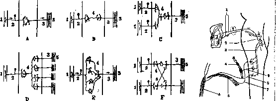
Figure 1.
Figure 2. Figure 1. Diagram of the relationship of neurons in various forms of reflex mechanism: 1-reflexogenic zone; 2-receptor; 3-effector; 4-associative (correlative) neuron; 5-muscle. Figure 2. Diagram of the cough reflex. The impulse due to irritation of the laryngeal mucous membrane along the sensory fibers of the X nerve spreads to the sensory nucleus, and from there to the motor fibers of the X nerve, as well as via the internuncial neuron (tr. solitario-spinalis) to the spinal cord centers corresponding to the diaphragm, intercostal and abdominal muscles: 2-motor and sensory fibers (X nerve); 2-motor and sensory nuclei of the X nerve; 3-associative neuron between the sensory nucleus of the X nerve and the spinal cord (tr. solitario-spinalis); 4-intercostal muscles; 5-abdominal muscles; 6-diaphragm; 7-p. phrenicus; 8-nerve to abdominal muscles; 9-nerve to intercostal muscles. Reflexes (R.) are involuntary responses to irritation of receptors, carried out through the nervous system. The simplest reflex arc consists of a receptor, afferent neuron, efferent neuron, and effector. More complex reflexes involve the participation of associative neurons. The reflexogenic zone is the area of irritation that causes a reflex. The reflex arc is the path along which the nerve impulse travels during a reflex. The reflex arc may consist of one nerve or segment; the reflex arcs of most R. correspond to the II-III spinal segments; in addition, the extent of the reflexogenic zone is subject to certain individual variations; it also changes in pathological conditions, increasing with increased R. and decreasing with their decrease. The accompanying table presents the most important clinical R. with indications of the methods for eliciting them and the level of passage of their corresponding reflex arcs through the spinal cord. Pathological changes in R. of somatic musculature can be quantitative (changes in intensity) and qualitative (changes in form). As for quantitative changes, it should be borne in mind that the intensity of R. is subject to certain individual variations under physiological conditions. Therefore, the absolute intensity of R. by itself does not have significant value for the diagnosis of organic disease of the nervous system, provided there is complete uniformity in the intensity of R. on both sides. Only extreme degrees of decrease or increase in R. can serve as a basis for suspicion of organic disease. On the contrary, unevenness of tendon or skin R. is always an indication of the presence of organic disease, the nature and localization of which can be established on the basis of comparing the unevenness of R. with other symptoms. For the graphic reproduction of the reflex act and the digital determination of the intensity of R., various instruments have been proposed: the reflexograph, or Weiler's reflexometer, is designed to record on a kymograph the curve of the knee R., more precisely-the change in the angle between the shin and thigh during the performance of the knee R. The essence of the apparatus consists of a thread thrown over pulley-like wheels, which is connected, on one side, with the pen of the kymograph, and on the other side-thrown over a wheel connected by an axial rod with a bandage encircling the calf. The blow to the tendon of the quadriceps muscle is produced by a special hammer, the handle of which is attached crosswise to a cylinder rotating on an axis. When the end of the handle is bent, the striking end of the hammer is raised, and when the pressure on the handle is suddenly stopped, the striking end of the hammer drops and strikes the tendon; thus, the force of the blow does not depend on the will of the investigator. In addition, it can be determined exactly in numbers by the movement of a weight that moves along the handle of the hammer between its striking part and the point of attachment of the hammer to the cylinder. The apparatus is designed in such a way that when the angle between the thigh and shin changes by 1°, the writing pen of the kymograph draws a line of 1 mm. With the help of Weiler's reflexograph, it is possible to determine the size of the reflex movement, the time of the reflex act and its individual parts; thus, Weiler managed to establish the time of the latent period of the knee R. (0.045-0.05 sec.). Bechterev's reflexometer is an instrument for the digital determination of the range of motion of the shin during the knee R.; the instrument consists of an arc-shaped graduated plate fixed across a vertical stand. The range of motion of the shin can be read off the divisions of the arc, parallel to which the foot moves when eliciting the knee R. The described apparatus, like many others proposed for the same purpose, have not found application in clinical practice mainly because the data on the state of R. necessary for clinical conclusions can be obtained without these instruments, which are complex and often inaccurate. As for the dependence of quantitative changes in R. on various pathological conditions, all kinds of disorders of conductivity of the reflex arc lead to weakening (hyporeflexia) or loss of R. (see Areflexia). Therefore, loss or weakening of R. is an important sign of peripheral paralysis. In paralyses depending on the lesion of the central motor neuron, changes in R. are more complex and varied. Some of the R. are found to be increased (hyperreflexia) due to the loss of inhibitory influences of the cortex on the reflex apparatus of the spinal cord; other R. are found to be weakened; this concerns those R. which are not inhibited but stimulated by the cortex. In addition to these quantitative changes in R., central paralyses are accompanied by qualitative changes in R.: for this type of paralysis, the appearance of such reflex reactions is characteristic, which are neither an increase nor a decrease in R., but are specific reactions not inherent in the normative adult. The totality of these reactions is denoted by the term pathological R. The essence of quantitative changes in R. in central paralyses is usually formulated as follows: in these paralyses, deep R. are increased, and skin R. are decreased. This position cannot be considered a rule: in severe degrees of central paralysis, the so-called "protective R." is observed on skin irritations; in the area of lip musculature in central paralyses (pseudobulbar paralysis), a completely parallel increase is observed in both skin and deep R. (see below); in the area of abdominal musculature in central paralyses, a parallel weakening is observed in both skin and deep R. All these facts show that the weakening or increase of R. in central paralyses cannot be dependent on their belonging to the group of skin or deep R. The factor determining the quantitative changes in R. in central paralyses is biological. With a lesion of the central motor neuron, the spinal cord is separated from the cerebral cortex; as a result, the automatic activity of the spinal cord, which is inhibited in the norm by cortical influences, is restored; but at the same time, all those reactions of the spinal cord that are developed under the influence of the cerebral cortex and are stimulated by it are lost or weakened. The cerebral cortex is a phylogenetically late superstructure over the spinal cord, and the phenomena observed with a lesion of the central neuron, i.e., with the isolation of the spinal cord from the influences of the cerebral cortex, can be considered as a regression to an older stage of evolution. In the spinal cord, returning in this case to its autonomous state, those reactions that are stimulated by the cortex disappear, and those that are inherent to its own mechanism and which are inhibited by the cortex are strengthened. Thus. if Table of reflexes. Name of R. Method of detection Motor effect Level of reflex arc Anal R. ! Insertion of a finger into rectum Contraction of the sphincter Sv ' Achilles R. Tapping on the Achilles tendon Contraction of the calf muscles Si and Sn j 3 4 Femoral-Rémak (observed in pathological cases-lesion of the upper-i parts of the spinal cord) Biceps, P. with biceps muscle Irritation of the antero-inner part of the upper third of the thigh Flexion of the first three fingers and foot to the sole, extension in the knee joint Lower lumbar and upper cross-sacral segments Tapping on the tendon of the biceps muscle of the arm Flexion in the elbow joint Cv-VI Upper abdominal (syn. supraumbilical) Irritation of the skin of the abdomen above the umbilical line Displacement of the abdominal wall toward the irritation DVIi-VIii Hypochondriac Irritation in the area of V-VI intercostal spaces Contraction of the upper part of the rectus muscle Dv-VI Pharyngeal Touching the posterior wall of the pharynx Cough movement Sensory and motor nuclei of the IX nerve.'
Infraspinatus, P. from the periosteum of the spine of the scapula Tapping on the scapula below the spine of the scapula Adduction of the shoulder, rotation outward, flexion in the shoulder joint Cv-VI Knee (syn. patellar) Tapping on the tendon of the quadriceps muscle Extension in the knee joint Ln-iv Cochleo-palpebral (syn. auditory) Sudden sound Closing of the nuclei of the cochlear and facial nerves Scapulo-humeral Tapping on the inner edge of the scapula Adduction of the shoulder, its rotation outward, sometimes flexion in the shoulder joint i Civ-v Scapular, s.' spino-scapularis (syn. scapular) Tapping on the inner edge of the scapula or superficial linear irritation inward from the inner edge of the scapula Adduction of the scapula Cv-VI н Masseter-reflex (syn. mandibular) With the mouth open (without tension) of the subject, a short tap is made on the lower incisors Contraction of the masticatory muscles Sensory and motor nuclei of the trigeminal nerve Supraorbital Tapping on the inner edge of the supraorbital arch Closing of the eyelid Nuclei of the trigeminal and facial nerves Palatine, R. with uvulae Touching the soft palate Lifting of uvulae Sensory and motor nuclei of the vagus nerve Lower abdominal (syn. infraumbilical) Irritation of the skin of the abdomen below the umbilical line Displacement of the abdominal wall toward the irritation Dx-xn Finger (Mayer's reflex) Sharp flexion of the middle finger toward the palm, produced by the researcher Opposition of the thumb, its flexion in the metacarpophalangeal and extension in the interphalangeal joint Cvn-VIn and Their Plantar Linear irritation of the skin of the sole Flexion of the toes toward the sole Si and Sn Forearm (Leri) Forceful simultaneous flexion of the fingers and hand Reflexive flexion of the upper limb in the elbow joint Cti R. with the cremaster, R. t. cremasteris, R. with the testis Linear irritation of the inner surface of the upper thigh Elevation of the scrotum Li-п £ % Name of R. | Method of detection Motor effect Level of the reflex arc P. m. tensoris fasciae latae (P. Brissaud) Linear irritation of the skin Contraction of m. tensoris fasciae latae Si and Sn 22 23 24 25 R. with the ulna (syn. ulnar, cubital) Tapping on the styloid process of the ulna Pronation and flexion in the elbow joint CVI R. with the radius (syn. carpo-radial) with processus styloideus radii Tapping on the styloid process of the radius Flexion in the shoulder joint, pronation, flexion of the fingers Su DO Dx Corneal, R. with cornea Touching the cornea Closing of the eyelid Nuclei of the trigeminal and facial nerves Sucking (normally present only in infancy) Touching the lips Sucking movements Nuclei of the trigeminal and facial nerves Sternal R. When irritating the skin of the epigastric area Contraction of the abdominal muscles 27 28 Triceps, R. with the triceps muscle Tapping on the tendon of the triceps muscle of the arm Extension in the elbow joint CVI-vn Femoral (Guillain u. Barre): a) Tibio-femoral b) Peroneo-femoral Tapping on the tendons of mm. semimembr. and semitend. (the patient is lying on his back with the lower limb slightly abducted and slightly bent) Tapping on the tendon (the patient is lying on his side, opposite to the side of research) Contraction of mm. semimembranosi and semitendinosi Contraction of m. biceps femoris Ljv-v Si I 29 i Sneezing Tickling the nasal mucosa Sneezing Nuclei of the trigeminal and vagus nerves j 30 Gluteal (syn. gluteal) Linear irritation of the skin of the gluteal area » Contraction of the gluteus maximus i Liv-v and Si In central paralysis, the plantar R. is weakened or disappears, this is explained not by the fact that this R. is cutaneous, but by the fact that this R. is phylogenetically and ontogenetically new: it is absent in a child and is acquired only during individual life in the process of adaptation of the foot to the function of walking in a vertical position. Similarly, abdominal R. disappear or weaken not because they are cutaneous, but because they are acquired during individual life in connection with the vertical position of the body; similar to plantar R., abdominal R. represent a reaction stimulated by the cortex; they are absent in animals, as well as in children before they acquire the function of standing and sitting. On the contrary, the increase in the knee R. in central paralysis depends not on its belonging to the group of deep R., but on the fact that the function of extension at the level of the knee joint is phylogenetically old, inherent to all mammals and therefore automated, innate form of reflex reaction, not stimulated but inhibited by the cerebral cortex. The biological facts presented should be kept in mind to explain the exceptions and contradictions that exist in relation to the widespread position that in central paralysis, deep R. are increased and cutaneous R. are weakened or disappear. The area of quantitative changes in R. can also include the fact of an increase in the reflexogenic zone with increased R. and its limitation with weakened R. A particularly important role in the diagnosis of lesions of the central neuron belongs to the pathological R. mentioned above. Under this term should be understood reflex reactions that do not exist in a normal adult and appear only with a lesion of the central motor neuron. To this characteristic of pathological R. one could add another important feature for explaining their essence: all these R. are characteristic of infancy or childhood; this circumstance can serve as a reason for explaining pathological R. by the biological data presented above in relation to quantitative changes in R. The presence of reactions identical to pathological R. in early childhood, i.e., before the full development of the cortex, their disappearance in the adult as the cortical functions form, and finally their restoration with lesions of the pyramidal paths, i.e., with the isolation of the spinal cord from cortical influences,-all this indicates that pathological R. represent automatic reactions of the spinal cord, which in the normal adult are in a latent state under the influence of cortical activity. In their biological meaning, they are rudiments of functions, representing phylogenetically old, fixed and automated reactions. In the formation of cortical functions, these innate automatisms are transferred to a latent state and replaced by new forms of reactions corresponding to cortical functions. Among individual forms of pathological R., the following deserve mention. In the area of facial musculature with bilateral lesions of central neurons (tr. cortico-bulbaris), there is a tendency to reflex contraction of the lip musculature (m. orbicularis oris); the combination of various methods of eliciting these reactions from the lip musculature can be designated by the term "oral R.". When tapping on the root of the nose ("naso-labial R." of Astvatsaturov, glabellar R. of Moro), when touching the lips (Fressreflex of Oppenheim), when tapping on the upper lip (reflex of Toulouse and Vurpas, s. r. buccalis or trunk-like), when tapping on the upper or lower incisors, when touching the hard palate, there is contraction of the orbicularis oris muscle, which in severe cases leads to trunk-like closure of the lips; sometimes chewing and swallowing movements are added to it. In their physiological meaning, the mentioned oral R. represent a rudiment of sucking movements. This reaction of sucking movements is a phylogenetically very old function, automated in the course of evolution; the "sucking R." as an innate "unconditioned" R. exists in humans in infancy, but as the cortex develops and automatic segmental reactions of the lip musculature are replaced by voluntary cortical ones, the sucking R. passes into a latent state. With lesions of the pyramidal paths (pseudobulbar paralysis), there is a regression to a lower stage of evolution and the sucking R. is restored from its latent state. A very large number of pathological reactions have been described in the area of the foot and toes of the foot. The main type of them is the tendency to dorsal flexion of the big toe and to spreading of the other toes with lesions of the pyramidal bundle. For the first time, these symptoms were described by Babinski in the form of dorsal flexion of the big toe (see Babinski's reflex) and spreading of the toes ("fan symptom") with linear irritation of the skin of the sole. A similar reflex to Babinski's is the trochmotor reflex, elicited by tapping the back of the foot and the area of the metatarsophalangeal joints outward from the tendon of the long extensor of the big toe. Since the first description by Babinski of the dorsal flexion of the big toe (1895) with the method of irritation now indicated, other methods of obtaining the same phenomenon have been described: strong pressure from top to bottom on the posterior surface of the tibia (see Oppenheim's reflex), pressure on the mass of the muscles of the posterior surface of the calf (see
Gordon's sign, reflex, phenomenon), compression of the Achilles tendon (Schaefer's symptom), pressure on the sole (Tremmer's symptom), etc. It should be noted that with different degrees of pyramidal damage, the tendency for dorsal flexion of the big toe is sometimes so sharply expressed that this phenomenon can be caused by the most diverse irritants. However, it must be admitted that the most reliable method for detecting this symptom is the classic method recommended by Babinski; this is stroking the outer edge of the foot with a sharp object. In this form, dorsal flexion of the big toe represents the earliest symptom of developing damage to the central motor neuron; at the same time, it often remains the only sign of pyramidal damage after complete restoration of motor function.--The second group of pathological reflexes of the foot consists of phenomena of reflex flexion of the II-V toes; the most appropriate methods for detecting this symptom are: Mendel-Bechterev reflex (see) (flexion of II-V toes when tapping the outer part of the dorsum of the foot) and Rossolimo's symptom (flexion of II-V toes with a short blow to the end of the third toe). In their biological meaning, pathological reflexes of the foot represent a rudiment of the grasping function. As is known, features of the grasping function are quite clearly expressed in infancy. The grasping function of the lower (posterior) extremity is also characteristic of all the closest phylogenetic ancestors of man. In an adult, these features of the grasping function pass into a latent state under the influence of adaptation of the foot to the static function. With damage to the pyramidal tract, the inhibitory influence of the cortex falls out, and the features of the grasping function that were in a latent state are restored: mobility of the toes appears, a tendency to flex the II-V toes and extend the big toe, as well as a tendency to rotate the foot inward (see Gadorberger's reflex),-all these phenomena are characteristic of the foot in infancy. Among the pathological reflexes of the upper extremity, the so-called 'grasping reflex' deserves mention; The essence of this symptom is that patients, unable due to paralysis to perform a voluntary grasping movement, automatically grasp objects placed in their hand, and this reflex grasping cannot be voluntarily stopped by the patient. The grasping reflex is observed with damage to the frontal lobe and thus is not a sign of damage to the pyramidal tract; it represents an automated function of the motor cortical area (anterior central gyrus); the grasping automatism exists, as is known, in infancy, later it is inhibited from the frontal lobe, and with damage to the latter it is restored from a latent state. In addition to the above, there are other forms of manifestation of the grasping reflex: for example, sometimes patients suffering from damage to the frontal lobe show an involuntary tendency to grasp with the affected hand an object located in the healthy hand. A mild degree of grasping reflex is represented by the so-called 'tonic innervation'; this phenomenon consists in the inability to voluntarily stop a grasping movement while preserving the voluntary act of grasping; the patient can grasp an object but is unable to release it from the hand voluntarily. Among the pathological changes of reflexes, the so-called paradoxical reflexes should be mentioned. The essence of the phenomenon denoted by this term is that in some pathological cases, an attempt to cause a certain reflex leads to a reflex reaction from the antagonists of those muscles whose reflexogenic zone the irritation was directed to; for example, when irritation is applied to the tendons of the flexors, reflex contraction of the extensors occurs. Paradoxical reflexes can be observed both in central and peripheral paralysis. In central paralysis, paradoxical reflexes are caused by uneven increase in reflex excitability in different muscle groups. For example, in spastic paralysis, the reflex excitability of the knee extensors increases much more than that of the flexors, and at the same time the reflexogenic zone also increases enormously. This predominance of the increase in excitability of the extensors compared to the flexors, as well as the spread of the reflexogenic zone of the extensors can be so significant that irritation of the tendons of the flexors results in reflex contraction of the extensors. Paradoxical reflexes in peripheral paralysis are rarely observed, and there are some disagreements regarding the explanation of their mechanism. However, it should be admitted that the main cause of paradoxical reflexes in this case is also the irradiation of irritation to an inappropriate segment due to a sharp disturbance of the equilibrium in the reflex excitability of two reflex centers: the more excitable center reacts to irritations applied to the receptors of the less excitable one.-Reflex tone, i.e. the degree of tension of the musculature at rest, the degree of its preparedness for contraction even under physiological conditions, appears to be different in different muscle groups; in pathological cases this difference can reach even more acute degrees. Reflexes of the autonomic nervous system have not acquired and apparently cannot acquire that degree of diagnostic accuracy and objectivity which is characteristic of somatic reflexes. This fact is explained by many reasons: first, the reflex arcs of the autonomic system do not represent such isolation and autonomy as is characteristic of the somatic nervous system; therefore, autonomic reflexes appear more diffuse and widespread; second, the reflex excitability of the autonomic nervous system is subject to very significant individual fluctuations; third, the entire apparatus of the autonomic nervous system represents a very unstable mechanism, the equilibrium of which can fluctuate sharply under physiological conditions: such moments as mental experiences, the state of the digestive organs, fatigue or physical vigor and a whole series of other circumstances can influence autonomic reflexes. Finally, one cannot ignore the fact that the study of the autonomic nervous system belongs to recent times. The receptor apparatuses of the reflex autonomic arcs are, first, afferent fibers of the sympathetic nervous system, and second, afferent fibers of the somatic nervous system. Further, it should be borne in mind that all somatic nerves contain sympathetic fibers in their composition. Therefore, the source of autonomic reflexes can be irritations that are very diverse both in their nature and the place of their action. For example, painful irritations acting on the skin can cause various reactions not only from the autonomic effectors of the skin but also from the internal organs due to the abundance of anato-physiological connections between somatic afferent fibers and sympathetic centers. - No systematic classification of autonomic reflexes, based on any definite principle, has been established, and various types of autonomic reflexes are grouped either according to the irritants causing them (for example 'pharmacological reflexes'), according to the organ from which the reaction is obtained (for example 'vasomotor reflexes'), or according to the nature of these reactions (for example pressor and depressor reflexes, etc.). In addition to the described (see Autonomic nervous system, afferent pathways and reflexes of the autonomic nervous system, as well as visceroreflexes) autonomic reflexes, we mention the following forms of them. Skin-heart reflexes-changes in the pulse in the form of slowing or acceleration with various skin irritations: painful, tactile, faradic, etc. Palatal-heart reflex-acceleration of the pulse when pressure is applied to the hard palate. Orthostatic reflexes (Prevel)-acceleration of the pulse when transitioning from lying or sitting to standing. Orthocardiac reflex, observed only in pathological cases-expansion of cardiac dullness when transitioning from horizontal to vertical position. Erben's symptom-slowing of the pulse (irritation of n. vagi) when the subject is squatting with the head sharply bent forward. Chermak's reflex-slowing of the pulse and respiration when pressure is applied to the upper third of the sternoclavicular-muscle. Abdominal-heart reflex-increase of cardiac dullness when tapping the middle line of the abdomen between the xiphoid process and the navel. Cochlear reflex on the heart-irritations of hearing have as their result somatic reflexes of eye rotation and blinking (see table-cochleo-palpebral reflex); with stronger irritations, the spread of the impulse to the nucleus of the vagus nerve can lead to changes in the rhythm of respiration and pulse. Reflex of the solar plexus (syn. solar reflex)-pressure along the middle line of the abdomen to the sensation of pulsation of the abdominal aorta above the navel-slowing of the pulse. The general term pressor reflexes denotes phenomena of increase in blood pressure and narrowing under the influence of various effects. The opposite changes are denoted by the term depressor reflexes Mamidlo (areolar)-areolar reflex-see wrinkling of the breast nipple with skin irritations.
Testicular reflex - compression of the testicle causes a series of phenomena on the part of the heart, blood vessels, respiratory tract, and pupils. See also Adductor reflex, Antagonistic reflexes, Achilles reflex, Aschner phenomenon, Barré reflex, symptom, Bettiger reflex, Bechterev reflexes, Abdominal reflexes, Bulbar reflexes, Bulbocavernous reflex, Wartenberg reflex, Vodak reflex, Gnudi reflex, Lip reflex, Glabellar reflex, Hirschberg reflex, Haab reflex, Hering reflex, Danielopolu reflexes, Zhukovsky reflex, Pupillary fibers, reflexes, centers, Cutaneous reflexes, Contralateral reflexes. Corneal reflex, Conjunctival reflex, Knee reflex, Cremaster reflex, Magnus-Klein reflexes, Marie-Foix reflexes, Periosteal reflexes, Pirotrovskoy reflex, Pilomotor fibers, reflex, Postural reflexes, Rossolimo reflex, Tendon reflexes, Conditioned reflexes. Lit.-see lit. to art. Reflexology.
Related articles
Mentioned in
Cite this page
“REFLEXES.” Soviet Medical Encyclopedia. English translation of Bolshaya Meditsinskaya Entsiklopediya, 1st ed. (Moscow, 1928–1936), ed. N. A. Semashko. https://sovietmedicalencyclopedia.pages.dev/article/reflexes/