Wandering Cells
Historical document, translated for reference. It reflects medical knowledge of the 1920s–30s and is not medical advice.
Summary
Wandering cells are mobile cells found in connective tissues throughout the animal kingdom, capable of amoeboid movement. They play a key role in inflammation and phagocytosis, though their exact origin and relationship to other cells remain debated.
Encyclopedia article (1928–1936)
WANDERING CELLS (German Wanderzellen), a name applied to cells capable of moving, 'wandering,' in the tissues of the organism. As is known, movement is characteristic of many cellular elements; but the name W. c. is applied only to cells moving in the tissues of the animal organism by means of so-called amoeboid movement (see), and moreover, those in which this ability is a permanent property. Their main place of residence and movement is loose connective tissue. W. c. are elements distributed throughout the animal kingdom, starting from the lowest animals, where they are represented by cells called amoebocytes (see), and ending with humans. In vertebrates, including humans, in the resting state of connective tissue, W. c. are found everywhere in it in the form of rounded mobile elements of various sizes, having a round or kidney-shaped, chromatin-rich nucleus and a basophilic, grain-free protoplasm, arranged either in a thin or wider layer. These cells on sections of connective tissue are found partly singly, partly in small groups. When the resting state of connective tissue is disturbed, especially during its inflammation, the number of these W. c. already in the first hours increases significantly: they W. c.
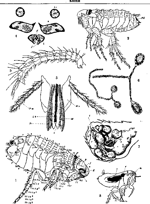
Figure 1. Dog flea Ctenocephalus canis: I-III-upper mesonotum; 1-3-dorsal half-rings of abdomen; I'-3'-ventral half-rings of abdomen; a-front, b-posterior half of head; 1-antennae; d-eye; e-frontal ctenidium; f-prothoracic ctenidium; g-palp; h-oral parts; i-trochanter; j-femur; k-tibia; ta-tarsal segments; Ts II, III-spiracles of thorax; Ts 1-3-spiracles of abdomen; Gi-spermatheca (I and III near the letters indicate the 1st and 3rd legs). Figure 5. Rat flea Ceratophyllus fasciatus, pygidium. Figure 3. Oral organs of flea: Lb-upper lip; M-upper jaws; Mx-lower jaws; Pt-lower jaw palps; L-lower lip; P-lower lip palps. Figure 4. Cross-section of flea oral organs: d-upper lip; j-upper jaw; k-lower jaw; ht-lower jaw palp; l-lower lip. Figure 5. Larva of W. Leptopsylla Pawlowskyi. Figure 6. Salivary glands of flea. Figure 7. Opened abdomen of rat flea Ceratophyllus fasciatus with cysticercoids of Hymenolepis diminuta: a-stomach; b-intestine; c-cysticercoids. Figure 8. Flea with stomach plugged with a mass of multiplied plague bacteria (s); p-crop; m-sucking muscle.
from the lowest animals, where they are represented by cells called amoebocytes (see), and ending with humans. In vertebrates, including humans, in the resting state of connective tissue, W. c. are found everywhere in it in the form of rounded mobile elements of various sizes, having a round or kidney-shaped, chromatin-rich nucleus and basophilic, grain-free protoplasm, arranged either in a thin or wider layer. These cells on sections of connective tissue are found partly singly, partly in small groups. When the resting state of connective tissue is disturbed, especially during its inflammation, the number of these W. c. already in the first hours increases significantly: they W. c.
W. c. W. c. transform due to hypertrophy of the nucleus and protoplasm into larger, protoplasm-rich and very mobile W. c. One of the properties of these elements both in resting and inflamed connective tissue is phagocytosis (see), i.e., the absorption of any colloidal particles, thanks to which Metchnikoff called them macrophages (see). Regarding the origin of W. c. and their connection with other cells of connective tissue and blood elements, to this day there is still no complete unanimity. Saxer considered them a product of mesenchymal elements, preserved in connective tissue from the embryonic period of its development, in the form of primitive W. c. (primitive Wanderzellen von Saxer). Marchand found that in resting connective tissue, in the closest neighborhood of vessels, in their adventitia, there are cells with macrophage properties: according to Marchand, these cells, which he called adventitial cells, originate from the endothelium of capillaries (endotheliocytes, in Marchand's later term); they can round up and transform into W. c. of connective tissue, which Marchand initially characterized as leucocytoid cells. According to Maximow, Weidenreich and some others, W. c. of connective tissue are related to lymphocytes and partly result from emigration from blood vessels, partly are formed in place (see below); in view of their ubiquitous distribution in the organism and mesenchymal origin, Maximow calls them ubiquitous W. c. mesenchymal amoebocytes, as well as mobile, indifferent mesenchymal cells. The mentioned lymphocytes of connective tissue, in case of an inflammatory process, are characterized by progressive development and transformation into larger W. c., called by Maximow polyblasts. On the other hand, a lymphocyte or polyblast wandering in the tissue can stop its amoeboid movement, flatten on the surface of connective tissue fibers, and, changing its shape, transform into a temporarily immobile cell; such cells that have lost mobility and become sedentary are called by Maximow and his student Chashin W. c. at rest (ruhende Wanderzellen). These W. c. at rest can later regain mobility and transform into polyblasts. Maximow points out,
f.
f.
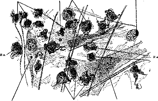
W.c.—wandering cells with pseudopodia; F.—fibroblasts of connective tissue; C.b.—collagen fibers. That the cellular elements called wandering cells by him in a resting state are the same adventitial cells of Marchand; they had been previously described by Ranvier under the name of clasmatocytes (see), and by Renaut—as rhagiocrine cells (cellules rhagiocri-nes), the distinguishing feature of which is the vital staining with neutral red. These same cells, accumulating in large groups in the omentum and serous membranes, form the so-called milk spots (taches laiteuses). Thus, in Maximov's doctrine of wandering cells, the first feature is terminology: he calls macrophage-type wandering cells 'polyblasts,' and the elements designated by Marchand as adventitial cells, he qualifies as 'wandering cells in a resting state.' Maximov's terminology, although quite common in the USSR, is objected to by many Western scientists (Marchand, Aschoff, and others). On the other hand, the identification of these cells with lymphocytes, presented by Maximov and some others, is also not universally accepted: as shown by experiments with vital staining on animals (Goldmann, Marchand, Aschoff, Kiyono and many others), dye grains are always deposited in large quantities in the cytoplasm of macrophages (polyblasts) and adventitial cells (wandering cells in a resting state) and are not deposited at all in lymphocytes. Incidentally, Goldmann, wishing to emphasize the tendency of macrophages to take up dyes, especially pyrrhol dyes, designates them as pyrrhol cells (Pirrholzellen), and Aschoff, in order to highlight their tissue origin in contrast to cells emigrating from the blood, proposes the term histiocytes. In general, it must be said that in light of modern views, the considerations regarding the origin of wandering cells, tissue macrophages, put forward by Marchand, are the most acceptable: in particular, their genetic connection with adventitial cells, which many authors derive from endothelium, easily explains the morphological and functional similarity of tissue macrophages with blood monocytes, which in turn may originate from reticulo-endothelium. Detailed studies by Carrel and Ebeling on in vitro cultures confirm the origin of macrophages from monocytes under appropriate nutritional conditions, and especially phagocytosis. Some (Weidenreich and others) admit the possibility of wandering cells forming from fixed connective tissue cells (fibroblasts). It should be noted that such a possibility has been proven for lower vertebrates (amphibians); as for higher vertebrates, even in regenerative and inflammatory multiplication of fibroblasts with their transformation into free, moving elements—fibroblasts do not transform into true wandering cells, macrophages. The matter is different with cells of reticular connective tissue: the possibility of their transformation into true wandering cells, macrophages, upon detachment from the matrix and rounding, can be considered fully established. The physiological role of wandering cells is manifested in their ability to phagocytize and digest phagocytized material. This is most clearly manifested in embryonic histolysis (see) of tissues, in any disintegration in tissues, especially—inflammatory changes; in all these cases, wandering cells, accumulating in large quantities, phagocytize and process various products of tissue and blood disintegration, which is morphologically noticeable in the form of various inclusions and vacuoles appearing in the cytoplasm of wandering cells. But not only in the indicated states of tissues, but also in resting connective tissue, both adventitial cells and mobile macrophages exhibit phagocytosis and digestion of various products circulating in the tissues and belonging to proteins, fats, carbohydrates, iron, pigments, etc. This role of macrophages in tissue intermediate metabolism is the basis for recognizing them as part of the body's reticulo-endothelial system (macrophage apparatus or reticulo-end. system of connective tissue). This is also confirmed by the fact that apparently wandering cells also have an excretory-secretory function with respect to captured and processed materials, which gives some researchers the right to classify wandering cells among the so-called nephrocytes (see). In addition, wandering cells, moving partly by amoeboid movement in the tissues, partly with the flow of tissue lymph, as well as in lymph vessels and lymph cavities, carry the captured material from place to place. Finally, it can be noted that the mother cells of macrophages, i.e., adventitial cells (wandering cells in a resting state), and reticular cells can not only transform into wandering macrophages, but under certain conditions, according to some authors, can differentiate into granulocytes (histiogenous leukocytes), lymphocytes, and erythrocytes (see Hematopoiesis). All of the above refers to the main type of wandering cells, namely, connective tissue macrophages. However, the latter are not the only cells deserving this designation. The term wandering cells can be fully applied to granular polymorphonuclear leukocytes in cases when they are outside blood vessels, among the tissue. To a special degree, neutrophilic leukocytes, to a lesser extent—eosinophils and basophils (mast cells), are characterized by amoeboid movement and phagocytosis, which manifest especially intensely during inflammation (see Leukocytes). To a lesser extent, plasma cells (see) play the role of wandering cells, to which apparently only a low degree of mobility is inherent. It should also be mentioned that in the central nervous system, wandering cells have significance as cells detaching from the neuroglia complex, possessing both amoeboid mobility and phagocytosis. The mechanism of movement of wandering cells—see Amoeboid movements.
SPb, 1913; March and F., Die. ortlieh reakti-ven Vorgange (Krenl L., Marchand F., Handbuch d. allg. Pathologie, B. IV, AM. 1, Lpz., 1024); Saji-moff A., Expcrimentelle Untersuchungen ttber die entziindliclie Neubildung von Bindegewebe, Jena, 1902; K i y o n o K., Die vitale Karrninspeichcrung, Jena, 1914; Aschoff L., Ein Beitrag zur Lehre von den Makrophagen, Yerhandlimgen der deutschen pathologischen Gesellsebaft, 16 Tagung, 1913; W e 1-d e n r e i c h F., Blutkorperchen und Wanderzellen, Jena, 1911; Carrel A. and Ebeling A., The fundamental properties of tbe libroblast and the macrophage, Journal ol experimental medicine, V. XLIV, 1926.
Related articles
Mentioned in
Cite this page
“Wandering Cells.” Soviet Medical Encyclopedia. English translation of Bolshaya Meditsinskaya Entsiklopediya, 1st ed. (Moscow, 1928–1936), ed. N. A. Semashko. https://sovietmedicalencyclopedia.pages.dev/article/wandering-cells/