Micelles
Historical document, translated for reference. It reflects medical knowledge of the 1920s–30s and is not medical advice.
Summary
Micelles are the smallest crystalline particles that form the basis of colloidal substances and many biological structures according to Nageli's theory. This article discusses the historical development of micellar theory, its application to biological structures, and the methods used to study micellar formations in various tissues.
Encyclopedia article (1928–1936)
Micelles (from Latin micella - particle), the smallest crystalline particles that, according to Nageli, form the basis of colloidal substances and many biological structures. The botanist Nageli noted that many organic formations, particularly various plant fibers, exhibit double refraction, which is a characteristic optical property of crystals. He therefore concluded that organic substance consists of the smallest, microscope-invisible crystals, or micelles. Their proper sequential fusion gives rise to the semi-crystalline structure of organic fibers, whereas in the case of disordered, chaotic combination of crystalline M., externally amorphous bodies are obtained. Similar considerations were applied by Nageli to colloidal solutions, which he conceived as consisting of the smallest M. and, in contrast to true molecular solutions, called "micellar solutions." Here, confirmation of his views was the possibility of obtaining certain protein substances, including hemoglobin, in a crystalline state during slow precipitation, whereas during rapid precipitation they yield externally amorphous gels. Nageli's micellar theory was developed in 1858, that is, before the research of Graham, which laid the foundation of the science of colloids. Graham considered one of the most important distinguishing features of colloids to be their amorphous nature, absence of crystalline structure. Under the influence of these ideas, which long dominated colloidal chemistry, the micellar theory was abandoned and did not gain recognition. Only in recent years, due to improved methods for studying crystal structures, it has been revived again through the research of Weimarn, Ambronn, Scherrer and others (see Aggregate state, Colloids, Colloidal chemistry). The most important of these methods is X-ray, which allows detection of the characteristic arrangement of atoms in crystals ("atomic crystal lattice") (see Crystals) even with completely disordered combination of individual crystals. The application of this method has shown that many colloids actually have a crystalline structure. It can also be detected directly by optical methods if the individual crystals are in the same position, arranged parallel to each other. For example, in a strong electromagnetic field, a colloidal solution of iron oxide behaves like a uniaxial crystal and exhibits double refraction. M., having an elongated rod-like shape, assume the same parallel position - under purely mechanical conditions - in flowing sols. Similarly, in a gel built from rod-like M., their parallel orientation can be obtained as a result of uniaxial tension. However, the existence of double refraction in the body under investigation is not always sufficient proof of the crystalline nature of its M. If the dispersed particles themselves are uniaxially birefringent crystals, having their own double refraction ("Eigen-doppelbrechung"), then when they are oriented parallelly, the entire liquid will behave like a single crystal. However, non-crystalline particles, having an elongated shape and identically oriented in space, also exhibit the same double refraction as a crystal whose optical axis coincides with the longitudinal axis of the particles. For this, it is only necessary that the refractive index of the latter differs sufficiently strongly from their dispersion medium. Such double refraction, depending on the position and shape of the particles ("Formdoppelbrechung", or "Stabchendoppelbrechung"), is in its external effect quite similar to the former. Both phenomena, however, can be unmistakably distinguished by means of a method developed by Ambronn and finding application in the study of biological structures. It consists in that the organic fiber under investigation is impregnated with liquids having different refractive indices. If the double refraction depends only on the longitudinal arrangement of the structural elements of the fiber, then it disappears when the fiber is impregnated with a liquid having the same refractive index. On the contrary, the double refraction is fully preserved under these conditions if the M. themselves are birefringent crystals. By means of this method, the crystalline nature of many organic fibers has been indisputably proven. It should be noted, however, that unlike true crystals, whose elements are strictly oriented in the direction of all their axes, individual micellar crystals in such fibrillar structures are arranged parallel only to their main, longitudinal axis, while maintaining a disordered arrangement of their transverse axes. Such semi-crystalline structures are called "mesomorphic". Various crystalline and mesomorphic structures are much more widespread in the living organism than was previously assumed.
D. Rubinstein* In relation to biological objects, the micellar hypothesis has now grown into a coherent doctrine. With the help of Ambronn's technique, we have become able to discover M. and judge their shape, and X-ray spectroscopy methods allow us to penetrate their structure. The best objects are fibrillar structures (metaplastic and paraplastic). Thus, it was found (Mering; 1922) that chitin (the substance forming the shell of crustaceans and insects) consists of rod-like M., which possess their own negative uniaxial birefringence, while chitin as a whole is anisotropic, which depends on the periodically regular arrangement of its M. In contrast to chitin, silk thread possesses positive intrinsic and positive rod-like birefringence. Collagen fibers also exhibit the same optical properties as silk, i.e., they consist of birefringent M., and their arrangement determines the birefringence of the entire fiber. In contrast to collagen fibers, elastic fibers are optically inactive, but when stretched or dried they become uniaxially birefringent. This indicates that in normal conditions the M. in the elastic fiber are arranged disorderly, whereas when stretched they are arranged in periodically parallel rows. During its development, tooth enamel has the same optical properties as chitin (Schmidt; 1928); when compacting during final development, the M. forming the enamel are arranged so densely that only their own negative uniaxial birefringence remains. According to the research of Stübel (Stübel; 1923), the anisotropic disks of muscle fibers consist of positive uniaxial M., their rod-like refraction is also positive, but between them are M. of lipoid substances possessing negative birefringence. The basic substance of bone also consists of micelles, and it can be shown that not only the osteonic fibers consist of micelles, but also the alkaline earth complex salts are also arranged in the form of micelles between the osteonic fibers. Furthermore, micellar structure has been found in horn substances, in tunica, in plant cell cellulose, in nerve fibers, etc. Recent research has shown that the flagella of protozoa, the cilia of epithelial cells, and the finest supporting fibrils of axopodia also consist of micelles, which, however, are not crystalline in themselves. Among the latest research, the most interesting works are Schmidt's (1928-29) on the micellar structure of nuclear chromatin. It turned out that the chromatin of the head of various sperm consists of micelles possessing uniaxial birefringence, while the tail of the sperm possesses positive uniaxial birefringence. Such rod-like structure of M. chromatin and their tendency to orient periodically and parallelly when compacting and forming thread-like structures explain many phenomena in the dynamics of nuclear processes: the thread-like form of chromosomes, their longitudinal splitting, increase in diameter in certain stages, etc. Birefringent chromosomes have not yet been discovered; since in many cases fibrillar structures are reversible, i.e., can disappear and reappear, it must be admitted that they are formed from micelles pre-existing in the cell plasma or in the intercellular substance, although they cannot be detected in either place, as they are in disordered motion. In short, almost everywhere where there are dense fibrous structures, their micellar structure is discovered. These studies are extremely precise and allow not only to detect M., but also to measure their anisotropy. X-rayscopy methods have made it possible to go even further into the realm of invisible structures. As in crystals, X-rays interfere when passing through micellar structures, giving typical X-ray photographs. On the basis of these X-ray photographs, by means of a special technique it is possible to'
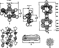
Figures 1-6. Submicroscopic structure of cellulose, starting from atomic compounds and ending with the micelle according to Meyer's research (all figures are made partly on the basis of calculations, partly on the basis of X-ray-
(of spectroscopic research). Figure 1 and 2. Schemes of the spatial model of a glucose particle, constructed as is customary in crystal chemistry, i.e. each atom or atomic group corresponds to a certain space, which is represented as a sphere of a certain radius, expressed in angstroms (for aliphatic compounds, a carbon atom corresponds to a space of 5.5 Å, for aromatic compounds=1.45 Å; an oxygen atom corresponds to a space of 2.7 Å. Hatched circles are carbon atoms. Figure 1 demonstrates the width, Fig. 2-the thickness of the glucose particle. Figure 3. Spatial arrangement of atoms in two residues of glucose-the particle of cellobiose. Joining in long chains, cellobiose residues form chains of main valences. The length of the cellobiose particle should be equal to 10.2 Å, which exactly corresponds to the length of the elementary particle found by X-ray spectrography. Figure 4. The elementary particle of cellulose crystallites; its dimensions are calculated on the basis of measuring the distances between the dark interference bands and the vertical plane of symmetry. Its dimensions are as follows: a:b:c=8.35:10.3:7.9 Å. On the basis of the intensity of the dark bands of the X-ray spectrogram, one can calculate how densely the atoms are arranged in the different planes of this body; the width of the dark interference bands allows one to calculate the size of the particles. Such studies allow us to state that the elementary particle of cellulose consists of 5 cellobiose residues and has the form of a parallelepiped. Figure 5. Approximate size (the form may vary) of the cellulose micelle in millimicrons. Calculations show that the micelle should consist of 60-100 chains of glucose residues, each of which in turn consists of 100 glucose particles. The thickness of the micelle is equal to 40-50 glucose residues. Figure 6. Cross-section of the micelle. To calculate the crystal lattice formed by submicroscopic particles, of which M. consist, to find out how densely the atoms are arranged in the different planes of this lattice, and finally to calculate the dimensions of these particles. Cellulose has been best studied with the help of X-rays (Herzog, Meyer; 1924-29). Cellulose consists of C6H10O5 glucose residues of so-called cellobiose. The dimensions of this particle, calculated in angstroms from the constitutional formula, turned out to exactly coincide with the dimensions of the elementary particle found by X-ray spectrography. Consequently, the elementary particle is the cellobiose particle. In cellulose, these glucose residues are connected to each other in the direction of the fiber. The schematic drawings given well demonstrate the relationships of the structural elements of cellulose. On the basis of such data, Meyer gives the following measurements for the cellulose elementary particle: a = 8.35 Å; b = 10.3 Å; c = 7.9 Å. Consequently it consists of five cellobiose particles. Joining in long chains, these glucose residues form a crystallite, or micelle. One can calculate that the length of the micelle will correspond to 50-80 glucose residues, the width-to 40-60. The dimensions of M. are determined with no less accuracy for other substances of biological origin. For example, M. of chitin with a long period equal to that of cellulose, 10.4 Å, contain from 1,000 to 2,000 acetyl-glucosamine residues. The elementary particle of silk gives the following periods: a=9.3 Å; b = 10.4 Å; c=7.0 Å. The main elements of which it is built-4 glycyl-alanine residues. Rubber has been well studied; it consists of isoprene residues. The length of its micelle is 300-600 Å, which corresponds to 75-150 isoprene residues; the thickness of the micelle is from 30 to 50 Å. According to Clark (Clark; 1927), the elementary particle of rubber stretched by 75% gives the following dimensions: 8.1x12.3x8.3 Å. The keratin of hair according to the research of Oestri and Woods (Oestri, Woods; 1930) consists of peptide chains characterized by a periodicity of 5.15 Å, whereas in the stretched state the periodicity is 3.4 Å. This is explained by the fact that hexagonal chains turn into zigzag chains upon stretching. These examples are quite sufficient to show how accurate X-ray spectrographic analysis is. In all cases where it is feasible, it can be established that the micelle is formed from chemically connected chains consisting of elementary basic particles. These chemically closed chains - possibly not always similar - Meyer proposes to call "chains of main valences" (Hauptvalenzketten); from 30 to 100 such chains form the M. At present all the data at our disposal force us to recognize that even highly polymeric substances of a colloidal nature, such as: albumins, lignins, starch, and also probably the protoplasm of cells, are built on the same type from "chains of main valences". It should be mentioned that M. are held near each other by intermicellar cohesive forces. These forces can be calculated; their measure can be the thermal energy that needs to be expended to separate one micelle from another. According to the calculations of Meyer and Mark, from 1 to 2 million calories are required to separate two cellulose M., whereas for example to break a simple double carbon bond (primary valence) only 75,000 calories. A chain of 100 isoprene residues corresponds to a force of 500,000 calories. This also explains why in the distillation of micellar substances the substance is destroyed faster than it turns into vapor, since to turn into vapor it is necessary to apply such an amount of thermal energy which is quite sufficient to break the chemical bonds inside the molecule. This also explains the strength of M. As for the intermicellar spaces, their properties have been studied very little, but undoubtedly they also play a large role in the various states of micellarly built colloidal systems.
Related articles
Mentioned in
Cite this page
“Micelles.” Soviet Medical Encyclopedia. English translation of Bolshaya Meditsinskaya Entsiklopediya, 1st ed. (Moscow, 1928–1936), ed. N. A. Semashko. https://sovietmedicalencyclopedia.pages.dev/article/micelles/