Hemoglobin
Historical document, translated for reference. It reflects medical knowledge of the 1920s–30s and is not medical advice.
Summary
The article discusses hemoglobin as a respiratory enzyme and its medicinal applications. It covers the biochemical properties and physiological functions of hemoglobin in the human body.
Encyclopedia article (1928–1936)
529 Hemoglobin as a respiratory enzyme...
531 Hemoglobin as a medicinal substance...
Hemoglobin (often denoted by the symbol Hb), the coloring matter (pigment) of blood, is contained in the red blood cells, or erythrocytes, constituting 90% of the dry matter of the latter. Upon hemolysis (see) of the blood, hemoglobin is separated from the stroma of the red blood cells and passes into solution. Hemoglobin plays a very important role in the animal body, namely, the supply of oxygen to all parts of the body: hemoglobin has the ability to enter into combination with oxygen, turning into oxyhemoglobin. The latter, losing oxygen, turns back into hemoglobin. The combination of hemoglobin with oxygen occurs during the passage of blood through the capillaries of the lungs; in the capillaries of the systemic circulation, the splitting off of oxygen from oxyhemoglobin and the supply of oxygen to all parts of the body take place. Hemoglobin is contained not only in the blood of vertebrates, but also in the tissue fluids of invertebrates, and in the latter it is only in rare cases contained in cellular elements (as in vertebrates in erythrocytes), but is usually dissolved in the plasma. Among invertebrates, hemoglobin is found in the blood of various worms, in many lower crustaceans; conversely, it is rarely found in mollusks and insects. Content and quantitative determination of hemoglobin. The hemoglobin content in human blood can, depending on various conditions, fluctuate, but on average is equal to 14% in the blood of men, 13% in women, and 20-21% in newborns. In the first years of a child's life, the hemoglobin content in their blood drops, reaching 11%, but then rises again, reaching 13-14% by the twentieth year of life. In men, an average of 8.5 g of hemoglobin accounts for 1% of body weight. The hemoglobin content in the erythrocytes of various animals is different; however, when calculated per unit surface area of erythrocytes, the hemoglobin content in different animals turns out to be the same (which is connected with the different sizes of erythrocytes of different animals); specifically, an average of 30.1·10-14 g of hemoglobin accounts for 1 sq. μ of the surface of erythrocytes of different animals, as can be seen from the following table: Human and animals Average surface of erythrocytes in sq. μ Hemoglobin content in one erythrocyte in 10-12 g Amount of hemoglobin per sq. μ of erythrocyte surface in 10-14 g Man 98.4 30 30.1 Dog 81.2 24 27.1 Rabbit 75.2 20 17 (average 23) Horse 49.1 18 30.1 Rat 78.7 18 29.1 Sheep 27.2 8 - Normal hemoglobin content in erythrocytes can change in either direction under the influence of pathological states (see Hyperchromasia, Hypochromasia). Methods for the quantitative determination of hemoglobin content in blood. Quantitative determination of hemoglobin content in blood with an accuracy sufficient for clinical purposes is usually performed colorimetrically, using special devices such as hemometers or hemoglobinometers (Fleischl, Sahli, etc.; see Hemometers). More accurate figures are given by determination using the Bürker-Leitz colorimeter. Completely accurate quantitative determinations of the content of hemoglobin and its compounds with gases are performed using spectrophotometers. Chemical nature. Hemoglobin is a high-molecular protein substance belonging to the group of proteids (conjugated proteins), specifically chromoproteins. Its composition, according to Hüfner, is expressed by the formula: C636H1025N164FeS3O181; in its molecule, thus, one iron atom is surrounded by 2,000 other atoms. However, this single iron atom plays the main role in the ability of hemoglobin to enter into combination with oxygen, forming unstable, easily dissociating oxyhemoglobin. Hemoglobin as a chromoprotein is constructed of two components. Iron is part of one of the components of the hemoglobin molecule, namely, the pigment (upon which depends the color of hemoglobin, the color of erythrocytes, and the color of blood)—hemochromogen. The other constituent part of the hemoglobin molecule is a protein substance—globin, belonging to the group of histones; globin contains a lot of the diamino acid histidine and therefore has a clearly basic character. Hemoglobin contains 4% hemochromogen and 96% globin. The splitting of hemoglobin into the components of its molecule occurs very easily; heating with acetic acid is sufficient for this. If the splitting of hemoglobin is carried out without the access of air, then after hydrolysis we obtain globin and hemochromogen. If, however, the hydrolysis of hemoglobin is carried out with the access of air, the splitting-off hemochromogen will immediately attach oxygen and turn into hematin; in this case, after the hydrolysis of hemoglobin, we will have globin and hematin. These same substances are obtained upon the hydrolysis of oxyhemoglobin, since hemoglobin and oxyhemoglobin differ from each other only in that hemochromogen is part of the hemoglobin molecule, and hematin is part of the oxyhemoglobin molecule; the protein component of their molecules is the same (globin): Hemoglobin -> globin + hemochromogen. Oxyhemoglobin -> globin + hematin. Hemoglobin, by attaching oxygen, turns into oxyhemoglobin, the solutions of which have the beautiful light red color characteristic of arterial blood. Reduced hemoglobin. Upon the reduction of oxyhemoglobin (removal of O from it), it turns into hemoglobin, which is also called restored or reduced hemoglobin, since it is obtained by the reduction (restoration) of oxyhemoglobin. Solutions of reduced hemoglobin have a dark, cherry-red color characteristic of venous blood. Properties of reduced hemoglobin. Hemoglobin crystallizes in the form of dark purple-red crystals, which, however, are not easy to obtain; therefore, for analytical purposes, the much more easily obtainable crystals of oxyhemoglobin are usually used. Hemoglobin is easily soluble in water, insoluble in alcohol, ether, chloroform, benzene. Solutions of hemoglobin in a thick layer have a dark cherry-red color, in a thin layer—greenish. Its spectrum at a 0.1-0.3% solution is characterized by a broad absorption band lying between the lines D and E; the middle of this absorption band coincides with a wavelength of 559 mμ. The second absorption band of hemoglobin lies in the ultraviolet part of the spectrum (its middle coincides with a wave at 429 mμ) and can be detected only by photographing the spectrum. Hemoglobin has the character of a weak acid; under the influence of acids, alkalis, and certain inorganic salts, it undergoes decomposition. Characteristic of hemoglobin is its ability to form more or less easily dissociating compounds with various gases, such as oxygen, carbon monoxide, nitric oxide. Such compounds of hemoglobin with gases are oxyhemoglobin, methemoglobin, carbon monoxide hemoglobin, nitrosohemoglobin. Oxyhemoglobin is an unstable, easily dissociating compound of hemoglobin with oxygen and is formed upon simple shaking of hemoglobin solutions with air (or oxygen). Oxygen is chemically bound to the hemoglobin molecule through the agency of the iron located in this molecule (in hemochromogen). 1 g of hemoglobin at 760 mm of air pressure binds 1.34 cubic cm of oxygen, from which it follows (according to Hüfner) that in the oxyhemoglobin molecule there is 1 molecule of oxygen per 1 molecule of hemoglobin and that the molecular weight of oxyhemoglobin is 16,700. Upon the removal of oxygen, oxyhemoglobin dissolved in water turns back into hemoglobin. Oxygen can be removed 1) with the help of a vacuum, 2) by passing an indifferent gas through the solution, 3) with the help of various reducing agents (for example, ammonium sulfide, Stokes' reagent, which is an ammoniacal solution of iron tartrate, etc.). The formation of oxyhemoglobin from hemoglobin, in other words, the binding of oxygen by hemoglobin, is a reversible reaction: Hb + O2 ⇄ HbO2, oxyhemoglobin. Oxyhemoglobin crystallizes much easier than hemoglobin, and by "crystalline hemoglobin" one usually understands crystals of oxyhemoglobin. From the blood of certain animals, crystals of HbO2 are very easily obtained: it is often sufficient, by adding water to the blood, to turn it into lac blood and (adding a little alcohol) place it in the cold. One can use the same method as in obtaining protein crystals (see). Crystals of oxyhemoglobin from the blood of different animals have various shapes; most often these are needles, prisms, and plates of the rhombic system. Upon repeated recrystallization, the crystals can change shape; here, therefore, we are dealing with typical heteromorphism, testifying to a certain lability of the molecule. The spectrum of solutions (0.1-0.3%) of oxyhemoglobin is characterized by two absorption bands lying between lines D and E. The middle of the left (darker and narrower) absorption band coincides with a wavelength of 576 mμ, and the middle of the right (wider and lighter)—with a wavelength of 541 mμ. In the ultraviolet part of the spectrum lies a third broad absorption band, the darkest part of which coincides with a wavelength of 415 mμ and which can be detected only by photographing. Physiological role of hemoglobin and oxyhemoglobin. As already stated, oxyhemoglobin is an unstable, easily dissociating compound of hemoglobin with oxygen. If the hemoglobin solution is in contact with a gas mixture containing oxygen, then, depending on the amount of oxygen in this mixture, in other words, depending on the magnitude of its partial pressure, a greater or lesser part of the hemoglobin will be in the form of oxyhemoglobin.
The following table shows what percentages of oxyhemoglobin and reduced hemoglobin will be present in a solution at various partial pressures of oxygen. Partial pressure of oxygen Hemoglobin in % Oxyhemoglobin in % 0 mm mercury . . . 10 » » » ... 20 » » » ... 40 » » » ... 100 » » ... 72 84 92 The amounts of oxyhemoglobin and hemoglobin in the blood depend also on the magnitude of the partial pressure of oxygen in the gas mixture with which the blood is in contact, and on the temperature. At a normal oxygen content in the air (21%), about 99% of hemoglobin can be converted into oxyhemoglobin; at 10% oxygen, 92% of hemoglobin is converted into oxyhemoglobin; at 2.5% oxygen, 50% of hemoglobin, and so on. If the dependence of hemoglobin oxygen binding on its partial pressure is depicted graphically, a curve called the oxyhemoglobin dissociation curve is obtained (if the figures of the second column of the table are plotted on the ordinate axis and the partial pressure values of oxygen on the abscissa), since it shows what percentage of oxyhemoglobin is dissociated at a given partial pressure of oxygen. An oxygen saturation curve of hemoglobin can be obtained (if the figures of the third column of the table are plotted on the ordinate axis), as it will show what percentage of hemoglobin is bound to oxygen at a given pressure.
This property of hemoglobin and oxyhemoglobin determines their physiological role in the transport of oxygen from the lungs to all parts of the animal body. Only a small amount of oxygen can dissolve in blood plasma; therefore, without the participation of hemoglobin, the blood could not deliver the amounts of oxygen necessary for all body tissues. Hemoglobin, on the other hand, can bind large amounts of oxygen, and both the binding of oxygen and its release (dissociation of oxyhemoglobin) occur extremely easily and are determined almost exclusively by the magnitude of the partial pressure of oxygen in the medium surrounding hemoglobin. Therefore, as soon as, due to the diffusion of oxygen into the blood of pulmonary capillaries, the partial pressure (tension) of O2 in the blood plasma (in the pulmonary capillaries) increases, oxygen is immediately bound by hemoglobin, and part of the Hb is converted into oxyhemoglobin. Because of this, the O2 content in the blood plasma decreases again, and conditions are created for the transfer of new quantities of oxygen from the air of the pulmonary alveoli into the blood. Conversely, as soon as blood flows through the capillaries of the systemic circulation, part of the O2 leaves the blood plasma into the tissues and the partial pressure of O2 in the blood plasma consequently drops, the dissociation of oxyhemoglobin immediately begins; the oxygen that previously entered its molecule passes into the blood plasma, and thus conditions are created for the diffusion of new quantities of oxygen into the tissues.
Hemoglobin plays an important role not only in the transport of oxygen, but also of carbon dioxide, which is bound by the protein component of the hemoglobin molecule (globin). Very important for the implementation of gas exchange both in the lungs and in the tissues is the circumstance that oxyhemoglobin turns out to be a stronger acid than reduced hemoglobin. Therefore, in the lungs, upon oxygen absorption, the formed oxyhemoglobin as a stronger acid takes away alkali from bicarbonates, displacing free carbonic acid. In tissue capillaries, the acidic properties of oxyhemoglobin, as it turns into reduced hemoglobin, weaken, alkali is released and, binding the carbon dioxide coming from the tissues, goes again to the formation of bicarbonates.
Methemoglobin is also a compound of hemoglobin with oxygen, only this compound is much more stable than oxyhemoglobin and cannot be decomposed, like the latter, by means of a vacuum. Methemoglobin is formed upon the action of potassium permanganate, amyl nitrite, pyrogallol, potassium ferricyanide, and others on oxyhemoglobin. Methemoglobin is most easily obtained as follows: a concentrated solution of potassium ferricyanide is added to a solution of oxyhemoglobin, the mixture is cooled to 0°, mixed with a quarter of its volume of cold alcohol, and left to stand in a vessel with a cooling mixture. Methemoglobin precipitates in the form of brown crystals. Methemoglobin can be formed not only outside the organism, but also within the organism—during internal hemorrhages or under the influence of a number of substances such as antifebrin, phenacetin, antipyrine, etc. Since oxygen is tightly bound in its molecule and cannot be cleaved under the conditions that cause the dissociation of oxyhemoglobin in the organism, methemoglobin can therefore no longer serve for oxygen transport. If a significant part of hemoglobin is converted into methemoglobin in the organism and consequently ceases to participate in oxygen transport, this can threaten severe consequences. Methemoglobin can be converted back into hemoglobin only with the help of reducing agents like ammonium sulfide, Stokes' reagent, etc.
According to the latest data, the methemoglobin molecule contains half as much oxygen as the oxyhemoglobin molecule. If the structure of oxyhemoglobin is depicted by the scheme: Hb<--O, then (according to Küster and Haurowitz) the structure of methemoglobin should be represented by the scheme: Hb-OH, or alkaline methemoglobin acidic methemoglobin.
The absorption spectrum of an aqueous solution of methemoglobin depends on the concentration of the solution and its reaction; especially characteristic of a neutral solution is the absorption band lying in the red part of the spectrum. Acidic solutions of methemoglobin are colored brown. Under the influence of hydrocyanic acid, methemoglobin is converted into red cyanmethemoglobin. This circumstance is of importance in the medico-legal detection of hydrocyanic acid poisoning.
Carbon monoxide hemoglobin (carboxyhemoglobin) is a compound of one hemoglobin molecule with one carbon monoxide molecule, wherein carbon monoxide, like oxygen, binds to the hemoglobin molecule by means of iron. In aqueous solution, carbon monoxide hemoglobin dissociates to a much lesser extent than oxyhemoglobin. Therefore, to saturate hemoglobin with carbon monoxide, a many times smaller partial pressure of carbon monoxide is required than the partial pressure of O2 required to saturate hemoglobin with oxygen. On the other hand, at equal partial pressures of oxygen and carbon monoxide, the dissociation of carbon monoxide hemoglobin is many times less than the dissociation of oxyhemoglobin, namely: Partial pressure of O or CO Dissociation (%) Oxyhemoglobin Carbon monoxide hemoglobin 70.0 35.3 18.4 4.6
0.15 For this reason, H. will only bind equal amounts of O and CO from a mixture of these two gases if in this mixture O is 100 times greater than CO. When there is 1% CO and 16% O in the inhaled air, 95% of H. will combine with CO, and only 5% with O. When there is 0.1% CO in the air, 80% of hemoglobin combines with it. This is the basis for the poisonous effect of carbon monoxide; with even a small content in the air, it displaces O from oxyhemoglobin and, combining with most of the H., makes it unsuitable for transporting O. This makes normal tissue breathing impossible, and if 70% of H. is found to be combined with CO (converted into carbon monoxide H.), death usually occurs. Carbon monoxide H. can be decomposed by nitric oxide, which displaces carbon monoxide and combines with H., forming nitric oxide H. If the combination of H. with CO is stronger and more stable than the combination of H. with O, then the combination of H. with NO is even more stable. Products of hemoglobin decomposition. The structure of hemoglobin has been the subject of numerous studies. These studies in regard to clarifying the structure of the coloring component of the hemoglobin molecule (hemochrome or hematine) have already been successful; as for the protein component-globin, we know very little about the structure of its molecule. When acids and alkalis act on H., it breaks down into the protein globin and the iron-containing pigment (hemochrome, hematine). Hemochrome can be obtained from H. by splitting it with 32% alkali at room temperature without access to air. Hemochrome combines with O and CO. Hematine is obtained by the hydrolysis of H. by acids or alkalis in the presence of air; according to Küster and Fischer, it has the composition C34H35O5N4Fe; Willstätter believes that there are 33, not 34, carbon atoms in the H. molecule. Hematine combines with hydrochloric acid, forming hydrochloric acid hematine, or hemin, which easily crystallizes. The obtaining of hemin crystals is the basis of the Teichmann test, which serves as a qualitative reaction for blood and plays a major role in forensic medical practice. Hematine is insoluble in water, alcohol, ether, and chloroform; it dissolves in dilute alkali and acidified alcohol. The structure of hematine was long unknown. The starting point for recent research on the structure of hematine was the obtaining in 1901 by Nencki from H. of hematoporphyrin. Recent research has shown that hematoporphyrin is a mixture of various substituted pyrroles. The possibility of obtaining crystalline hemin also facilitated the work of clarifying the structure of H. As a result of studying the decomposition products of hematine and hemin and their structure, it became possible to construct structural formulas for hematine and hemin. The formulas proposed by Küster, H. Fischer, and Willstätter differ somewhat from each other, but all researchers agree that the hematine molecule is built from four pyrrole rings connected together and having side chains; in the center between the pyrrole nuclei lies an iron atom. Example formula of Küster for hemin:
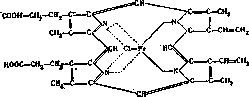
Hematoporphyrin, C34H38N4O4, is obtained by the action of sulfuric acid on hematine; in this process, iron is cleaved off from hematine and water is added. Hematoporphyrin is easily soluble in alcohol, alkalis, and acids; acidic solutions of hematoporphyrin are colored purple-red. The spectrum of hematoporphyrin solutions varies depending on the reaction and concentration of the solutions. Acidic solutions give two absorption bands: one-between the C and B lines, the other-between D and E. Hematoporphyrin can be present in negligible amounts in normal urine; in large amounts it is excreted in acute poisoning with sulfonal, with prolonged use of sulfonal or similar compounds, in chronic lead poisoning. Hematoporphyrin is a sensitizing substance, because after its injection into animals, the skin's sensitivity to light is greatly increased and the skin shows marked signs of irritation, which disappear if the animals are protected from sunlight and placed in dark rooms. Hematoporphyrin is close in structure to bilirubin (the pigment of bile).-Mesoporphyrin, C34H38N4O4, is obtained from hematoporphyrin upon its reduction. In color and spectrum it is similar to hematoporphyrin. With further reduction of the latter, a colorless porphyrinogen is obtained, very similar to the corresponding decomposition product of chlorophyll. With further decomposition of hematoporphyrin, etioporphyrin, C34H38N4, can be obtained. Connection between hemoglobin and chlorophyll. If more profound decomposition of hematine, hemin, or hematoporphyrin occurs, then bonds between the pyrrole rings break in their molecules and products are obtained that contain one pyrrole nucleus in their molecule. For example, upon decomposition and oxidation of hematine or its derivatives, derivatives of hematoporphyrinic acid can be obtained; upon decomposition and reduction, derivatives of pyrrole-phyllopyrrole, methyl-ethyl-pyrrole, etc., are obtained. All these substances are interesting because they or similar substances are also obtained upon the decomposition of chlorophyll-the pigment contained in the green parts of plants, thanks to which in plant cells the synthesis of organic substances from carbon dioxide and water takes place. The chlorophyll molecule is built from the alcohol phytol and chlorophyllide-a pigment containing magnesium in its molecule, with which it decomposes upon hydrolysis with the help of alkalis. With further hydrolysis, chlorophyllide turns into chlorophyllin and then into etiophyllin. From the molecules of the latter, Mg can be removed and porphyrin obtained. With further decomposition of these porphyrins (with oxidation or reduction), the above-mentioned decomposition products of hematine are obtained, namely-derivatives of hematoporphyrinic acid, derivatives of pyrrole (phyllopyrrole, etc.). In addition, etioporphyrin can be obtained from etiophyllin (replacing Mg with two hydrogen atoms), which, as we saw above, is also obtained from hematoporphyrin and is very similar to it in structure. All these data undoubtedly speak for the similarity in the structure of H. and chlorophyll, for the fact that in the bodies of animals and plants the synthesis of both pigments originates from the same material and initially proceeds in the same way; but then the paths diverge: iron enters the H. molecule, Mg enters the chlorophyll molecule; one pigment (hematine) combines with globin, the other (chlorophyllide)-with phytol. Therefore, one cannot conclude that H. and chlorophyll are similar substances and that they perform the same function. Hemoglobin as a respiratory enzyme. New, very interesting data about hemoglobin are revealed by the recent research of Warburg. Studying the respiration of various cells, Warburg established that it is inhibited in a specific way by carbon monoxide, and then came to the conclusion that carbon monoxide reacts with the respiratory enzyme present in the cells. Studying the effect of light on the inhibition by carbon monoxide of the action of the respiratory enzyme, Warburg established that the respiratory enzyme must be a colored substance of red color (but not cytochrome). Then, comparing the specific properties of H. and the respiratory enzyme, Warburg came to the conclusion that the respiratory enzyme is a substance that has pyrrole rings and iron in its molecule, and that this substance is related to H. It then became clear that the respiratory enzyme has common properties with hemin and its derivatives, because the catalytic action of hemin (oxidation of cysteine) also changes under the influence of carbon monoxide and light, just like the action of the respiratory enzyme. Formation and decomposition of H. in the animal organism. The mechanism of H. formation in the bodies of animals is unknown. It is certain that hematine is formed not only from decomposition products of H. and chlorophyll that enter the animal body with food, but that it can also be formed from simpler substances, probably first of all from certain amino acids containing a pyrrole nucleus in their molecule, for example, from pyrrolidine-carboxylic acid and tryptophan. It is possible that from one or the other of them both the colored component of H. and the colored component of chlorophyll are formed. As for the fate of the decomposition products of hemoglobin, we know that from hematine the bile pigment bilirubin is formed, in whose molecule there is no iron, among other things. In places of former hemorrhages from H., under the influence of the vital activity of tissues, a series of decomposition products of still unknown structure (hematoidin, hemosiderin, etc.) are formed. These substances are responsible for the color of the so-called "bruises" and contusions.
The relationship between hemoglobin and chlorophyll. If more profound decomposition of hematine, hemin, or hematoporphyrin occurs, then bonds between the pyrrole rings break in their molecules and products are obtained that contain one pyrrole nucleus in their molecule. For example, upon decomposition and oxidation of hematine or its derivatives, derivatives of hematoporphyrinic acid can be obtained; upon decomposition and reduction, derivatives of pyrrole-phyllopyrrole, methyl-ethyl-pyrrole, etc., are obtained. All these substances are interesting because they or similar substances are also obtained upon the decomposition of chlorophyll-the pigment contained in the green parts of plants, thanks to which in plant cells the synthesis of organic substances from carbon dioxide and water takes place. The chlorophyll molecule is built from the alcohol phytol and chlorophyllide-a pigment containing magnesium in its molecule, with which it decomposes upon hydrolysis with the help of alkalis. With further hydrolysis, chlorophyllide turns into chlorophyllin and then into etiophyllin. From the molecules of the latter, Mg can be removed and porphyrin obtained. With further decomposition of these porphyrins (with oxidation or reduction), the above-mentioned decomposition products of hematine are obtained, namely-derivatives of hematoporphyrinic acid, derivatives of pyrrole (phyllopyrrole, etc.). In addition, etioporphyrin can be obtained from etiophyllin (replacing Mg with two hydrogen atoms), which, as we saw above, is also obtained from hematoporphyrin and is very similar to it in structure. All these data undoubtedly speak for the similarity in the structure of H. and chlorophyll, for the fact that in the bodies of animals and plants the synthesis of both pigments originates from the same material and initially proceeds in the same way; but then the paths diverge: iron enters the H. molecule, Mg enters the chlorophyll molecule; one pigment (hematine) combines with globin, the other (chlorophyllide)-with phytol. Therefore, one cannot conclude that H. and chlorophyll are similar substances and that they perform the same function. Hemoglobin as a respiratory enzyme. New, very interesting data about hemoglobin are revealed by the recent research of Warburg. Studying the respiration of various cells, Warburg established that it is inhibited in a specific way by carbon monoxide, and then came to the conclusion that carbon monoxide reacts with the respiratory enzyme present in the cells. Studying the effect of light on the inhibition by carbon monoxide of the action of the respiratory enzyme, Warburg established that the respiratory enzyme must be a colored substance of red color (but not cytochrome). Then, comparing the specific properties of H. and the respiratory enzyme, Warburg came to the conclusion that the respiratory enzyme is a substance that has pyrrole rings and iron in its molecule, and that this substance is related to H. It then became clear that the respiratory enzyme has common properties with hemin and its derivatives, because the catalytic action of hemin (oxidation of cysteine) also changes under the influence of carbon monoxide and light, just like the action of the respiratory enzyme. Formation and decomposition of H. in the animal organism. The mechanism of H. formation in the bodies of animals is unknown. It is certain that hematine is formed not only from decomposition products of H. and chlorophyll that enter the animal body with food, but that it can also be formed from simpler substances, probably first of all from certain amino acids containing a pyrrole nucleus in their molecule, for example, from pyrrolidine-carboxylic acid and tryptophan. It is possible that from one or the other of them both the colored component of H. and the colored component of chlorophyll are formed. As for the fate of the decomposition products of hemoglobin, we know that from hematine the bile pigment bilirubin is formed, in whose molecule there is no iron, among other things. In places of former hemorrhages from H., under the influence of the vital activity of tissues, a series of decomposition products of still unknown structure (hematoidin, hemosiderin, etc.) are formed. These substances are responsible for the color of the so-called "bruises" and contusions.
A. Palladium. Hemoglobin as a medicinal agent (Haemoglobinum) and its preparations consist of more or less pure hemoglobin of the blood and products of its cleavage. It is prepared from defibrinated bovine blood. Hemoglobin, taken orally, is not absorbed in the stomach, but is decomposed with the formation of hematine. In the intestine, only a part is absorbed, while the larger part is excreted from the body with the fecal masses. It is used as an organic preparation containing firmly bound iron, in anemia, chlorosis, and dyscrasias. It has no advantages over other iron preparations, all the more so that it often contains large amounts of serum protein. It is sold in the form of dry preparations (powders, chocolate-coated tablets, pills) and in liquid form (solutions and syrups). Dose: 0.5-1.0, several times a day.
Related articles
Mentioned in
- Adler Reaction on Blood Pigment
- Affinity
- Amyl Nitrite
- Anatomical Preparations
- Autenrieth Colorimeter
- Autodigestion
- Bacteriohemolysins
- Barcroft Apparatus
- Bilirubinemia
- Brown Induration of the Lung
- Chlorinated Hydrocarbons
- Circulatory System
- Copper
- Cyanide Compounds
- Cyanosis
- Decompression Sickness
- Drowning
- Erythroblast
- Erythrocytes
- Erythrocytoma
Cite this page
“Hemoglobin.” Soviet Medical Encyclopedia. English translation of Bolshaya Meditsinskaya Entsiklopediya, 1st ed. (Moscow, 1928–1936), ed. N. A. Semashko. https://sovietmedicalencyclopedia.pages.dev/article/hemoglobin/