Refraction
Historical document, translated for reference. It reflects medical knowledge of the 1920s–30s and is not medical advice.
Summary
Refraction refers to the optical focusing of the eye in a state of accommodation rest. The article explains the eye's optical structure, different types of refraction (emmetropia, hypermetropia, myopia), and discusses theories about the distribution of refractive errors in the population.
Encyclopedia article (1928–1936)
REFRACTION (from Latin refringere - to reflect, to break), the optical focusing of the eye in a state of accommodation rest. The eye as an optical apparatus is constructed on the type of a photographic camera. In the eye, as in a photographic camera, two main components are distinguished: the light-refracting apparatus and the light-sensitive screen. The light-refracting apparatus of the eye has a complex composition. It consists of the cornea, aqueous humor, lens, and vitreous body. Of these, the cornea and lens are of the greatest importance. The role of the light-sensitive screen in the eye is played by the retina. A ray of light on its path to the retina must pass through four refracting surfaces: the anterior and posterior surfaces of the cornea and the anterior and posterior surfaces of the lens. The refractive power of the eye's optical apparatus depends on the radius of curvature of the refracting surfaces, the distance between these surfaces, and the refractive index of the eye's media. All these quantities, with the exception of the refractive indices, can be determined on a living eye. The radius of curvature of the cornea is easily and quickly measured by Javal's ophthalmometer. The radii of curvature of the lens surfaces and the distance between the refracting surfaces - the depth of the anterior chamber and the thickness of the lens - can be measured by Helmholtz's ophthalmometer (see Ophthalmometer) or Tscheming's ophthalmo-facometer. The elements of the eye's optical apparatus are not some constant unchanging quantities; on the contrary, they vary greatly. The average values of the eye's optical constants obtained by direct measurement - see Eye, dioptrics. Corresponding to the strong variability of individual optical elements, the total refractive power of the eye also fluctuates greatly. It varies from 57.47 to 66.11 diopters, averaging 58.9 diopters (Tron). For the refraction of the eye, in addition to the refractive power of the various optical media of the eye, the length of the eye's axis is also of great importance, which is understood as the distance from the center of the cornea to the area of the yellow spot. This axis length, based on Tron's measurements, varies from 20.75 to 46.04 mm. For the refraction of the eye, however, the decisive factor is not the magnitude of each of these optical elements individually, but the ratio between the refractive power of the eye's optical apparatus, which is measured by its principal focal distance, and the length of the axis. This ratio determines the position of the principal focus in relation to the retina, which characterizes the optical setting of the eye. Three main types of eye refraction are distinguished: emmetropia, or "normal" refraction, hypermetropia, or farsightedness (see), and myopia, or nearsightedness (see). Myopia and hypermetropia, in contrast to emmetropia, are sometimes also designated by the term ametropia. Based on their optical foundations, all cases of ametropia can be divided into three groups: refractive ametropia, axial ametropia, and combinational ametropia. Refractive ametropia is caused by sharp deviations in the refractive power of the eye's optical apparatus, while axial ametropia is caused by sharp deviations in the length of the axis. Combinational ametropia should be understood as all those cases where both the length of the axis and the refractive power correspond to the values that are also found individually in emmetropic eyes, but their mutual combination is such that for a given axis length the refractive power is too great or too weak. In emmetropes, the refractive power varies from 52.59 to 64.21 D, and the axis length from 22.42 to 27.30 mm. The combination of a refractive power of 52.59 D with an axis length of 22.42 mm results in high hypermetropia. To which of these three groups a particular case of ametropia should be assigned can only be determined by measuring all the optical elements of the eye. Combinational ametropia is most common, and refractive ametropia is least common. The refraction of the eye is subject to age-related fluctuations. In newborns, hypermetropia of mild and moderate degree predominates. Emmetropia occurs in them significantly less frequently. Myopia is an exception. Subsequently, there is a shift in refraction toward myopia. In hypermetropic eyes, the degree of hypermetropia decreases. Mild degrees of hypermetropia may over time transition to emmetropia and even to myopia. Emmetropia, in turn, transitions to myopia. All these phenomena are due to growth phenomena, which to varying degrees manifest in the individual elements of the eye's optical apparatus.
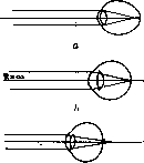
Figure 1. Path of rays: a - in a myopic eye; b - in an emmetropic eye; c - in a hypermetropic eye.
Special significance have age-related changes in the radius of curvature of the cornea and the length of the axis. There are no reliable data on age-related changes in the curvature of the lens surfaces. All these changes in refraction end with the growth period. In old age, a small shift in refraction toward hypermetropia begins again, which is due to changes in the lens. All age-related changes in refraction do not reach high degrees. Different types of refraction of the human eye occur with different frequency. A graphical representation of these relationships forms a refraction curve. The question of the shape and character of the refraction curve is one of the most current and controversial issues in the modern doctrine of refraction, which is due to the following factors. In the assessment of individual refractive states, there are currently two diametrically opposite views. The founders of the modern doctrine of refraction (Donders, Mauthner, Arlt, etc.) believed that between emmetropia, on the one hand, and ametropia, on the other, there are fundamental differences not only in relation to their optical essence, but also
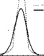
Betsch's Curve and Binomial Curve
in relation to their origin. They consider that the main factor determining the character of the eye's refraction is the length of its axis. In emmetropes, the axis length is normal, in hypermetropes it is pathologically shortened due to developmental lag, and in myopes it is pathologically lengthened due to the influence of external factors (work at close range) (fig. 1). Thus, a sharp boundary is created between "normal" emmetropia and "pathological" ametropia. In contrast to this, another doctrine, substantiated by Steiger, considers both emmetropia and ametropia as different members of a normal variation series. In this case, ametropia no longer bears the stamp of patho
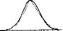
logy. This question, which ultimately comes down to comparing the refraction curve with a normal variation (binomial) curve, has been the subject of a number of works in recent years, which have basically clarified the following: the refraction curve differs from the binomial curve by two characteristics - asymmetry and high-peakedness. Asymmetry is due to the fact that high degrees of myopia occur significantly more frequently than high degrees of hypermetropia, while high-peakedness is due to the high frequency of emmetropia, which occurs much more frequently than corresponds to the normal variation curve. The latter circumstance is clearly seen from the comparison of Betsch's refraction curve with the normal refraction curve (fig. 2). On the other hand, measurements made on 170 eyes of various refractions showed that the main elements of the eye's optical apparatus - the refractive power of the eye and the length of the axis - vary in good agreement with the normal variation curve (fig. 3 and 4). From this, the following conclusions can be drawn. The idea that pathological deviation in the length of the eye's axis is the basis of hypermetropia and myopia is incorrect for most cases. At the same time, however, there are factors that lead to some combinations of the eye's optical elements occurring more frequently than others. As a result of this, such a distribution of the frequency of individual refractions is obtained, which gives the discrepancy in the refraction 57 6v "1 vy vy v7 v9 71 Ty 75 7", Figure 3. Refractive power of the eye.

Figure 4. Length of axis. 78 of the pathological and normal variation curve. The essence of these factors, although not yet fully elucidated, there are reasons to believe that it is a matter of the influence of heredity on the one hand, and the impact of the external environment on the other. Refraction can be determined by both objective and subjective methods. The subjective method is based on the study of visual acuity using lenses. In all anomalies of refraction, a decrease in visual acuity is observed due to the fact that the main focus of the refractive media does not lie on the retina. By applying appropriate lenses (see Glasses), it is possible to move the main focus closer to the retina, which leads to an increase in visual acuity. The best visual acuity will be obtained at the moment when, with the help of the attached lens, the main focus falls exactly on the retina. At this moment, the strength and nature of the refraction of the attached lens indicate the degree and nature of the refraction anomaly. The subjective method, however, has only a limited area of application. Its essential drawback is that it is entirely based on the statements of the person being examined. Therefore, it is insufficient in forensic medical examinations, determination of disability, acceptance for military service, and professional selection. It also does not give satisfactory results in astigmatism. In all these cases, the data from subjective research are supplemented by objective methods of determining refraction. The most common method of objective determination is skiascopy (see), or shadow test, which consists of the following. When the eye is illuminated by an ophthalmoscope (see Ophthalmoscope, ophthalmoscopy), the area of the pupil becomes pink-red due to the reflection from the fundus of the eye. When the ophthalmoscope is rotated, shadows appear in the illuminated area of the pupil, the direction of which, along with other factors, depends mainly on the refraction of the eye being examined. Observing the movement of these shadows in the area of the pupil, combined with the application of ophthalmic lenses to the eye, makes it possible to determine the refraction of the eye without any assistance from the person being examined. Skiascopy is a precise objective method for determining refraction, but it requires considerable skill for proper execution. In recent years, a number of foreign firms have produced optical instruments - refractometers, which make it possible to easily, quickly and accurately determine refraction. The best of these is the Thorner refractometer of the Busch firm. Refraction and professional selection. The professional suitability of the eye is determined mainly by its visual acuity. There is a whole series of standards for professional selection from the side of the visual organ, developed by various institutions. They all base professional selection on visual acuity, completely ignoring a number of other factors that play a very significant role in judging the professional suitability of the eye. One of these factors is refraction. The usual standards for professional selection are based on visual acuity for distance. Meanwhile, most work is performed at a working distance of 25-40 cm, and therefore from the point of view of professional selection, visual acuity for near becomes very important. With myopic refraction, often with low visual acuity for distance, visual acuity for near can be quite high. Therefore, if only visual acuity for distance is taken into account in myopes, a whole range of people who are in fact quite suitable for performing this work may be rejected during professional selection. No less importance than myopia have for professional selection other anomalies of refraction, namely hypermetropia and astigmatism. Good visual acuity is not yet a guarantee that the worker will actually be able to cope in practice with any very fine work. When performing work for a long time, in addition to clear vision of work objects and machine details, the absence of asthenopic phenomena from the eyes is also necessary. They are based on excessive work from the side of the accommodation and convergence apparatus. Accommodation, in turn, is closely related to the refraction of the eye. Since of all refraction anomalies the greatest strain of accommodation is observed in hypermetropes, significant degrees of hypermetropia, starting from about 3 D, even with good visual acuity, may be a contraindication for admission to a number of jobs that place high demands on the organ of vision, for example, work on spinning machines. However, it should be noted here that in addition to the degree of hypermetropia, in each individual case it is necessary to take into account a number of other factors, such as the state of nutrition, the state of the nervous system, etc., because in a healthy body and at a young age, even the strain of accommodation that is necessary for hypermetropes when working at close range can be tolerated for a long time without asthenopic phenomena. Astigmatism also deserves special attention from the point of view of professional selection. Many astigmatics, when wearing cylindrical lenses, give good visual acuity, but still cannot work in glasses for a long time and cannot overcome the side, distressing sensations for the patient that are associated with wearing cylindrical lenses. This is especially often observed in people who only start wearing glasses late in life.
Related articles
Mentioned in
Cite this page
“Refraction.” Soviet Medical Encyclopedia. English translation of Bolshaya Meditsinskaya Entsiklopediya, 1st ed. (Moscow, 1928–1936), ed. N. A. Semashko. https://sovietmedicalencyclopedia.pages.dev/article/refraction/