Birefringent Substances
Historical document, translated for reference. It reflects medical knowledge of the 1920s–30s and is not medical advice.
Summary
This article from the 1928-1936 Soviet Great Medical Encyclopedia details birefringent substances in biological tissues, explaining their optical anisotropy in muscles, connective tissues, and pathological conditions.
Encyclopedia article (1928–1936)
BIREFRINGENT SUBSTANCES, substances possessing optical anisotropy (see). Double refraction as an expression of an ordered molecular structure in animal tissues is very widespread, but it is not always detected clearly enough, depending 1) on the fact that tissue elements are arranged randomly, i.e., the optical axes of small elements run in different directions, and too weak an effect is obtained in the polarizing microscope, and 2) on the fact that the optical axis is located insufficiently favorably for the detection of anisotropy under the microscope. In addition to the anisotropy inherent in certain formations in connection with their structure, this phenomenon can also be observed depending on tension and compression, as was shown by Ebner in 1882. The phenomena of double refraction are most clearly observed in muscles; a classical example in this respect is striated muscles, in which one can distinctly discern the regular alternation of dark anisotropic disks Q (otherwise A) with light simply refracting disks J; the secondary bands Z and N are also anisotropic, but to a lesser degree than disk Q. Smooth muscles also exhibit the phenomenon of double refraction, but to a weaker degree (polarization is positive, uniaxial with the direction of the axis along the fiber, as in striated muscles). Among connective tissue formations, clear anisotropy is observed in bones, where on transverse ground sections of a tubular bone one can clearly distinguish the figure of crosses in Haversian lamellae. In connective tissue proper, collagen fibers are anisotropic, but in loose connective tissue this property is detected with difficulty due to the entanglement of the arrangement of bundles; in the case of a regular course of bundles (in tendons, fasciae), anisotropy appears more distinctly. Elastic fibers in the unstretched state show almost no double refraction. Tooth tissues are also anisotropic, especially dentin. In all [OCR error/fragment]
[OCR error/fragment]
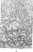
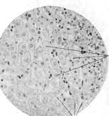
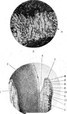
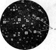
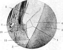
Figure 1. Premenstrual swelling of the uterine mucosa - decidua menstrualis: a - compact layer; b - spongy layer; c - surface columnar epithelium. (According to Aschoff.) Figure 2. Decidual cells (a) from the compact layer of the decidual membrane; b - connective tissue interlayers. Figure 3. Adrenal cortex (a - columns of cortical cells) under polarization. Figure 4. "Liquid crystals" (a) - anisotropic lipoids of the adrenal cortex. (Figures 3 and 4 from the Museum of the Pathologo-Anatomical Institute of the 1st Moscow State University.) Figure 5. Cervical region of the gum: 1 - tooth pulp; 2 - dentin; 3 - cementum; 4 - free space formed as a result of enamel removal during decalcification; 5 - cervical part of the gum; 6 - connective tissue basis; 7 - epithelial cover; 8 - connective tissue papillae; 9 - round-cell infiltrate at the free margin of the gum; 10 - epithelium lining the surface of the gum facing the tooth crown (absence of a papillary layer beneath it is noted); 11 - place where the enamel cover of the crown ends and the root cementum begins (here, according to previous assumptions, the bottom of the gingival pocket should have been located). Figure 6. Gingival pocket: 2, 3, 4, 10, 11 - correspond to the same designations in Fig. 5; 1 - Nasmyth's membrane; 5 - gingival pocket; 6 - true bottom of the gingival pocket. To the article Birefringent substances. Gum, Decidua. connective tissue formations, the axis lies along the length of the fiber. In nervous tissue, myelin fibers are anisotropic (positive uniaxial polarization with an axis running radially). During the development and death of nerve fibers, their optical properties change, which can serve for the purpose of studying these processes. In epithelial tissue, anisotropy is also observed, but very unclearly, and the arrangement of the polarization axis is very diverse. In addition to the indicated phenomenon, anisotropy can also be observed in various kinds of crystals, both protein, fatty, and inorganic. Many lipoids also give the phenomenon of double refraction depending on the cholesterol content in them, as do pure cholesterol deposits (for example, lipoids of the cortical substance of the adrenal gland, see separate table, figs. 3 and 4). So-called autolytic myelin is also referred to as birefringent substances, which appears in the form of so-called myelin figures during autolysis of tissue; the double refraction of this myelin differs in that it does not disappear upon heating, whereas the anisotropy of tissue lipoids is lost at temperatures of 50-60°, returning again upon cooling. Under pathological conditions, birefringent substances are observed in body tissues very frequently, and in most cases they belong to lipid substances possessing double refraction, i.e., anisotropic lipoids, among which the main importance belongs to cholesterol and its compounds. Such birefringent substances can either be deposited into the tissue (into its cells or into the intercellular substance) directly as such, or they appear sequentially as a result of the chemical metamorphosis of other fatty substances deposited in cells in the order of their fatty degeneration. Examples of pathological processes in which the appearance in tissues of a birefringent substance of the above type is observed are: atherosclerosis; the deposition of cholesterol compounds at an advanced age in the cornea of the eye, in tendons, in the renal papillae; fatty degeneration of alveolar epithelium cells in tuberculous pneumonia; the appearance during prolonged fatty decay in tissues of macrophages containing a mass of birefringent cholesterol droplets and called xanthoma cells in view of the fact that their presence imparts a reddish-yellow color to the tissue (Greek xanthos - yellow); finally, the development of special tumors called xanthomas. In degenerative changes of parenchymatous organs, birefringent substances in the form of cholesterol-containing lipoids may also appear in them, usually sequentially; in particular, this takes place in the kidneys in that form of their disease which Munk singled out into the concept of lipoid nephrosis. Upon the deposition of birefringent substances in the kidneys, the latter are also detected in the urinary sediment when examined by means of a polarizing microscope; one must, however, bear in mind that in urine, besides lipoids, double refraction can be caused by certain crystals and plant and tissue fibers, hairs, etc., accidentally trapped in it. Pityriasis rubra diathete
Related articles
Mentioned in
Cite this page
“Birefringent Substances.” Soviet Medical Encyclopedia. English translation of Bolshaya Meditsinskaya Entsiklopediya, 1st ed. (Moscow, 1928–1936), ed. N. A. Semashko. https://sovietmedicalencyclopedia.pages.dev/article/birefringent-substances/