Nail
Historical document, translated for reference. It reflects medical knowledge of the 1920s–30s and is not medical advice.
Summary
The nail is a skin appendage derived from the epidermis, forming a dense plate on the dorsal surfaces of finger and toe tips. This article details the anatomy, growth, chemistry, and various pathological conditions affecting nails.
Encyclopedia article (1928–1936)
NAIL, ungnis, an appendage of the skin, which, similar to the hooves and claws of animals, is a derivative of the epidermis. Nails are dense quadrangular plates consisting of a special kind of keratinized elements, located on the dorsal surfaces of the terminal phalanges of the fingers and toes. The outward-projecting part of the N. is the nail plate, which is smooth, shiny, convex in shape, especially in the transverse direction, and ends freely at the anterior edge [see separate table (pp. 519-520), fig. 3]. The posterior, hidden edge and the two lateral edges of the nail plate are embedded in a grooved depression of the skin, or nail groove, which is covered behind and on the sides by elevated folds of skin or nail folds, limiting the nail bed. The visible part of the nail forms the nail body, which represents the thinnest part of the nail and rests with its lower surface on the nail bed, the shape and size of which correspond to the shape and size of the N. The lower surface of the nail body is provided with thin ridge-like projections that extend between the corresponding ridges of the nail bed. The proximal part of the nail, immersed in the posterior nail fold, is called matrix unguis. Matrix together with the adjacent part of the nail is called its root (radix unguis), because from here the nail plate grows. Part of the nail root emerges from the fold in the form of a dull whitish strip of nail body, or lunula (lunula), i.e., a sharply delimited anteriorly by a convex line of white crescent-shaped space, which is clearly noticeable especially on the large and other fingers. The lunula and nail root owe their white color to the presence of a rich keratohyalin grains in the granular layer, through which vessels do not show through. The remaining surface of the nail plate, consisting of cells of the glassy layer, is transparent, and therefore differs in a pinkish tint due to the transparency of the blood vessels of the nail bed. The histological structure of the N. shows great similarity with the structure of the epidermis, and the nail bed and matrix-with the papillary layer of the skin. The N. consists of numerous flat, polygonal, transparent, densely interconnected keratinized cells, which still contain nuclei or their remnants and often air bubbles; these cells genetically correspond to the cells of the clear layer of the epidermis. Pressed against each other and provided with spines, these cells are arranged like tiles parallel to the surface of the matrix, so that the lower layers always extend further forward than the upper layers lying on them. They can be separated by acids or caustic alkalis. When the cells swell, the nuclei become clearly visible. On nails with longitudinal ridges, concentrically arranged horny plates are found. The nail bed, extending from the lunula to the free edge of the nail, consists of the germ layer of epithelium, which is formed in the area of the nail root from the cells of the matrix. In the proper skin of the nail bed, there is a dense plexus of connective tissue fibers. At the border with the epidermis, it forms a series of regular longitudinal ridges, which, as the nail becomes free, gradually pass into the connective tissue papillae of the skin. The ridges and the spaces between them are filled with epidermal cells, which here correspond to the basal and Malpighian layers of the rest of the epidermis. The nail bed does not participate in the formation of the N. [see separate table (pp. 519-520), fig. 2]. The development of nail primordia begins on the 7th week of embryonic life in the form of a thickening of the epithelial layer on the terminal phalanges (the so-called primary nail field of Kölliker 4a), on the inner and outer edges of which transverse folds are formed. By the time of the origin of the N., the nail groove already exists. By the end of the 3rd month of intrauterine life, the primordium of the posterior part of the nail fold and the nail matrix begin to form. In this place, the nail plate is formed later, consisting of very light horn cells, in the form of a sharply expressed primordium of the clear layer (perionychium), which gradually reaches the nail groove. When later the fully formed nail begins to grow forward (at the 8-9th month), most of the elements of the perionychium disappear; only its part at the free end and on the nail fold remains throughout life; bearing here the name of eponychium and hyponychium (epionychium et hyponychium). The growth of the nail occurs both in thickness and in length, generally quite quickly (from 1/3 to 1 mm per week). On the hands, the growth of the N. is faster than on the feet, in children faster than in the elderly (Berthold). In thickness, the N. grows due to the layer of epidermis located on the nail bed, in length-due to the epithelium of the generative layer of the matrix. The thickness of the N. is generally uniform (from 1/3 to 1/2 mm) and probably depends on the degree of inclination of the matrix. Throughout a person's life, the N. grow continuously and can reach considerable length (10-40 cm). According to Moleschott, in the course of a year, the fingers provide 2-3 g of nail substance; relatively more in summer than in winter. In pathological processes and injuries of the entire thickness of the matrix, the growth of the N. stops: this is often expressed by the appearance of a transverse groove on the surface of the nail plate. Long-term injuries to the matrix often lead to atrophy of the N. In destructive changes of the nail matrix by ulcerative or necrotic processes (burns, frostbite, suppurations, syphilis, lepromas), it can lead to complete and permanent loss of the N. Devoid of blood vessels and nerves, the N., as dense horny plates, are resistant to thermal and chemical influences, are adapted to protect the underlying tissues from all kinds of external injuries. The normal and pathological chemistry of the N. is still insufficiently studied. According to Moleschott, Hammarsten and others, the chemical composition includes water-13.74%, a significant amount of calcium phosphate, other mineral parts (carbonates, phosphates, calcium compounds 1% each), ash (1%), cystine (dry substance 5.15%), cholesterol and an abundant amount of keratin. The nail pulse was first described in 1646 (Francus de Frankenau). Caused by increasing and decreasing blood filling of the subungual arterial vessels, the nail pulse has some diagnostic value, especially in those pathological processes that are accompanied by stagnation phenomena. Herz (Max Neega) invented in 1896 for measuring the nail pulse an apparatus-sphygmograph (onychograph), with the help of which it is possible to obtain an onychogram, which shows a certain similarity with the pulse curve. The classification of diseases of the N., called onychia, onychosis (onychosis), onychopathy (onychopathy), is difficult, because the same clinical pictures of lesions can be caused by various causes and, conversely, identical causal factors can cause different symptoms. In general, with any lesion of the N., it is necessary first of all to determine whether it depends on a purely local disease of the nail root, nail bed or surrounding tissues, or is an expression of disorders of the whole organism or its individual systems. In the general aggregate, all diseases of the N. can depend a) on traumatic, thermal, chemical or parasitic changes of the substance of the N. or nail bed, often occurring as professional diseases; b) from congenital developmental abnormalities; c) from the influence of heredity, pregnancy, nervous and mental suffering; d) trophic and endocrine disorders of the nail; e) in connection with various chronic skin diseases; f) in connection with various febrile general infectious and chronic diseases, in particular of the lungs. Traumatic injuries (onychosis traumatica) most often occur as a result of foreign bodies getting under the N., ingrowth of the N., biting of the N., due to scratching with long-existing itchy dermatoses, constant pressure from ill-fitting shoes, etc. Biting the nail, onychophagia (onychophagia), is considered by most as a bad habit, occurring in school-age children, and often in adults. Bitten nails are sharply shortened; the free edges disappear; often such N. turn into stumps, in front of which the finger pulp of the fold rises. Biting nails is sometimes observed in persons who have certain signs of degeneration or nervous and mental disorders. In some schools where biting of N. is noted as a mass phenomenon, imitation apparently also plays a significant role. Biting of N. is often observed in subjects with acroasphyxia. A. I. Pospelov considered onychophagia a prodrome of Raynaud's disease. The rational methods of treatment are psychotherapy, elimination of local cyanosis (massage, warm baths, faradization).
Thinning and grooves of the free edges, shortening of the nail plates, sometimes along with artificial coloring, are often observed in workers from the most diverse industrial and labor processes, as well as from the action of chemical substances. - In many itchy chronic dermatoses (eczema, prurigo), the nails acquire a shiny polished surface from habitual scratching of the skin, and on the free edge, depressions form in the middle. Hangnails represent superficial small traumatic tears of the horny layer of the nail folds. They arise from "careless care of the nails" and can serve as portals of entry for infectious agents, especially pus-forming cocci, with subsequent paronychia, lymphangitis, etc. - Treatment: careful trimming of epidermal fragments, patching with adhesive tape, collodium, etc. - Pterygium unguis, wing-like membrane, an obsolete term for abnormal growth of the nail cuticle covering a significant portion of the proximal part of the nail plate; it may be congenital or acquired. In the latter cases, it can develop as a result of careless care of the nails or prolonged exposure to X-ray rays. - Under the name i i c u s unguium, Wilson described the shedding of the posterior nail fold and the nail cuticle up to the root, which, according to Rayer, is observed as a professional onychia among leatherworkers, and according to Wilson - in chronic dermatitis affecting the nail fold. In trophic processes after damage to nerve trunks, hypertrophic changes of the nail cuticle can also be observed. Ingrown nail, or incarnated nail (unguis incarnatus), - a common affliction of the foot, characterized by its lateral ingrowth into the nail fold due to abnormal or increased growth of the nail in width under the influence of mechanical pressure 61». It is observed mainly on the big toe. Usually starting at the upper edge of the lateral nail fold, it spreads to the lower edge of the nail fold, accompanied by painful ulceration and the development of granulations around the edge of the nail. In essence, this affliction represents a chronic purulent inflammation, which can be complicated by lymphangitis, phlegmon, etc. The diagnosis is usually not difficult and in doubtful cases is easily established with the help of a blunt probe, which is inserted under the nail plate. In differential diagnosis, tuberculous ulcer, chancre-like ulceration, paronychia, and gumma should be kept in mind. The etiology is not entirely clear. An ingrown nail usually arises under the influence of mechanical pressure or prolonged traumatic effects on the nail (e.g., during forced marching). Wearing tight shoes is a significant cause. Subjects aged 15-20 years are most predisposed. There is an indication that cutting the corners of the nail contributes to the ingrowth of nail plates into the tender parts of their edges. According to Konig, a pronounced convexity of the nail plate and deep indentures predispose to the development of this affliction. Chipault considers that stagnation of blood in the lower extremities, elephantiasis, chronic edema can lead to the formation of an ingrown nail. The prognosis with surgical intervention is favorable. - Treatment consists of inserting under the edge of the nail tampons soaked in 1% silver nitrate, iodoform, etc., in order to pull the inflamed fold away from the edge, and applying strips of adhesive tape. In severe inflammation - scraping of the middle part of the nail or radical excision of the lateral part of the nail or the entire nail along with the granulations and nail bed under local anesthesia. In mild cases - warm baths, anti-inflammatory and cooling remedies are used. Onychogryphosis (onycho-gryphosis, claw-like nails), a lesion of the nail, consisting in a sharp, and sometimes colossal, increase, thickening and curvature of the nail plate due to a change in the direction of growth [see separate table (pp. 519-520), fig. 5]. Rising and bending, the nail takes the form of a grayish-brown claw or a snail shell, forming hard horn-like masses of stone density due to the thickening of the subungual horn cells. In the maximum degrees, convex, disfigured, almost spirally twisted, ram's horn-like formations are obtained, traversed by longitudinal and wavy transverse stripes. Having a tendency to grow, the affected nail can reach a length of 10-15 cm. Due to intense pain, patients avoid shortening the affected nails. This affliction is observed more often on the lower extremities, mainly on the big toes, less often on the fingers, especially in the elderly. The diagnosis "does not present difficulties. Virchow attempted to explain the pathogenesis of onychogryphosis by mechanical irritation due to pressure of shoes on the 17 matrix of the nail, which is hardly acceptable. Inflammatory changes of the nail bed, noted by Unna, apparently occur secondarily. Hebra points out that in the development of onychogryphosis both the matrix of the nail, excessively producing abnormal horn substance, and the nail bed, the epidermal cells of which excessively proliferate, participate. As possible causes of symptomatic onychogryphosis, various authors mention traumatic injuries to the fingers, peripheral and central nerves, hallux valgus, syphilis of the nail bed, circulatory disorders, thrombophlebitis and aneurysma arterio-venosum, endocrine disorders, etc. Onychogryphosis is sometimes observed as a familial disease. Mikula saw a congenital-hereditary onychogryphosis of all fingernails and toenails in 3 generations. Heller considers idiopathic onychogryphosis as a trophic disorder of the nail matrix and nail bed, originating from the central or peripheral nervous system. In recent years, some authors speak in favor of a mycotic nature of this lesion, but the role of the fungi they found in the pathogenesis of onychogryphosis is not proven. -- Treatment: baths with potash for softening, salicylic plasters, scraping of the nail surface; in advanced cases, removal with a knife or file of softened areas or the entire nail; cautious use of X-ray rays or radium. Onychauxis (hypertrophia unguium, megalonychia), a simple expansion and thickening of the nail, often encountered in varicose dilation of veins or elephantiasis of the lower extremity, in acromegaly, and sometimes as a congenital developmental defect. Very hard hypertrophied nail plates of brownish or blackish color, usually furrowed; either one or several nails are affected. -- Expansion, convexity and curvature of bluish-colored nails as a result of hypertrophy of the fingertips are also a consequence of chronic diseases of the lungs or heart, Basedow's disease (see Hippocratic nail, Drummer fingers). - Subungual hyperkeratosis (hyperkeratosis subungualis Hebra), lifting the nail plate above its substrate, is a symptom of a process of increased keratinization of nail cells (onychosis), which is observed in various afflictions, namely - in eczema, psoriasis, pityriasis rubra, onychomycosis, neoplasms localized on the nail bed or so-called subungual tumors, onychogryphosis, etc. Heller classifies it as localized eczemas. In essence, this symptom should be considered as an expression of any chronic inflammation of the nail bed. - Disease of the nail bed and impairment of the function of the matrix can also manifest as softening of the nail plates (hapalonychia). This term was introduced by Kaposi for a rare form of atrophy of the nail, observed by him in one psoriatic patient suffering from hyperhidrosis: after a purulent paronychia, the nails turned into membranous plates. Softening of the nail substance is rare and usually associated with excessive sweating of the hands and feet. Heller uses this name in cases where it concerns softening of nails that appear normal. In softening of syphilitic origin (onychomalacia syphilitica Ed. Fournier), the nail plates become soft, pliable and easily bent; their surface appears uneven, and the posterior nail groove is smoothed. Under the term "egg-shell nail," J. Nevins Hyde described a peculiar lesion of the nail, characterized by the detachment of the free edges and a special transparency of the nail plate. Hyde observed in three women suffering from hyperhidrosis, sclerosis and local circulatory disorders, very soft, easily upward-bendable nails, showing a tendency to peel off, of a pinkish-whitish color with a shade remarkably similar to the delicate inner membrane of an egg. - Onychoschizia (onychoschizia, schizonychia), a term denoting loss of substance from the nail bed in the absence of true detachment of the nail; onychoschizia usually occurs as a symptom in psoriasis, eczema and syphilis. - In the so-called idiopathic shedding of the nails, there is repeated complete shedding of the nail plates (onycholysis totalis, onychoptosis), often without an apparent cause, apparently due to impairment of the function of the matrix cells of the nail.
This phenomenon is sometimes observed in alopecia areata, epilepsy, tabes dorsalis, diabetes mellitus, after concussion, etc. The restoration of destroyed nails, for example after removal of the nail in the surgical treatment of subungual hematomas, abscesses, fractures, and other injuries, usually occurs within 3-4 months, with the exception of cases where after surgical removal of the nail, scar tissue forms, preventing the restoration of the nail. Parasitic diseases of the nail-see Onychia mycotica. Congenital developmental abnormalities of the nail: complete absence of one or many nails, koilonychia, unguis epider'micus-see Anonychia. Congenital aplasias of individual or all nails, abnormally or not in the usual place developed nails, are often familial, manifesting in several generations, and are frequently observed both in congenital malformations of the limbs (syndactyly, microdactyly, polydactyly, etc.) and in the most diverse congenital developmental abnormalities and anomalies associated with other congenital-hereditary skin diseases (see Keratoses, Ichthyosis). Complete absence of one or several nails on the fingers of the hands and feet in combination with onycho-atrophy (see separate table, fig. 3) was observed by Jacob (P. Jacob) in 3 brothers and sisters. O'Neill described a case of anonychia in a brother and two sisters. Familial isolated absence of nails of the thumbs of the hands (anonychia pollicum familiaris) in a brother blind from birth and his sister (figure 1)
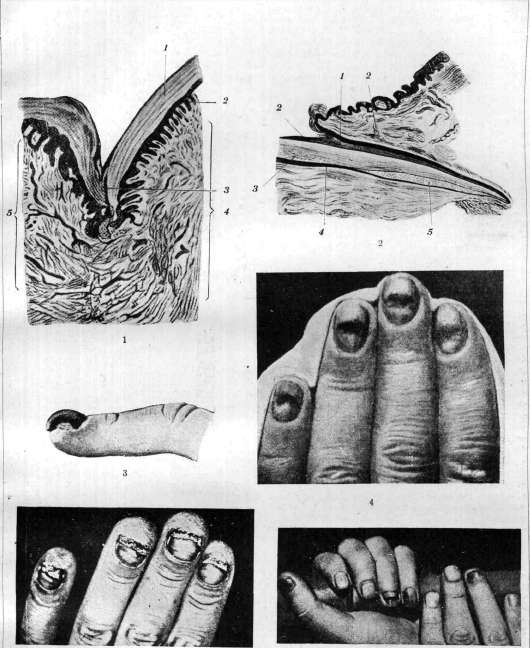
Figure 1. Cross-section of the nail: 1-nail; 2-epithelial outgrowths of the nail bed; 3-eponychium; 4-corium; 5-nail fold. -Figure 2. Longitudinal section of the nail: 1-smooth epithelial layer of the nail fold; 2-nail folds; 3-nail; 4-epithelium of the germ layer; 5-matrix. Figure 3. Onychogryphosis after nail injury. Figure 4. Nail disease in a worker of the mercury production. Figure 5. Nail disease in a worker of the lime production. Figure 6. Congenital onychoatrophy
of individual nails. To the article Nail 52S was noted by Ebstein (E. Ebstein). Familial anonychia in combination with atrophy of the nail was observed (figure 2) in a child, two t fig. 1.
fig. -g. Figure 1. Anonychia pollicum familiaris (Ebstein). Figure 2. Familial anonychia and onychoatrophy (Most). brothers and two sisters of which had identical changes (5th was healthy). The father of the child, his five brothers and mother suffered from a similar disease. Direct inheritance was established in the observation (N. Tobias) of 25 members in 4 generations, of which 12 people (5 women+7 men) had early changes in the nails on the fingers of the hands; on the thumbs instead of nails there were horny deposits in the form of a rough hyperkeratotic film, so-called epidermal nails, splitting and brittleness of the remaining nails (onychorrhexis) and "wart-like" growths of the nail bed under the free edge of the nail. Of particular interest is the extremely interesting familial picture of hereditary dystrophy of the nails, presented by Barrett. Of 61 members of families in 5 generations, 14 cases of disease of hair and nails were found. The change in hair varied sharply in intensity (loss and weak growth of hair, uniform deformation of nails). In some patients with nail lesions, as well as in subjects with healthy nails, there were signs of myxedema and hyperthyroidism. The change in nails was expressed in a defect of the anterior edge (by 1.2-0.6 cm), absence of the lunula and thickening and brittleness of the free edge. Traces of suppuration were found on the nail bed. The fingers of the hands and feet were affected to the same degree. Hereditary subungual hyperkeratosis was seen by Wilson in 7 members of one family in 3 generations. Complete hyperkeratosis of the nail was observed by Ebstein in 6 individuals in 3 generations. Koler described onychogryphosis of many nails in 4 women in 4 consecutive generations. As a hereditary familial disease, Eisenstadt observed in 3 brothers dystrophy of hair, eyebrows, and eyelashes, as well as intense hypertrophy of the nail bed. In favor of the possibility of inheritance of brittleness and splitting of the nail (onychorrhexis) speak the observations of Noble, concerning 4 members of one family in 2 generations. Other authors saw similar changes in a brother, sister and father, then in 5 brothers and sisters. Onycholysis partialis semilunaris was established in two sisters, in whose family congenital cataract could be established in four generations in 11 subjects. An interesting pedigree of dystrophy of the nails was published by Thompson (fig. 3). Drumstick fingers were sometimes observed as a familial disease with irregular dominant inheritance: in a father and two children, in a father and son; grandfather and grandmother, 6 sons, 1 daughter and 7 grandchildren were free from the lesion (Ebstein, Lewy). In the case of Fischer (H. Fischer), traced over five generations, it was possible to prove the connection of lesions of the phalanx bones with dystrophic onychosis, with palmar-plantar keratosis and hypotrichosis. Pregnancy and the associated change in metabolism cause very minor lesions of the nails. Foggie saw in one woman at the 5th month from conception during all 6 pregnancies changes in the nails, which he is inclined to consider as psoriasis. Another author observed trophic disorders of the nails in a woman during each pregnancy, seeing in this toxemia of pregnancy. Dystrophic onychoses, sometimes occurring during nervous diseases, both of peripheral and central origin, in nervous shocks, psych. excitations, psychoses, mental and moral shocks, are expressed mainly in the formation of transverse or arc-shaped groove or ridge on the nails. Papillon, who studied the question, distinguishes a) transverse grooves in mentally ill persons, arising under the influence of somatic-visceral diseases, b) transverse grooves in chronic mentally ill persons g developed as a result of acute excoriations (acute mania, delirium), c) transverse grooves in chronic mentally ill persons, appearing without external visible causes. Trophic lesions of the nails can be very diverse. These include various types of atrophy, leukonychia, hypertrophy of the nails, onychogryphosis, koilonychia, etc. All these are symptoms observed in many nervous diseases, neuroses, traumatic neuritis, syringomyelia, tabes dorsalis, hemiplegia, Morvan's disease, Raynaud's syndrome, erythromelalgia, acrodynia, etc. Under the name hyperaesthesia unguium, or onychalgia nervosa, Oppenheim described the clinical picture of a functional neurosis of apparently normal nails. Any touch to the subungual space or cutting of the nail causes severe pain. Patients show nervous symptoms and come from a nervous family. The disease does not respond to treatment. Treatment-improve the general condition, analgesic ointments (anesthesin), galvanic hand baths, etc. The role in onychopathology of endocrine-sympathetic disorders is discussed by Geller. Less skeptically is Darier, who asserts that he has achieved certain results with the application of appropriate hormonal therapy. According to him, various infections and intoxications (inheritance t

6 o o a o a o
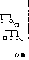
Figure 3. Familial dystrophy of the nails (Thompson).
Diseases of the nail. In various professional and industrial processes, discoloration, scaling, wearing down, deformation, and callus-like thickening of certain areas of the nail plates, as well as the formation of notches, tears, bruises, and roughness (so-called professional signs or stigmata) may be observed. There is no doubt that excessive sweating of the hands and generally impaired function of the glandular apparatus, along with other unfavorable working conditions, often give rise to the development of professional injuries to the nail. In workers handling arsenic acid and especially Schweinfurt green, in tanners, and in persons using depilatory agents containing arsenic, vesicular dermatitis and discoloration of the nails and hair are often observed. Chronic arsenic poisoning causes loss of nails and hair (Alopecia). Intense yellow discoloration of the nails from picric acid is observed in workers manufacturing erazite for projectiles (Oppenheim). When handling hydrofluoric acid, in addition to persistently non-healing burns and ulcers, significant damage to the nails is often observed in glass engravers and etchers, sometimes leading to complete loss of the nails. The most common changes in workers exposed to gaseous hydrofluoric acid are specific erosions of the nail substance in the area of the lunula. Deep acid cauterizations cause necrosis and shedding of the nail plate (Eulenburg). An almost identical lesion of the nails is encountered in glassblowers in the form of progressive resorption of the nail plates and insufficient regeneration of nail substance under the influence of prolonged cauterizing action of sodium hydroxide (Oppenheim). In workers in mercury production, severe damage to the nails often appears not only from direct contact with mercury during its processing but also from general poisoning (Girschfeld, Kussmaul) [see separate table (art. 519-520), fig. 4]. Typical erosions of the free edges of the nails, leading to eczematous changes in the nail bed and loss of nail plates, are found in workers in lime production due to the accumulation of chloride of lime in the subungual spaces (see separate table, fig. 6). A peculiar dermatosis of the nail is the professional disease described first under the name of confectioner's disease. It occurs primarily in workers engaged in the production of candied fruits and chestnuts, under the influence of thermal irritants and the harmful effect of sugar solution getting into the subungual spaces, which serves as an excellent nutrient medium for the growth of microorganisms, usually occurring during intensive work and failure to observe elementary rules of cleanliness and hygiene. Individual predisposition undoubtedly plays a major role. Clinical picture: inflammation of the nail folds; swelling of the nail bed; partial separation of the nail plate; formation of abscesses under the nail (paronychia). In 1925, the blastomycetic nature of this disease was experimentally proven (Kingery, Thienes). In workers engaged in wool sorting, damage to the fingertips is observed with a gradual tendency to spread to the surrounding parts of the nail (maladie des pelMirs; Ztipferkrankheit). Upon cessation of work, spontaneous healing occurs (Neisser). The professional dermatosis described first by Oppenheim in laundresses and cooks in the form of partial semilunar separation of the nail (onycho lysis partialis semilunaris), with sediment of dirt in the subungual spaces, arises from mechanical and chemical insults (mainly alkalis). Hot soda and soap solution causes softening and maceration of the horny substance of the skin of the nail. An identical process with the formation of semilunar notches along the edge of the nail and thickening of the terminal phalanges was observed in workers twisting tobacco stems for the manufacture of chewing tobacco, under the influence of permanent mechanical irritation that tears the plate from the bed, and the chemical action of tobacco mordant, which dissolves the nail substance. Microbial lesions of the nail-see Onychia. Changes accompanying various, most diverse chronic dermatoses, while generally extremely diverse, also have many features in common; the nail substance may lose its natural color and luster, becoming grayish or yellowish-brown in color, or, taking on a dull and matte appearance, becomes brittle and fragile due to loss of elasticity, showing longitudinal and transverse grooves. Partial or complete separation of the nail, atrophy or hypertrophy of the nail may also accompany various skin ailments, which often pass directly per continuitatem to the matrix of the nail. Among the most frequent skin diseases complicated by nail lesions are eczema and psoriasis. In eczema, the disease is characterized by inflammation of the nail bed and fold, clouding, transverse and longitudinal grooves, separation and deformation of the nail plate. Due to insufficient formation of horny substance in limited areas of the matrix, many dermatoses are accompanied by the appearance of numerous small, irregularly arranged, superficial cup-shaped or pinpoint depressions or erosions, sometimes filled with dirt, especially in eczematids and psoriasis, which externally resemble the surface of a thimble. Hebra associated these phenomena with syphilis. Sometimes, however, they also form independently of these diseases. Often they may precede skin manifestations. Correct diagnosis is possible mainly in the presence of accompanying skin lesions. In various acute, chronic, general infectious and febrile diseases and intoxications, the nails may undergo the most diverse changes due to impaired nutrition and circulation and metabolic disorders. General diagnosis of nail diseases. Recognition of nail lesions is often associated with great difficulties, since it is not always possible to establish the connection between nail diseases with certain causal factors. Among many physicians, there has become entrenched the habit of abusing the diagnosis of so-called trophic or trophoneurotic nail diseases in cases of onychopathies of unclear etiology, which usually does not withstand criticism. In doubtful cases, mycological studies with seeding on nutrient media should be performed to determine the true nature of the disease. Since the clinical picture of many nail changes is generally not very characteristic, the diagnosis should not be based on individual symptoms but on the results of thorough examination of the entire body. It is necessary to pay attention to the fact that many nail ailments often remain after the disappearance of skin manifestations. A carefully collected history and neurological examinations often have decisive importance in syphilitic nail lesions. Hygiene and treatment of nail diseases. Thorough systematic care of the nails, cutting the nails, and observing elementary rules of cleanliness and hygiene are essential requirements, especially in professional skin and nail injuries. To protect the nail substance and its growth from inevitable tears and damage to the terminal phalanges caused by mechanical insults, a finger cot, adhesive bandage, or application of a wax plate secured with adhesive tape (in atrophic processes) is usually used. Treatment of nail diseases is an extremely ungrateful task. In addition to local keratolytic agents and scraping of the nails, from which reliable success cannot always be expected, high-frequency currents or X-rays can be used, especially in chronic and neglected forms of various diseases. The nature of the underlying disease does not always allow for causal treatment. In new growths and tuberculous processes of the nail bed, vigorous surgical intervention is required.
Related articles
Cite this page
“Nail.” Soviet Medical Encyclopedia. English translation of Bolshaya Meditsinskaya Entsiklopediya, 1st ed. (Moscow, 1928–1936), ed. N. A. Semashko. https://sovietmedicalencyclopedia.pages.dev/article/nail/