Keratoses
Historical document, translated for reference. It reflects medical knowledge of the 1920s–30s and is not medical advice.
Summary
This article provides an overview of keratoses, a group of non-inflammatory skin diseases characterized by the thickening of the horny layer. It details the classification of these conditions into diffuse and limited forms, discusses the etiology of palmoplantar keratosis, and outlines symptomatic treatments including topical applications and X-ray therapy.
Encyclopedia article (1928–1936)
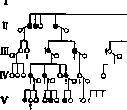
KERATOSES (keratosis, keratoma, keratodermia, keratodermatosis) (from the Greek keras - horn), a collective term for a whole series of skin diseases of non-inflammatory origin, which are morphologically and pathologically characterized by a moderate thickening of the horny layer. The degree of hyperplasia of the normal horny layer undergoes significant fluctuations, being in close dependence on the peculiarities of the process and its localization. One also distinguishes forms of keratinization anomalies where hypertrophy of the horny layer is revealed primarily in the form of increased shedding of horny scales. In dry seborrhea and the so-called xerosis of Darier (pityriasis + seborrhea), i.e., in phenomena accompanied by delicate branny desquamation, the thickening of the horny layer is expressed in a very weak form. Conversely, in ichthyosiform hyperkeratoses and especially in palmoplantar keratoses, it forms real dense, stubbornly incurable horny hardenings with a compact arrangement of horny fibers, closely fused with the underlying tissue, sometimes reaching significant thickness and height. Such powerful proliferation of the horny layer is called hyperkeratosis. The skin of the palms and soles, possessing a special structure by nature, is often predisposed to hyperkeratosis, frequently showing a tendency to form painful cracks in the flexion folds. Many dermatoses located in this area often acquire a similar character, causing significant diagnostic difficulties. Such a change in the horny layer reflects on the remaining elements of the skin and causes abnormal thickening of the granular and Malpighian layers with subsequent inflammatory hyperemia of the connective tissue. Classification. From the point of view of the topographic distribution of the affected foci, two groups of keratoses are distinguished. 1. Diffuse or universal keratoses, i.e., spread over almost the entire body or capturing large areas of the skin. This should include dry seborrhea, ichthyosis, and generalized or partial ichthyosiform hyperkeratoses. 2. Limited or local keratoses, consisting of separate, sharply defined spots or keratotic plaques, which can spread randomly over the entire body or be localized symmetrically or in certain favorite areas of the skin. This group includes systematized hyperkeratotic nevi, linear nevi, naevus keratodes, senile keratosis, cutaneous horn, porokeratosis, palmoplantar keratosis, etc. The most frequently encountered palmoplantar keratoses can be divided into two main groups: 1. Genotypic (k. palmaris et plantaris hereditaria - Meleda disease, k. symmetrica palm. et plant. adultorum). 2. Paratypic (symptomatic), arising as a result of trauma, constant irritation (k. professionalis), intoxication (k. arsenicalis), or in connection with severe infectious diseases. Individual forms of keratoses. Keratosis palmaris et plantaris hereditaria (keratodermia familiaris, ichthyosis palmaris et plantaris, acrokeratoma familiare). An anomaly of keratinization expressed in increased proliferation of the horny layer. It is located symmetrically on the palms and soles, as well as on the palmar and plantar surfaces of the fingers and toes in the form of hyperkeratoses of a yellowish or brownish color with an increase in the pattern of skin furrows. A characteristic sign of the disease is a persistently held erythematous or bluish-crimson border 4-5 mm wide around the circumference of the lesion. The horny masses consist of wide, tightly fused, dry, rigid plates of yellow-waxy or brownish color, sometimes reaching 1 cm in thickness. The lesion is usually sharply demarcated from normal skin. Its surface is speckled due to the loss of elasticity by numerous, sometimes bleeding cracks, penetrating to the Malpighian layer and even to the cutis, and resembles "honeycombs" in its external appearance (Jarisch). Sometimes the process begins to develop from the openings of the excretory ducts of the sweat glands. The underlying skin is tense, sclerosed, and atrophic. In the stage of maximum development, the skin changes acquire a blackish color, cause dulling of sensitivity, and create significant difficulty in movement. In the area of hyperkeratoses, increased sweat secretion is noted. Darier, Brooke, and Crocker observed deviations from the typical form in the form of erythematous redness and intracorneal vesiculation. The process continuously progresses and spreads, sometimes capturing the lateral and dorsal surfaces of the hands and feet, and also localizing on the knees, elbow flexures, and joint folds. Often the limbs show vascular phenomena in the form of local asphyxia. It is often observed in combination with generalized ichthyosiform hyperkeratosis; in its course, it is close to symmetric erythrokeratoderma, with which it can also be combined (Brocq, Dubreuilh). In many cases, the simultaneous development of baldness or various dystrophic trichoses and changes in the nails are noted. The latter become dull, irregularly curved, and dotted with grooves and point depressions. Histologically: powerful thickening of the horny layer; the transparent and granular layers are thickened. The papillae are elongated and thinned. The prickle-cell layer consists in places of 20-30 rows of cells. Around the sweat glands, the cells of the Malpighian layer form a light zone, which depends on the impregnation of the cells with sweat (Vorner). The ducts of the sweat glands are dilated and surrounded by keratinized masses and are sometimes filled with horny plugs. Etiology. Palmoplantar keratosis is often inherited as a dominant trait (see diagram). Dominant inheritance has been traced in one case to the 5th generation (Leven). In well-studied cases, no skips were observed. In exceptional cases, a connection with hypotrichosis, changes in the nails, fingers ("drumstick fingers"), and acneiform follicular keratosis was noted. In one case, inheritance was limited to the female sex (Ballantyne, Elder). The possibility of the existence of forms with recessive inheritance is also not excluded. Siemens observed the actual disease in two of 8 sisters and brothers whose parents were free from the disease. In some cases, the sick children were born from parents who were blood relatives. In addition, Siemens noted for the first time the genotypic occurrence of linear keratinizations (k. palmo-plantaris striata) with exclusive involvement of either the palms or the fingers. From the above, it follows that biologically, palmoplantar keratosis is by no means homogeneous and that with a more thorough analysis, it will likely be possible to discover clinical differences as well. Prognosis: unfavorable. Treatment is symptomatic: local wet wraps, warming compresses, warm baths, soap washings, 10% salicylic ointment; internally - ichthyol and arsenic; encouraging results have been achieved from the use of X-rays. A special form of palmar keratosis with the transition of keratinization to the dorsal side of the hands and feet, on the elbows and knees, is the so-called "Meleda disease," which is often found on the Dalmatian island of Meleda, where Honorka, Ehlers, and Neumann established the seemingly endemic nature of the disease. The nature of inheritance has not been sufficiently studied to date (Siemens). Keratosis symmetrica palmaris et plantaris adultorum (Besnier) (acrokeratoma, keratodermia, tylosis essentialis) - a form of keratosis developing by the period of maturity or even later, characterized not by diffuse involvement of the palms and soles, but by separate islands of keratinization with localization in the heel area under the heads of the metatarsal bones, as well as on the palmar and plantar sides of all fingers [see separate table (p. 583-584), fig. 12]. Often, various disorders in the form of increased sweating, urticaria, redness with exfoliative desquamation, acrocyanosis, etc., precede it for many years. Symptomatic keratoses include keratoses from mechanical and chemical causes, professional keratoses of toxic and medicinal origin (arsenic and tar). Keratosis professionalis, professional keratosis in the form of diffuse callosities, arises as a result of permanent traumatic irritation, friction, and abnormal pressure conditions. They are unilateral or symmetrical. By the character and location of professional keratosis, it is often possible to establish the type of profession and especially the kind of tool used at work. For example, in glassblowers and bottle blowers, callous thickenings of the horny cover form on the palmar surfaces of the hands over the heads of the metacarpal bones, sometimes reaching a height of 1 cm due to the tight gripping of the heavy iron blowpipe (see also Occupational dermatoses). Keratosis arsenicalis (arsenokeratosis) develops during poisoning or prolonged use of arsenic, being located symmetrically on the palms and soles. Often, paresthesia, loss of sensitivity, conjunctival irritation, diarrhea, increased excitability of the bladder (Hutchinson), and the appearance of itchy desquamative or bullous erythema precede it. Keratosis arsenicalis often arises in two forms, which can be combined.
Diffuse hyperkeratosis with cracks and fissures is encountered more frequently. The affected areas appear dry, hard, matte, as if slightly powdered, with the presence of papillary formations on the surface. The second form of keratoses is rarely observed independently and is expressed in the development of scattered or closely crowded wart-like horny elevations, sometimes grouping around the excretory ducts of the sweat glands, in the orifices of which horny plugs are embedded (Moreira, Pringle). In view of the fact that hyperhidrosis occurs simultaneously with the development of this form, many authors link it to the excretion of arsenic through the sweat glands. Sometimes keratoses spread to the upper and lower extremities, to the face and trunk. Simultaneously, other manifestations may arise, such as: swelling of the face, herpes zoster, and melanosis (see Dermatitis). Sometimes these keratoses undergo cancerous degeneration. Histologically: lymphocytic infiltrate around the openings of the follicles and excretory ducts of the sweat glands, intra
Keratosis palmo-plantaris diffusa (according to Siemens). The sex is not determined; the diagnosis was established by a physician as follicular and intraporal hyperkeratosis. Recognition does not present difficulties. The absence of a blue-purple peripheral border, noticeable peeling, and a more irregular arrangement of hyperkeratotic masses, as well as the later development of the lesion and anamnestic data, distinguish arsenical keratosis from hereditary palmo-plantar keratosis. Usually, keratosis arsenicalis stubbornly resists local remedies, persisting for many months and even years. Sometimes it disappears spontaneously following the cessation of arsenic intake. Keratosis picea (tar keratosis) develops frequently in the form of warty growths and disks in chimney sweeps, plasterers, tar workers, roofers, and in workers at tile factories, in peat extraction, and generally in persons dealing with unrefined coal tar in their work. The smallest particles of tar, embedding themselves in the openings of follicles and orifices, initially form plugs in the form of black comedo-like dots. The lesion is more often encountered on the hands, genitals, scrotum, and forearms. According to Bayet, there is an undoubted analogy between tar and arsenical keratoses. He managed to establish the presence of arsenic in coal, coal dust, soot, and in by-products during the extraction of illuminating gas. Michaelis suggests calling skin diseases caused by tar, pitch, or coal 'coal-arsenic disease.' Herxheimer, Lewin, and Schamberg believe that coal tar possesses radioactive properties. On the basis of tar keratoses, a malignant epithelioma, and sometimes even a sarcoma, can also develop. (In chimney sweeps, neoplasms arising as a result of skin irritation by coal soot are called carcinoma asbolicum.) Keratosis pilaris simplex (keratosis follicularis, keratosis follicularis lichenoides, lichen pilaris, seu follicularis (Bazin), cacotrophia folliculorum (T. Fox), ichthyosis anserina scrofulosorum, xerodermia pilaris, keratosis suprafollicularis (Unna), pityriasis pilaris, ichthyosis pilaris (Kaposi)) is a chronic inflammatory change of the skin arising as a consequence of the accumulation of horny scales in the orifices of follicles around the hair shafts in the form of dense, conical, whitish-gray nodules, ranging in size from a pinhead to a hemp seed, from the center of which a hair often protrudes. The disease usually arises in early childhood and reaches its maximum development by the period of puberty. In intensely expressed forms, especially in female subjects afflicted with livedo calorica, the follicular elements acquire a bluish-purple or violet coloration at the base. The process is located predominantly on the extensor surfaces of the arms and thighs (especially in the region of the elbows), less often on the back and the skin of the abdomen. Grayish dense horny masses, embedding themselves in the openings of the hair follicles, prevent the hair from emerging outward, thanks to which their shafts twist spirally. With the passage of time, the hairs atrophy and disappear, and the papules turn into punctate scars. The disease persists stubbornly for many years. Histologically, the process reduces to a thickening of the horny layer in the upper part of the opening of the hair follicle. Obstruction of the latter leads to mechanical irritation and dilation of the vessels of the hair follicle, to moderate perivascular infiltration with subsequent atrophy of the follicle and its sebaceous gland. Etiology. In almost a third of patients, relatives were also afflicted with a similar disease (Darier). Dominant heredity is highly probable (Leven). A special form of follicular keratosis in combination with hypotrichosis in the occipital region of the scalp, intense inflammation of the eyelids, with photophobia and corneal opacity (degeneratio corneae), was described by Siemens under the name keratosis follicularis spinulosa decalvans. It was observed in 4 generations. In female individuals, this disease manifested in an abortive form. In them, the hair and eyes were also not involved in the process. Proceeding from the fact that all daughters of the affected men were also afflicted, while their sons were healthy, Siemens comes to the conclusion that this concerns dominant sex-linked heredity, which should be considered incomplete and irregular in view of the presence of only an abortive form in the affected women. A special form of keratosis follicularis, also combined with changes in the hair and eyes, was described by Lameris and traced in one family over 5 generations with 12 affected men, where it appeared to be apparently recessive, sex-linked. This harmless affliction of the skin is in essence a cosmetic defect. Treatment: soap washings and fatty exfoliating ointments (e.g., 5% salicylic or sulfur ointment). Keratosis circumpilaris is a change of the skin on the extensor sides of the limbs in the form of limited foci, representing (in the opinion of Audry) a variety of follicular keratosis. In this form, the cornified layers form a sheath around normally developing hairs, without embedding into the orifice of the follicle. Adamson believes that this disease should be included in the group of keratosis spinulosa. Keratosis verrucosa is a peculiar symmetrical lesion, described by Weidenfeld, characterized by the formation on a background of unchanged skin of intensely itching, hemispherical, round or polygonal flat nodules of a pinkish or dirty-grayish color. Histologically: powerful thickening of the horny layer (parakeratosis), expansion of the transparent layer and thickening of the granular and prickle-cell layers, as well as an increase in the papillary layer. Weidenfeld is inclined to attribute these changes either to chronic urticaria with secondary sharp cornification or to the group of limited keratoses with a secondary proliferation of the papillary layer of the dermis. Keratosis pilaris rubra atrophicans faciei (ulerythema ophryogenes, s. superciliaris Taenzer-Unna, folliculitis rubra Wilson, xerodermia pilaris erythematosa, sive ichthyosis rubra Besnier) is a skin lesion arising in early childhood (after 3 years) and adolescence in the form of persistent diffuse redness with a fine-grained rough surface, with localization in the region of the supraorbital arches, eyebrows (especially on the outer third), which later (but not always) can spread to adjacent areas of the skin of the forehead, cheeks, chin, less often to the scalp, lateral surfaces of the neck, and extensor sides of the arms. Against the background of redness, miliary follicular pointed papules of a bright pink or brownish-red color form in abundant quantity, located predominantly in isolation. (Thinning of hair on the eyebrows and beard. The hairs are mostly thinned and curled.) The matter gradually leads to the formation of atrophic whitish spots and small delicate reticular scars (Fig. 1). According to Darier and Brocq, the disease more often affects male individuals. Galewsky, Jordan, and others have seen, on the contrary, more often in young women. In the initial erythematous stage, the disease histologically differs little from follicular keratosis. Unna pays special attention to the rarefaction of the perifollicular tissue and sclerosis of the connective tissue, as well as to the absence of sharp perifollicular cell proliferation. The etiology is unclear. Joseph, Sternthal, Jordan, and others consider this form as an atypical lupus erythematosus. In the opinion of Galewsky, this disease belongs to hereditary dermatoses. He managed to observe several familial cases of this disease. The disease is distinguished by extraordinary stubbornness. In treatment, it is advisable to use sulfur (3–5%) and keratolytic agents (resorcinol, salicylic acid, green soap). American authors praise mountain sun, which, according to the observations of Pehr, does not give an effect. Jordan recommends applying the method of Hollander—administration of quinine internally and smearing with iodine tincture. Keratosis follicularis contagiosa Brooke (keratosis follicularis Morrow-Brooke, ichthyosis sebacea cornea Wilson, ichthyosis follicularis Lesser) is a rare symmetrical skin lesion first described in 1892 by Brooke, encountered in the majority of cases in children. The favorite localization is the occiput, forehead, ears, lips, cheeks, trunk, posterior folds of the axillary cavity, and especially the extensor surfaces of the limbs. Initial changes are characterized by follicular hyperkeratosis with subsequent strengthening and thickening of the normal reticular pattern of the skin. On these polygonal protruding areas of the skin, numerous black comedo-like dots arise, which subsequently transform into small awl-shaped outgrowths ranging in size from 1/2 mm to 3 mm, of a gray or yellow color. Later, these cornified thread-like outgrowths, growing, turn into papules of a brownish color, up to the size of a millet seed, the tops of which are provided with black punctate horny formations, resembling miliary warts.
The papules are located in isolation or in groups against a background of unchanged or slightly reddened skin, sometimes merging to form dry, extremely dense, hard, rough, yellowish-brown plaques. The surrounding skin is intensely pigmented, acquiring a dark brown, sometimes dirty-yellow hue. The hard horny spines are extracted with great difficulty and, upon falling, produce a metallic sound (Riecke). Subjective sensations are usually absent. Histologically: obliteration of the sebaceous-hair follicles by horny plugs. Hyperkeratosis, limited exclusively to the mouth of the follicle. Hypertrophy of the prickle cell layer; in the skin proper—perifollicular infiltration. The sebaceous glands atrophy; the hairs disappear. The etiology is unclear. Brooke established the contagiousness of the disease in many cases. Peyri-Rocamora (Peyri-Rocamora) observed a suddenly occurring epidemic of follicular keratosis in 14 children aged 7 to 13 years with the simultaneous presence in some of them of streptococcosis of the type angulus infectiosus (perleche) and dry seborrhea on the face, which testifies (according to Brooke) to the contagiousness of the disease. Upon bacteriological examination, a gray staphylococcus was found in pure culture, which, possibly, plays an etiological role. The prognosis is favorable. The disease disappears spontaneously. For treatment purposes, it is useful to apply rubbings of fatty ointments. Keratosis follicularis acneiformis is a skin disease, externally similar to keratosis follicularis contagiosa Brooke, accompanied by the formation of keratosis on the palms and soles with changes in the nails. Siemens distinguished this skin lesion as an independent genotypic disease. Jadassohn and Lewandowsky saw this disease in two out of 8 sisters and brothers, Siemens—in a mother and son. Keratosis follicularis spinulosa [lichen spinulosus (Crocker-Adamson), keratosis spinulosa, keratosis follicularis villosa, acne cornee, acne keratique], described for the first time by Crocker, is characterized by small, dry, thread-like horny spines of a yellowish-flesh or white-grayish color, resembling nettle hairs and sitting in the mouths of sebaceous-hair follicles, the openings of which rise in the form of flat-elevated or pointed papules, the size of a millet grain, pale pink in color. The nodules are located in groups, forming plaques. After the removal of the epidermal outgrowths, conical depressions remain. When stroking the plaques with a sheet of paper, a characteristic scratching noise is heard. Eruptions occur especially in children (somewhat more often in boys), as well as in young subjects, on the neck, nape, torso, abdomen, limbs, in the axillary and inguinal regions, sometimes on the face. Subjective sensations
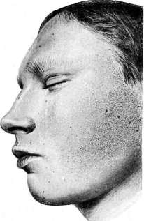
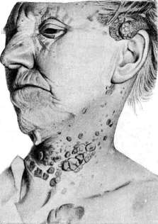
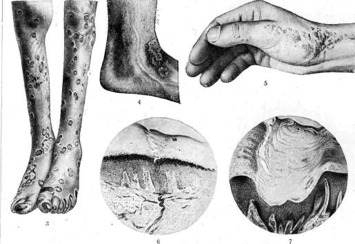
Figure 1. Ulerythema ophryogenes. Figure 2. Keratosis senilis. Figure 3. Gonorrheal keratosis. Figure 4 and 7. Porokeratosis Mibelli. Figure 5 and 6. Keratoma disseminatum naeviforme. (Figure 1, 4-6—from the collection of the State Venereological [Institute])
institute; figs. 2, 3 and 7 from Sutton.) B. M. E., To the art. Keratoses. They are very weakly expressed. The process may exist for many months and even years. In adults, it sometimes disappears spontaneously after a few weeks. Histologically: follicular hyperkeratosis, very insignificant perifollicular inflammation, congestive hyperemia of the follicle vessels, and hypoplasia of the epidermal cells surrounding them. The disease is similar to hair K.; clinically it sometimes resembles lichen scrofulosorum and lichenoid trichophytids. In the opinion of Brocq, in view of the absence of inflammatory changes, the process represents not a lichen, but a K. It can be combined with folliculitis decalvans, with lichen ruber planus, etc. Many authors have observed the development of spikes (i.e., the phenomenon of 'spinulosis', according to Darier) in peripilar syphilides, pityriasis rubra pilaris Devergie, in symmetric keratodermia and seborrheic eczema, in alopecia, and in ichthyosis. Etiology. The majority of authors believe that k. spinulosa is an expression of a peculiar reaction of the skin to various external irritations. Treatment is not required in the majority of cases; sometimes arsenic internally, exfoliating ointments, fractional doses of X-rays. Poral K. Porokeratosis (porokeratosis Mibelli, keratodermia, s. hyperkeratosis excentrica, hyperkeratosis figurata centrifuga atrophica, hypereleidosis excentrica atrophicans, hypereleidosis) is an extremely rare chronic skin lesion, described in 1893 by Mibelli and Respighi independently of each other. It is characterized initially by the appearance of very small, sharply defined, conical, brownish, wart-like papules with a comedo-like horny plug in the center. The rash shows a tendency toward slow peripheral growth and fusion, subsequently forming partly round, partly irregular circinate plaques the size of a coin and larger, with a smooth normal or squamous, callous, or atrophic surface, bordered by a peripheral, strongly tortuous papular ridge of grayish or brownish color, in places covered with powerful horny layers up to 1 cm high. On the crest of the ridge of some efflorescences, a narrow linear or ring-shaped groove is noticeable in places, from which a thin brownish horny plate protrudes like a prismatic crest. The horny plugs may fall out, leaving depressions or pit-like indentations. The central part of the plaques lies at the level of normal skin or slightly lower due to atrophy, sometimes it becomes pigmented or acquires a brownish-red color. The disease is encountered mostly in men at any age. The eruptive elements are located on the back of the hands, especially on the extensor surfaces of the lower extremities, on the feet (fig. 4) and fingers, sometimes on the scalp, the back of the head, the face, the genitals, the buttocks, and in the lumbar region. The oral mucosa is rarely affected. Subjective symptoms are usually absent. Histologically: significant hyperkeratosis, especially near the excretory ducts of the sweat glands, with which there is, apparently, a close connection. Sharp thickening of the transparent layer (Mibelli, Pavlov); increase, atrophy, or absence of the granular layer, significant acanthosis; the orifices of the sweat ducts are filled with horny masses and obliterated; the lumina of the sweat gland glomeruli are dilated (fig. 7). The process apparently begins with dystrophy of the epithelium of the excretory ducts and pores of the sweat glands, as well as the openings of the follicles. In a diagnostic respect, this dermatosis can sometimes show a slight similarity to ring-shaped lichen planus; it should not be confused with punctate keratodermia. -Etiology. Porokeratosis Mibelli is a genotypic dominant disease, traced to the 4th generation. Men fall ill significantly more often. Siemens' student, Fulde, managed to establish on the basis of literary data the following ratio of men to women: 88♂: 35♀. Apparently, it concerns irregular dominant heredity, partly limited to the male sex. The prognosis in terms of recovery is unfavorable. The process may sometimes exist for decades. Keratolytic agents have a weak effect. Surgical intervention, freezing with CO2 snow give only a temporary effect, as do X-rays, radium, mesothorium, and quartz lamp rays. Keratosis punctata (porokeratosis punctata) is a punctate keratodermia of the Besnier type, or porokeratosis according to some authors, a peculiar lesion of the soles and palms, characterized by miliary transparent and dense horny formations embedded in the thickness of the epidermis, on the surface of which numerous small well-like depressions are noticeable. This form of K. with small punctate disseminated or grouped horny projections is encountered at any age, affects both sexes to an equal degree, and can exist for a long time. Etiology - Hallopeau and Claisse found a connection of the disease with the excretory ducts of the sweat glands and assumed that it concerns a special form of nevus. The disease stubbornly does not yield to treatment. Keratolytic agents (in combination with the careful use of X-rays) can apparently give the most satisfactory results. - Porokeratosis symptomatica. Porokeratotic depressions are encountered not infrequently in various painful processes accompanied by increased keratinization, e.g., in syphilis, Darier's disease, juvenile warts, lichen planus, arsenic K., follicular K., etc. - Keratosis plantaris sulcata. Under this name, Castellani and Mendelson described a chronic skin lesion of the soles in soldiers in the form of diffuse callosities, riddled with deep cracks, which are often infected, serving as entry gates for the introduction of pyogenic microorganisms. Keratotic masses are sloughed off in places, leaving characteristic pit-like depressions. The cause is permanent irritation of the skin. Bed rest and keratolytic agents constitute the best method of treatment. - Keratosis periporalis, described by Gans in 1924, is a disease of the skin of the palms and soles where, with a previously existing hyperhidrosis, there were hyperkeratotic formations sharply rising above the skin level with the presence of funnel-shaped depressions filled with horny plugs. Histologically: hyperkeratosis around the openings of the excretory ducts of the sweat glands with subsequent atrophy of the adjacent part of the epidermis and disappearance of the papillary layer. Gans is inclined to include this disease form in the group of punctate K. The etiology is unknown; it is possible that it concerns a late nevus. Local keratolytic agents and X-rays have almost no effect on the process. Keratodermia maculosa disseminata symmetrica palmaris et plantaris (Buschke-Fischer), porokeratosis papillomatosa palmaris et plantaris; keratoma dissipatum (naeviforme) (Brauer; figs. 5 and 6); keratodermia verrucosa nodularis, described in 1910 by Buschke and Fischer, is a skin disease in the form of numerous intradermal miliary callous nodules of a yellowish or brownish color, which initially were located at the general level with the epidermis, and then gradually increased and rose above the surrounding skin, reaching the size of a lentil, sometimes with the subsequent formation of pit-like depressions and a bluish-red coloring of the center of the nodules [see separate table (art. 583-584), fig. 13]. Subjective sensations are absent. The course of the disease is chronic. Histologically: limited hyperplasia of the horny layer, thickening of the granular and transparent layers; the papillae of the skin are flattened or elongated. Inflammatory infiltrate is usually absent. The etiology is unknown. A hereditary predisposition is possible. Therapy - keratolytic agents and X-ray treatment give only a temporary effect. -To keratosis macul. dissem. sym. palm. et plant. are also attributed the cases described by Brauer of hereditary palmar-plantar disseminated keratodermia in the form of numerous 'parakeratotic horny inclusions' of various sizes, bordered by a wide hyperkeratotic ridge. Kyrle's disease (hyperkeratosis follicularis et parafollicularis in cutem penetrans) is an extremely rare anomaly of keratinization, distinguished by Kyrle in 1916. The primary efflorescences appear as limited nodules the size of a pinhead, of a yellow-brown color, located folliculary and parafolliculary, with a small dense scale or crust on the apex, which as they develop further become more compact and more darkly colored. Showing a tendency toward rapid peripheral growth and fusion, the horny keratinized nodules acquire a warty character due to the proliferation of epidermoidal cells in the periphery, forming larger plaques with polycyclic outlines, in places riddled with cracked horny masses. Around the affected foci, inflammatory redness is absent. Upon forcible removal of the crusts from the isolated elements, openings and depressions of a round shape with regular edges are noticeable, secreting an insignificant amount of serous fluid.
Localization—hairy skin of the head, face, torso, upper and lower extremities. The process is close to Darier's disease, but not identical to it. Histologically: follicular and parafollicular hyperkeratosis, here and there parakeratosis and significant thickening of the keratohyalin layer; acanthosis; in the excretory ducts of sweat and, especially, sebaceous glands and even in the glands themselves—an accumulation of cornified masses. Due to the rupture of thinned epidermoidal walls, powerful horny plugs penetrate through the basal layer directly into the skin proper (in cutem penetrans) and cause significant inflammatory changes in the dermis. According to Kyrle and others, the microscopic picture presents certain diagnostic difficulties, resembling psoriasis, contagious follicular keratosis, lichen verrucosus, psorospermosis follicularis vegetans, pityriasis rubra pilaris. The etiology has not been clarified. Keratosis senilis (verrucae seborrhoicae seniles, keratosis praecancerosa senilis, epitheliomatosis multiplex senilis)—a disease occurring in elderly persons (more often at the age of at least 50 years), on the forehead, temples, nose, cheeks, back, less often—on the neck, shoulders and forearms, as well as on the elbows and the back of the hands. It is characterized by multiple, sharply defined, flat, greasy to the touch hyperkeratotic and wart-like formations; the latter reach the size of a 10- or 20-kopeck coin. They have a yellow-brown or dirty-gray color and are incorrectly called seborrheic warts, with which this keratosis has only some resemblance (Fig. 2). The central part of the elements shows signs of cicatricial atrophy, and therefore it is sometimes difficult to distinguish them from lupus erythematosus (Brocq). These formations can exist for a very long time, continuously increasing in number and often representing a complication of senile skin atrophy. In essence, keratosis senilis is a precancerous dermatosis and often serves as a starting point for the development of relatively benign epitheliomas. Histologically: in the initial stage of development, the horny layer is sharply thickened and provided on the inner side with conical extensions penetrating deep into the Malpighian layer, which in these places is thinned and atrophied, sometimes permeated with infiltrate cells; in places—parakeratosis, acanthosis and perivascular infiltration (Breudenthal found phenomena of dyskeratosis in the horny layer); in the papillary layer—edema. Etiology: according to Siemens, keratosis senilis can sometimes be observed as a familial disease, occurring even at a less advanced age.
Actinic rays and sudden atmospheric fluctuations are auxiliary factors. Treatment—keratolytic agents (resorcinol, green soap, salicylic acid, white precipitate of mercury). Sometimes an effect is achieved from X-rays, scraping with a sharp curette, and galvanocauterization.
Keratosis of mucous membranes (keratosis mucosarum). The mucous membranes of the oral cavity and lips, as well as the genital organs, normally have a histological structure similar to the skin, with the only difference that the granular layer is absent here and, furthermore, the superficial layers of the epithelial covering of the mucous membranes do not undergo complete cornification in a normal state. However, with pathological changes, it can lead to the development of true keratinization due to the formation of keratohyalin and eleidin in the epithelial layer (so-called prosoplasia). Darier, Brocq, and others include various chronic painful processes in this group: leukoplakia (leukokeratosis), syphilitic glossitis, furrowed tongue, glossitis rhombica mediana, black hairy tongue, etc.
M. Per. Syphilitic keratoses are an eruption of syphilitic papules or tubercles located on the palms and soles and covered on their free surface with significant hyperkeratotic layers. This eruption also bears the not entirely successful name psoriasis syphilitica palmaris et plantaris or lues psoriasiformis secundaria et tertiaria palmaris et plantaris. These eruptions rarely accompany fresh syphilids of the skin and mucous membranes, but are more often encountered in the form of late relapses of the 2nd period, as well as in the tertiary period of syphilis. In the secondary period, it is a matter of an eruption of individual, or in a later period, grouped, slightly brownish papules, the size of a lentil or less, semi-spherical or conical in shape, deeply embedded in the skin and therefore almost not protruding above the level of the skin, but well noticeable to the touch due to their significant density. The papules are either covered with thickened horny layers or these latter occupy only the center, leaving a reddish or bluish-crimson border along the periphery. In plantar syphilids, especially in the heel area, such significant horny layers can accumulate that they make the entire formation look like corns (clavi syphilitici). The eruptions are as a rule symmetrical, do not show a particular tendency to coalesce, and, arising in places of deep folds, cause the formation of painful, difficult-to-heal fissures. The regression of the elements, having begun with the peeling of their center, such that they appear as if surrounded by a wreath of scaly layers, gradually passes to the periphery, after which the eruption disappears without leaving any trace. In the tertiary period, on the contrary, eruptions on the palms and soles are as a rule not symmetrical, very rarely grouped, but usually appear in the form of serpiginous plaques covered with horny masses, with irregular arcuate edges, convex outward. The infiltrate constituting the plaques in this case is deeper and denser than in the elements of the 2nd period and has a tendency to ulcerate. Histologically, in the first case, we are dealing with an ordinary lenticular papule, in the second with a syphilitic tubercle, with the only difference that the horny layer in both cases is hyperplastic and contains areas of parakeratosis. The horny nature of palmar and plantar syphilids is conditioned on one hand by the anatomical structure of the skin of these areas, and on the other by the influence of external factors. That is why these lesions are encountered more often in persons engaged in physical labor. In diagnosis, one should keep in mind first of all psoriasis and calloused chronic eczema, however, with them there is no such dense infiltrate and a brownish peripheral border rising above the scaly edge. Among other diseases, one should keep in mind lichen ruber, gonorrheal and arsenical keratoses. Syphilitic keratoses are difficult to treat. In addition to specific therapy, one often has to resort to local remedies in the form of local hot baths, lubrication of fissures with a 3-5% silver nitrate solution, etc. Gonorrheal keratoses are a very rare complication of gonorrhea (about 70 cases have been published), encountered predominantly in men; it was first described by Vidal in 1893; among Russian authors, a detailed description was given by Bogrov in 1916. Clinically, it is a peculiar eruption starting either from a spot in the center of which a papule or vesicle arises, or immediately from a vesicle. The latter quickly becomes cloudy and is covered with a dirty-yellow, dense scaly crust, upon the removal of which an uninflamed red, non-bleeding base is revealed. The described elements resemble calloused nodules. Often a hyperemic rim arises around them. The size of the nodules varies from a millet seed to a 20-kopeck coin and more. Flat scaly brown formations are compared to the heads of upholstery nails, and larger layered ones to an oyster shell. In general, depending on the structure and color of the horny crusts, they resemble now impetigo and ecthyma-like, now eczematous and even seborrheic crusts (Fig. 3). The elements of the rash are either scattered diffusely or, merging and increasing along the periphery, form large plaques the size of a palm or more. Sometimes the layers grow in height and resemble cutaneous horns. Upon forcible removal of the horny masses, they grow back; in cases heading toward recovery, they easily fall off by themselves without leaving a scar. Usually, gonorrheal keratoses are located symmetrically. They are divided into two forms (Baermann): localized (more often) and disseminated. The favorite site of the lesion is the hands and especially the feet (the edges of the feet and toes are involved), as well as the genitals. In the disseminated form, the rash is located on the limbs and trunk. Lesions of the mucous membranes are a great rarity. Histologically, it comes down to an exudative-infiltrative inflammation of the papillary layer and epidermis, with the growing crust representing an accumulation of alternating layers of parakeratosis and fibrinous exudate with a mass of leukocytes included in it. Findings of gonococci in the elements of the rash (initial and keratodermic) are rare and not always indisputable. The course of gonorrheal keratoses is chronic. Subjective sensations are absent or insignificant. There is no increase in temperature, or it depends on complications. Gonorrheal keratoses appear only with a general gonorrheal infection, which can be expressed by polyarthritis, endocarditis, etc., and in accordance with the state of these complications, the keratoses can worsen, pass, or recur. The diagnosis of gonorrheal keratoses is usually not difficult. One has to keep in mind the favorite localization, symmetry, and direct connection with the course of the general gonorrheal infection. Gonorrheal keratoses differ from palmar and plantar syphilitic keratoses by the onset with a vesicle or pustule, a pink hyperemic zone around the elements, and the absence of an infiltrate under the horny crust. The mechanism of the origin of gonorrheal keratoses has not been finally clarified. Of the three theories—reflex, trophic, and infectious—the latter is the most recognized; at the same time, gonorrheal keratosis should be considered as a genuine metastasis of gonorrheal infection to the skin (Scholtz). Local treatment brings little benefit. Often Tinctura Iodi, ointments with chrysarobin, salicylic acid, etc., even caused the appearance of new eruptions. On the contrary, rational therapy of the underlying suffering (gonorrheal urethritis and its complications) led to the most rapid disappearance of the skin eruptions.
N. Efron. Keratosis follicularis—see Darier's disease.
Related articles
Mentioned in
Cite this page
“Keratoses.” Soviet Medical Encyclopedia. English translation of Bolshaya Meditsinskaya Entsiklopediya, 1st ed. (Moscow, 1928–1936), ed. N. A. Semashko. https://sovietmedicalencyclopedia.pages.dev/article/keratoses/