Keratoplasty
Historical document, translated for reference. It reflects medical knowledge of the 1920s–30s and is not medical advice.
Summary
This article from the 1928–1936 Soviet medical encyclopedia details the history, types, and techniques of keratoplasty, a surgical procedure to restore the cornea. It covers tectonic and optical keratoplasty, the use of autografts, homografts, and heterografts, and discusses the challenges of corneal transparency and graft survival.
Encyclopedia article (1928–1936)
KERATOPLASTY, an operation for restoring the cornea destroyed by a pathological process. Tectonic and optical keratoplasty are distinguished. Tectonic keratoplasty aims to fill a defect in the cornea (for example, following ulcers, trauma, during operations to remove the pterygium, symblepharon, staphyloma, thin scars, etc.) with some suitable, even if opaque, tissue: neighboring conjunctiva, mucous membrane from the lip, tendon or fascial tissue. Thus the anatomical integrity of the eyeball is preserved, and the danger of various complications, for example infection, is eliminated. At present, conjunctival keratoplasty is especially widely applied in the treatment of corneal wounds and ulcers: covering defects in the cornea with the movable conjunctiva of the eyeball, properly cut out and separated. The technique of this operation and its variants have been developed in particular detail by Kuhnt (N. Kuhnt).-True keratoplasty, long of interest to ophthalmologists because of the brilliant prospects it holds for preserving and restoring vision to those blinded by corneal destruction (leucoma, staphyloma), is optical keratoplasty. This is truly an operation of the future, although much has already been done in this area. Its aim is to replace the opaque tissue of a leucoma or staphyloma of the cornea with a transparent graft, a cornea from an animal (rabbit, dog, cat—heteroplasty), from another person (homoplasty), or from the patient himself (autoplasty keratoplasty, for example in replacing a central leucoma with a surviving transparent corneal area on its periphery). The possibility of a cornea from an animal taking root in man has been fully proved, but the transplanted cornea inevitably becomes cloudy; therefore heteroplastic keratoplasty for optical purposes is not applied. Full keratoplasty is spoken of when the whole cornea is transplanted (attempts were even made to transplant the entire anterior segment of the eyeball), and partial keratoplasty when a part of the cornea is replaced by a transparent graft. Furthermore, penetrating keratoplasty and lamellar keratoplasty (Fig. 1) are distinguished. In the first the entire thickness of the cornea is excised and replaced, and the anterior chamber is opened. In the latter only the anterior, cloudy layers of the cornea are excised with the preservation of the deep, presumed transparent layers, and the defect is replaced by an equally non-penetrating graft. Partial keratoplasty offers the greatest chances of success in terms of graft survival and preservation of its transparency. Various methods of keratoplasty exist. In all methods, for the success of the operation it is necessary to observe the following basic conditions: the conjunctival sac must be sterile; the piece cut from the leucoma of the "host" and the transparent piece of graft cut from the cornea of the "guest" must be of the same size; the graft must not be subjected to trauma; its transfer to the cornea of the "host" must be performed as soon as possible, and its attachment in its new place must be flawless. Equality of size of the excised piece and the graft is achieved best by using a corneal trephine with a clockwork mechanism (Fig. 2) (v. Hippe1; 1877). Partial penetrating keratoplasty is performed as follows. Local anesthesia; general anesthesia is avoided because of the possibility of vomiting after the operation ejecting the graft with the prolapsing vitreous body. The eyelids are separated with Demar's elevators. The eye is firmly fixed with 2 forceps or 2 ligatures passed through the tendons of the external and internal rectus muscles. At this time, a circle 3–4.5 mm in diameter (rarely larger) is excised from the eye of the "guest" (in situ or enucleated—if this eye is soft, it is compressed with the fingers or stretched by injection of novocaine or NaCl 0.75% into the vitreous body) and preserved in dry sterile gauze for 3–4 minutes until the moment of transplantation. After this, the same trephine crown excises a round penetrating hole in the leucoma of the "host" and the graft is immediately inserted into this hole. The eyelids are carefully closed over it; a bandage is applied; immobile bed rest; the first dressing in 2–3 days. Fixation of the graft is usually unnecessary. But it is sometimes necessary, for example in cases of prolapse of the vitreous body, etc., and is accomplished with sutures or by covering the graft with the conjunctiva of the eyeball.-Partial non-penetrating keratoplasty is also performed according to Hippel or by other methods (Diirr, Lohlein; Fig. 3), in which a strip of conjunctiva connected with it is also transplanted together with a piece of cornea. For the evaluation of the results of keratoplasty there is extensive material from the Prague clinic (A. Elschnig), where keratoplasty has been intensively developed and studied since 1908. Of 93 cases of partial penetrating keratoplasty (statistics of Ascher up to 1921), loss of the eye occurred in 4 cases; loss of the graft in 20 cases; survival of opaque grafts in 35, semi-transparent in 20; in the remaining cases the graft survived with preservation of transparency (of these, in 9 cases transparency was preserved for more than 9 months). Visual acuity increased noticeably in only 9 cases, of which the majority of effective results relate to keratoplasty after parenchymatous keratitis in eyes with poor vision but still seeing. The latest data from the same clinic (Stanka; 1927) note similar results. Unfortunately, keratoplasty has proved unsuccessful so far in eyes, especially those in need of it: in solid leucomas and staphylomas fused with the iris, after ophthalmoblennorrhea, smallpox, severe purulent keratitis in general; in flattened scars, usually always fused with the iris and lens, and with increased intraocular pressure. - Full penetrating keratoplasty (Fig. 4), as well as transplantation of the entire anterior segment of the eyeball, have been described as isolated cases; they have proved the possibility of even such extensive grafts taking root, but preservation of corneal transparency has not been achieved. The fate of the graft is not yet fully elucidated. Some authors (Salzer, Bonnefon, Lacoste) deny true survival of the cornea and prove that the graft is absorbed and gradually replaced by opaque or transparent tissue of the host; others (Fuchs, Elschnig, Filatov) consider that true transplantation is possible in man during keratoplasty. In the USSR keratoplasty is especially studied by Filatov.
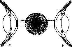
Figure 1. Keratoplasty according to Hippel: a-penetrating; b-lamellar.
Figure 2. Corneal trephine according to Schr6der. The eye is firmly fixed with 2 forceps or 2 ligatures passed through the tendons of the external and internal rectus muscles.
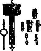
Figure 3. Keratoplasty according to Lohlein. Figure 4. Full keratoplasty according to Elschnig.


Figure 3. Keratoplasty according to Lohlein. Figure 4. Full keratoplasty according to Elschnig.
Related articles
Cite this page
“Keratoplasty.” Soviet Medical Encyclopedia. English translation of Bolshaya Meditsinskaya Entsiklopediya, 1st ed. (Moscow, 1928–1936), ed. N. A. Semashko. https://sovietmedicalencyclopedia.pages.dev/article/keratoplasty/