Pterygium
Historical document, translated for reference. It reflects medical knowledge of the 1920s–30s and is not medical advice.
Summary
This article defines pterygium as a growth of the conjunctiva onto the cornea, distinguishing between the true form (often associated with environmental exposure and pinguecula) and the false form (pseudopterygium, resulting from scarring). It details the pathological anatomy, clinical progression, and surgical treatment methods prevalent in the early 20th century.
Encyclopedia article (1928–1936)
PTERYGIUM (pterygium, s. pterygion), a duplication of a part of the conjunctiva of the eyeball, fused to a greater or lesser extent with the cornea. Depending on the mode of origin of this peculiar eye affliction, two varieties are distinguished: true and false pterygium. Both somewhat resemble the wing of an insect in appearance, from which they received their name. True pterygium. Its etiology and pathogenesis still remain unclear. It usually develops in middle-aged and elderly persons without any visible external cause, not accompanied by any phenomena of eye irritation. The fact of the constant (with very rare exceptions) development of pterygium in the area of the palpebral fissure, where the conjunctiva is most exposed to unfavorable influences—atmospheric, temperature, radiant energy, physical and chemical agents—suggests that it is precisely these factors, acting over a long period, in connection with age-related changes in the tissues, that play the main role in the origin of pterygium. And indeed, observations show that pterygium, like pinguecula (see Conjunctiva), is most often encountered in persons who are most exposed to these harmful influences: bakers, millers, stonemasons, farmers, coachmen, workers in chemical factories, sailors, those working near hot furnaces, and residents of hot countries. The circumstance that the development of pterygium is often preceded by the development of pinguecula, their identical localization, as well as the fact of finding remnants of pinguecula in the stroma of pterygium during histological examinations, gave many authors, led by Fuchs, reason to consider pterygium a further stage of development of pinguecula and led to the peculiar notion that the degenerated tissues of pinguecula are capable, under the influence of unknown causes, of shifting in the direction of the cornea, pulling the conjunctiva of the eyeball behind them, whereby the pinguecula as such disappears, turning into pterygium. But such a notion about the pathogenesis of pterygium is denied by other authors.
In the pathological-anatomical respect, pterygium consists in general of the same elements as the conjunctiva of the eyeball. The epithelium of the latter passes directly onto the pterygium, passing further into the epithelial covering of the cornea, sometimes containing an abundant quantity of epithelial cells in a state of mitosis, and goblet cells. Depressions of the epithelium are noted, leading to the formation of glands; the latter sometimes serve as the starting point for the development of retention cysts. The stroma of the pterygium corresponds to the stroma of the conjunctiva and is quite richly supplied with vessels; sometimes the development of cysts from dilated lymphatic vessels of the stroma is observed (Motolese). Galenga draws attention to the hyaline degeneration of the stroma. Rarely are remnants of pinguecula tissue found in the stroma. Bowman's membrane in the area of the fusion of the head of the pterygium with the cornea turns out, as Fuchs' studies have shown, to be destroyed, with the stroma of the pterygium passing directly into the stroma of the cornea.
True pterygium, developing most often from the inner or lower-inner side, predominantly in a horizontal direction, in the area of the palpebral fissure, represents a fold of the conjunctiva of the eyeball, usually in the shape of a triangle, the base of which is directed toward the equator of the eye, and a somewhat rounded apex is tightly fused with the cornea [see separate table (Vol. XIV, art. 231-232), fig. 4]. The part lying on the sclera is called the body, and the part fused with the cornea is called the head. The body and head are connected to each other by means of the so-called neck. Pterygium, smooth on the surface, appears to be permeated by vessels converging toward the head. Sometimes small nodules and spots are noted in the tissue of the pterygium, grayish in the area of the head, yellowish in color on the neck, considered as remnants of the so-called pinguecula. The boundaries of the latter appear in the area of the anterior edge of the head mostly in the form of a grayish, translucent, gelatinous-looking, avascular border. Its upper and lower boundaries are formed by a fold of the conjunctiva, under which there is a blindly ending pocket, such that a probe inserted under the fold cannot be brought out from the opposite side without violating the integrity of the walls of the pocket. The base of the pterygium, facing the equator of the eye, does not have sharp boundaries and often passes directly into the semilunar fold. Pterygium is encountered in men approximately twice as often as in women, and more often in both eyes. Cases of the development of pterygium on the same eye simultaneously from the nasal and temporal sides are not so rare.
The course of the process is very slow, and at a certain moment, it passes from a progressive stage into a stationary one without visible causes, in the majority of cases not yet having reached the most central parts of the cornea. The disorders caused by pterygium, apart from cosmetic defects, reduce to a decrease in vision due to clouding of the central parts of the cornea or astigmatism arising sometimes on the basis of pterygium, to a violation of the normal mobility of the eyeball, which can sometimes be accompanied by diplopia, and to difficulty in the outflow of tears due to the tension of the semilunar fold and the violation of normal relationships in the area of the lacrimal lake. The prognosis is in the majority of cases favorable. However, in each individual case, we are not in a position to resolve the question of when the pterygium will pass from its progressive stage into a stationary one. Treatment is exclusively operative. In this, it should be kept in mind that to eliminate the possibility of recurrences and to restore the mobility of the eyeball, a thorough removal of the marginal border of the head and an expedient backward displacement of the conjunctiva, stretched onto the cornea, with possible sparing of the conjunctiva, are necessary. All the diverse operative methods undertaken for pterygium regarding the separation of the head from the cornea do not differ essentially from one another; the entire difference lies only in the methods of closing the defect of the conjunctiva on the sclera. The oldest, simplest, and sufficiently satisfactory method is that of Arlt, which consists in the fact that after thorough separation of the head and neck of the pterygium, two incisions converging toward the equator of the eye are made, by which the head, neck, and part of the body are excised, and the defect is closed by tension of the conjunctiva with the help of sutures. At the basis of many other methods of operations proposed by different authors lies not excision, but displacement of the pterygium. On this are based the operations of Desmarres, Knapp, Czermak, and others. To avoid recurrence, it is important, besides thorough separation of the head, to change the direction of the fibers of the stroma of the pterygium. This side of the matter turns out to be realized in the operations proposed by Russian authors (Golovin and Palimpsestov).
False pterygium (pseudopterygium, pterygoid) represents a fusion of the conjunctiva of the eyeball with the cornea, which arose as a result of close contact of the swollen, edematous conjunctiva with the scarring ulcerous surface of the cornea. Thus, the genesis of false pterygium is definite, clear, and completely different from the genesis of true pterygium. The lesion of the cornea preceding the formation of the false pterygium can be primary or secondary, arising in the latter case as a result of a lesion of the conjunctiva, or the lesions of the cornea and conjunctiva arise simultaneously. Among the causes of false pterygium, injuries and burns of the conjunctiva and cornea stand in the first place, as well as those of the ulcerous keratitis which are themselves a consequence of inflammation of the conjunctiva. In this respect, blennorrhea presents the greatest interest and importance. In rarer cases, the cause of false pterygium is diphtheria of the conjunctiva. Differing from true pterygium in genesis, the false pterygium also differs in some features in the clinical respect. Since the lesion of the cornea can occur in any of its parts and be of the most diverse form and prevalence, the localization, position, and direction of the false pterygium do not differ in constancy. Since the ulcerous process in the cornea may not reach its edge, which often happens in practice, the fusion can have the form of a bridge stretching across the limbus, which allows one to insert a probe under the fold of the conjunctiva in this area, bring it out freely from the opposite side, and lift the entire fold on it. The latter, however, is not mandatory, since even with false pterygium, fusion can take place in the area of the corneoscleral boundary, corresponding to the neck of the pterygium. While with true pterygium, the parts of the cornea surrounding the head are completely transparent, with false pterygium they can turn out to be scarred to a greater or lesser extent. Forming often as a result of a burn of the conjunctiva and cornea, false pterygium is frequently in such cases combined with symblepharon (see Conjunctiva). Finally, false pterygium does not progress; once formed in the acute period of the lesion, it then remains stationary. The treatment of false pterygium is also operative, having some features depending on the peculiarities of the process. Operative intervention is usually undertaken either for cosmetic reasons or with the aim of eliminating violations of the mobility of the eyeball and visual impairment caused by the advancement of the pterygium toward the center of the cornea.
V. Chirkovsky. PTERYGOPALATINE FOSSA (fossa pterygopalatina) is located medial to the infratemporal fossa and is situated between the posterior edge (tuberosity) of the body of the maxilla and the orbital process of the palatine bone anteriorly, and the pterygoid process and part of the greater wings of the sphenoid bone posteriorly. Its average dimensions are: anteroposterior 6.2 mm, transverse 9.1 mm, height 18.6 mm (Ivanitsky). Its medial wall is formed by the outer surface of the vertical plate of the palatine bone. There is no lateral wall; here the pterygopalatine fossa communicates with the infratemporal fossa via a fissure that is wider at the top and narrows downward. The pterygopalatine fossa itself, being more voluminous in its upper section, also narrows downward and passes into the canal [canalis pterygopalatinus (Fig. 1)], which continues into the canals of the palatine
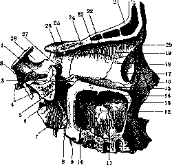
Figure 1. Right pterygopalatine fossa: 1-probe in for. rotund.; 2-for. ovale; 3-proc. orbit, os. palat.; 4-probe in canal, pteryg.; 5-for. spheno-pal.; 6-fossa pterygopalat.; 7-lam. lat. proc. pteryg.; 8-proc. pyram. os. palat.; 9-probe in canal, palat.; 10-pars perpend, os. palat.; 11-sin. maxill.; 12-proc. maxill. conchae nas. inf.; 13-pr. ethmoid, conchae nas. inf.; 14-pr. uncin. os. ethmoid.; 15-fac. orbit, max.; 16-os nas.; 17-fossa sacci lacrim.; 18-os lacrim.; 19-pars orbit, os. front.; 20-spina trochl.; 21-sin. front.; 22-lam. papyr. os. ethmoid.; 23 and 24-for. ethmoid.; 25-for. opt.; 26-corpus os. sphen.; 27-fiss. orbit, sup.; 28-sella turcica. (From Spalteholz.)
bone, opening on the lower surface of the hard palate—foramen palatinum majus et foramina palatina minora. On the medial wall there is an opening (foramen sphenopalatinum) leading into the nasal cavity. Between the vertical plate of the palatine bone and the corresponding surface of the body of the maxilla, small canals are located, also leading into the nasal cavity. Furthermore, the pterygopalatine fossa communicates through the foramen rotundum with the middle cranial fossa, through the inferior orbital fissure with the orbital cavity, through the infraorbital sulcus and the canal of the same name it opens onto the anterior surface of the facial skull, and through the pterygoid canal [canalis pterygoideus Vidii (Fig. 2)], passing in a horizontal direction from front to back at the base of the pterygoid process, it communicates with the region of the foramen lacerum anterius. The foramen sphenopalatinum and the axis of the pterygoid canal lie in the same horizontal plane. In brachycephals, the pterygopalatine canal runs more vertically than in dolichocephals, in whom its upper end deviates posteriorly.
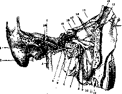
Figure 2. Right pterygoid canal of Vidius: 1-n. carotico-tymp. inf.; 2-n. facialis; 3-a. carot. et plex. carot. int.; 4-n. petros. superf. major; 5-n. petros. prof.; 6-n. canalis pteryg. Vidii; 7-gangl. spheno-palat.; 8-nn. palat.; 9-nn. spheno-palatini; 10-rami alveol. sup. post.; 11-ram. alveol. sup. med.; 12-os zygom.; 13-ram. zygom.-tempor.; 14-gl. lacrim. sup.; 15-ram. anastom. c n. zygomat.; 16-ram. zygomatico-facial.; 17-n. lacrim.; 18-n. zygom.; 19-n. maxill.; 20-pl. tympan. Jacobsoni. (From Spalteholz.)
Vessels and nerves pass through the openings and canals. Arteries (branches of the internal maxillary artery): the infraorbital artery passes through the inferior orbital fissure into the canal of the same name; the descending palatine artery is located in the pterygopalatine canal, gives a branch to the Vidian canal, and divides into the greater and lesser palatine arteries (Fig. 3), which run in the canals of the same name; the sphenopalatine artery exits through the foramen sphenopalatinum into the nasal cavity. Veins merge into a well-developed venous plexus (plexus venosus pterygoideus), located corresponding to the pterygopalatine fossa, with its main part on both sides of the external pterygoid muscle and with thinner branches along the inner surface of the internal pterygoid muscle, along the membranous part of the Eustachian tube, and along the course of the internal maxillary artery. From the plexus, blood is drained partly through the deep branch of the anterior facial vein, but predominantly through the posterior facial vein. Nerves: the maxillary nerve, the second branch of the trigeminal nerve, exits through the foramen rotundum into the pterygopalatine fossa, heading forward and downward into the infraorbital sulcus and canal. From its lower periphery, fibers branch off—the sphenopalatine nerves, which partly enter the sympathetic sphenopalatine ganglion located here, but for the most part, merely passing along its outer surface, transition into orbital (orbital branches), nasal (posterior superior and inferior nasal branches), and palatine (palatine nerves) branches. The nerve of the pterygoid canal (nerve of the pterygoid canal), composed of the greater superficial petrosal nerve and the deep petrosal nerve, enters the posterior corner of the ganglion. Among diseases of the pterygopalatine fossa region, the so-called Langenbeck's "retromaxillary tumors" occupy the first place in frequency and severity of course, the site of origin of which is the periosteum of the base of the pterygoid process and the circumference of the foramen sphenopalatinum. These tumors possess significant growth energy and spread in the direction of least resistance. Filling first the pterygopalatine fossa, then the zygomatic, temporal, and infratemporal fossae, the tumor penetrates through the foramen sphenopalatinum into the nasal cavity and through the inferior orbital fissure into the orbit; during its growth, the tumor can press inward the outer wall of the maxilla or destroy its posterior wall and penetrate into the maxillary sinus; finally, having destroyed the base of the skull, it can penetrate into its cavity. With such a strong destructive effect of the tumor, the obstacles to its spread are fascial and aponeurotic formations. Thus, for example, the growth of a tumor or its penetration toward the large vessels of the neck and the parotid gland, separated by the processes of the pterygoid fascia, is rarely observed; even less frequently is the spread of the tumor observed behind the pterygoid process into the region of the base of the skull, known as the pars paragutturalis, delimited by the strong pterygostylomastoid aponeurosis. Through the indicated pathways, the spread of tumors is possible and is observed in the reverse direction, for example, from the nasopharyngeal cavity (guttural fossa) through the foramen sphenopalatinum into the pterygopalatine fossa ("polyp of the base of the skull"). According to the microscopic picture, these tumors are fibromas in the stage of development, where
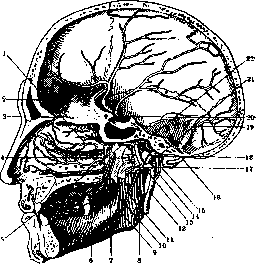
Figure 3. Arteries of the skull and nasal cavity: 1-aa. nasal, post, lat.; 2-a. mening. ant.; 3-a. ethmoid, ant.; 4-m. pteryg. ext.; 5-canal. pteryg. Vidii; 6 and 10-a. palat. major et min.; 7-a. spheno-pal.; 8-a. maxill. int.; 9-ram. mylo-hyoid.; 11-a. alveol. inf.; 12-a. carot. ext.; 13 and 20-a. mening. med.; 14-a. auric, prof.; 15-a. auricul. post.; 16-a. occip.; 17-ram. mening. accessor.; 18-a. tymp. ant.; 19-ram. mastoid. a. occip.; 21-a. tempor. superf.; 22-a. carot. int. (From Spalteholz.)
alongside fibrous tissue, clusters of large young connective tissue elements are encountered. Besides such tumors, cases of aneurysms of the maxillary artery and carotid artery (Clarke), tumors (fibrous degeneration) of the Gasserian ganglion spreading along the course of the branches of the trigeminal nerve have been described in the region of the pterygopalatine fossa; finally, a number of authors have described cases of syphilomas of the zygomatic fossa and the region of the choanae, which penetrated into the pterygopalatine fossa and simulated a "retromaxillary tumor." In cases of retromaxillary tumors or fibroma of the base of the skull penetrating into the pterygopalatine fossa, surgical intervention usually boils down to resection of the maxilla. Through the pterygopalatine fossa, one can penetrate with a needle to the foramen rotundum for puncture of the maxillary nerve in cases of neuralgia (80% alcohol is injected) or for the purposes of anesthesia (5 cm³ of a 1-2% novocaine solution is injected). Puncture is possible by two methods: 1) the injection site is located directly under the lower edge of the zygomatic bone, along a line passing vertically through the outer edge of the orbit. Along the outer wall of the maxilla, the needle is directed toward the maxillary tuberosity, and immediately behind it, turning the tip of the needle slightly inward, one penetrates into the pterygopalatine fossa; 2) the injection point is located at the place where the lower wall of the orbit transitions into the outer one. In view of the fact that the lower-outer wall is curved, the injection is performed almost vertically and then, penetrating deep and sliding along the wall, the needle is carefully moved into a horizontal plane. At a depth of 5 cm, through the inferior orbital fissure, one penetrates into the pterygopalatine fossa. To reach the sphenopalatine ganglion, it is necessary to use a slightly curved needle. The ganglion is located from the lower edge of the zygomatic arch on average at 52 mm, from the middle of its upper edge—at 50 mm, from the outer-lower corner of the orbit—at 55 mm, from the foramen palatinum majus—at 29 mm (Feldman and Ivanitsky).
Related articles
Mentioned in
Cite this page
“Pterygium.” Soviet Medical Encyclopedia. English translation of Bolshaya Meditsinskaya Entsiklopediya, 1st ed. (Moscow, 1928–1936), ed. N. A. Semashko. https://sovietmedicalencyclopedia.pages.dev/article/pterygium/