Spring Catarrh
Historical document, translated for reference. It reflects medical knowledge of the 1920s–30s and is not medical advice.
Summary
Spring catarrh is a distinctive inflammation of the conjunctiva characterized by seasonal exacerbations in spring and summer. The condition presents as chronic inflammation with specific proliferative changes in the conjunctival stroma and epithelium, with a characteristic milky-white appearance of the conjunctiva.
Encyclopedia article (1928–1936)
SPRING CATARRH (conjunctivitis vernalis s. aestivalis), a distinctive inflammation of the conjunctiva of the eye, first described in 1846 by Arlt, but most fully detailed in 1872 by Saemisch. A characteristic feature of this process is its periodicity associated with the seasons - exacerbation of the disease in spring and summer and its weakening during the cold months of the year. In its clinical and pathoanatomical picture, spring catarrh presents as chronic inflammation of the conjunctiva of the eyelids or the limbus of the eyeball (or both together), expressed in particular proliferations resulting from proliferation of the conjunctival stroma and its epithelial covering. Pathological anatomy. Pathological-anatomical studies have provided a detailed picture of the changes occurring in the conjunctiva of the eye in spring catarrh, but even to this day they have not clarified what in the pathological-anatomical picture is primary and what is secondary and sequential. Some authors, seeing mainly proliferation of the epithelial covering of the conjunctiva and other changes, consider the epithelial lesion in spring catarrh to be the fundamental phenomenon. Other authors, on the contrary, consider changes in the epithelium to be secondary, and regard proliferation of the conjunctival stroma as the most important characteristic changes for this process; this opinion is the prevailing one. In general, the process amounts to the fact that polypoid, flat proliferations of the cartilaginous conjunctiva represent the result of marked proliferation of the conjunctival stroma, with changes in the subconjunctival tissue being particularly typical - its early sclerosis and hyaline degeneration, and infiltration consisting mainly of plasma cells with a high content of eosinophils. The sclerosis of the subepithelial tissue explains the white, milky color of the conjunctiva, so characteristic of spring catarrh. Limbal proliferations also consist of greatly proliferated connective tissue (here a marked thickening of the epithelium is also usually noted). Changes in the epithelium explain the presence of so-called Horner-Trantas points - whitish-yellow spots, which are often noticeable in the limbal proliferations at the height of the process. In the regressive period, widespread hyaline degeneration appears, and according to Hoffmann, fatty degeneration of the connective tissue; as a result, the proliferations become flattened, the vessels are obliterated, and the epithelial thickenings undergo reverse development. Cytological examination of the conjunctival secretion in spring catarrh gives a very characteristic picture: here we encounter an abundant number of eosinophilic cells. The pathogenesis of spring catarrh still does not appear to be sufficiently clarified and, apparently, it is not homogeneous. Attempts to explain the etiology of spring catarrh by microbial findings have not been successful, although much, in the opinion of some researchers (Fuchs, Axenfeld, etc.), indicates a parasitic origin of this condition or, as Gabrielides thinks, a toxic one. Furthermore, the so-called light hypothesis, put forward by the dermatologist Kreibich as early as 1905, has many supporters. This hypothesis connects the origin of spring catarrh with the influence of ultraviolet light rays on the conjunctiva of the eye, by analogy with other so-called 'light' diseases, such as prurigo aestivalis, hydroa vacciniforme, etc. However, such an interpretation of the origin of spring catarrh encounters a number of serious objections: on the one hand, in observations where elimination of the influence of ultraviolet light rays did not prevent the manifestation of spring catarrh, and on the other, in the extremely rare occurrence of spring catarrh in some northern regions (such as Sweden), where one would expect, due to the abundance of light and richness of ultraviolet rays, particularly favorable conditions for the development of this process. It has been repeatedly noted, and particularly emphasized in recent years by Italian ophthalmologists, the influence of constitutional predisposition on the development of spring catarrh (the importance of lymphatic constitution is especially noted), as well as the importance of disorders of the internal secretion glands (there are indications of hypofunction of the adrenal glands). The clinical picture of this disease in its typical forms is manifested by the appearance in the area of the cartilaginous conjunctiva of the eyelid, especially the upper one, of flat, roundish, closely adjacent to each other bridge-like proliferations. These proliferations do not involve the area of the transitional folds and at most occupy the upper edge of the cartilage in the form of polypoid elevations. The conjunctiva appears of a delicate white color, as if flooded with milk. In severe cases, large mushroom-like, polypoid proliferations are observed, which never ulcerate or disintegrate. This form is called the palpebral form (see table, fig. 2). Along with it, the so-called limbal or bulbar form usually also arises, which is characterized by the development, usually in the area of the palpebral fissure, near the limbus, of individual nodules, which then merge into gelatinous, jelly-like elevations of yellow-gray or waxy, lifeless color (see table, fig. 3). The proliferations in the area of the limbus of compact consistency also never ulcerate. Combined involvement of the conjunctiva of the eyelids and the eyeball is called the mixed form of spring catarrh (most common). At the height of the process, when there are marked proliferations in the conjunctiva of the cartilage of the upper eyelid, the patient suffering from spring catarrh usually
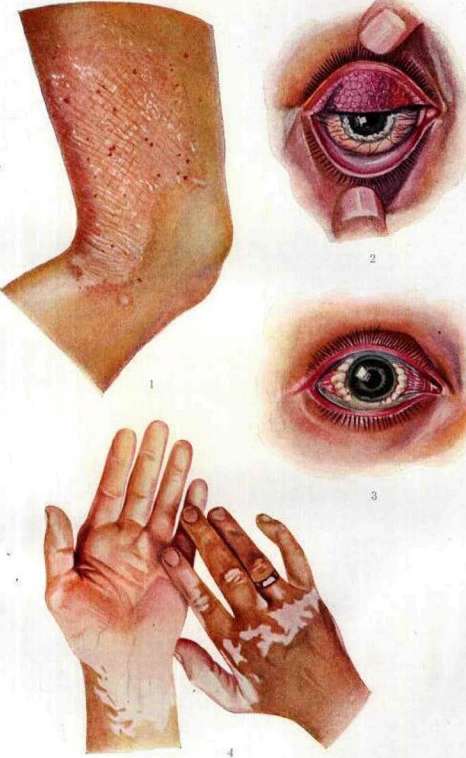
U L«sh*11 Nmsh* (But mulyazhu Kliniki chazpatk yidanti \ MGU), 2 -Ъ &shsh*tj iat*p^ * USHko (no ftrfcfce K ST. /ft'ft'rfJifJU AlUHu^V 'IWAM* IrJip.1 L- some drooping of the eyelids (ptosis), which, together with the pale, lifeless color of its conjunctiva, gives a kind of sleepy expression to his face. The process is accompanied by greater or lesser catarrhal phenomena (secretion is often observed in the form of threads). In some cases, the cornea is involved in the process,-cloudiness appears in it, very similar to the development of gerontoxon. The most characteristic feature is the periodicity in the development of the process. The disease usually begins in the spring or summer months and lasts throughout the summer period, and then, with the onset of cold weather, it subsides, while subjective sensations usually disappear completely, but the objective picture of the lesion remains to one degree or another even in winter. Spring or summer again cause an exacerbation of the process, and the disease again lasts throughout the warm period of the year, only to subside again in the winter months. Thus it repeats sometimes for many years, on average - for 4-6 years, but there are cases of exceptional duration. Thus, Zemish observed V. k. for 23 years, Trantas (Trantas)-31 years, and Gaspartini even 52 years. V. k. affects in most cases in childhood or adolescence, on average, from 6 to 20 years, but there are observations concerning children from 1 year to 4 years (Desmares, Terson), on the one hand, and of advanced age, on the other (Emmert saw V. k. in a 51-year-old, Gaspartini-in a 64-year-old). In atypical cases, V. k. lasts all year round,-this especially applies to hot countries; sometimes there are cases when the process begins or exacerbates not in spring, but in winter. V. k. resolves after a series of exacerbations and remissions by restoring the normal state of the conjunctiva,-traces of the transferred process are only detected upon examination with a magnifying glass in the form of a peculiar glassy thickening of the subepithelial layers of the conjunctiva of the tarsus or the conjunctiva of the sclera. The diagnosis of V. k. in typical cases, at the height of the process, presents no difficulties, and only at the beginning of the process and in deviations from the usual type may the question arise of confusing V. k. first of all with trachoma. An important differential sign, besides the absence of follicles in V. k., the absence of involvement of the transitional folds, so characteristic of trachoma, are the data of cytological examination: the abundance of eosinophils in the secretion of the conjunctiva in V. k., their absence or small amount in trachoma. Difficulties arise when trachoma and V. k. exist simultaneously, which has been noted more than once in the literature (May, Meyerhof, Khortsev and others.). The bulbar form of V. k. can sometimes be confused with conjunctival phlyctens and eczematous rashes at the limbus, but their fundamental difference lies in their rapid disappearance, in their tendency to ulceration, which is never observed in the limbal proliferations of V. k. - The prognosis for V. k., as is evident from the course of the process and its outcome, should be considered favorable. - Treatment. The proposed methods of treating V. k. are diverse due to the difference of views on the origin of the process. Undoubtedly useful in a number of cases is the use of glasses that protect the eye from light, its ultraviolet rays (conserves with Euphos, Hallauer glasses, etc.), or wearing a bandage or glasses with devices hermetically sealed from the effects of external air (Kreibich, Dimmer, Aksenfeld and others.). But, on the other hand, sometimes an effect is obtained from moving the patient to areas rich in ultraviolet rays of light (mountainous regions). A beneficial effect has been noted in some cases of radiotherapy. As for symptomatic treatment, the basic rule is to avoid the use of cauterizing agents, such as Arg. nitr., Cupr. sulfur. Moderate adstringentia are indicated (Zinc, sulf., Plumb. acet., etc.); if the patient is bothered by itching in the eyelids, burning-Cocain+Adrenalin, Acidi acet. diluti (1 drop to 15,0 - 20,0 water), etc. In marked proliferations, surgical treatment is also indicated - cutting off the proliferation, but this intervention does not prevent recurrences. As for the geographical distribution of V. k., this suffering is, in general, rare within the USSR (more often in the south); thus, Prof. Ochapovsky noted it often in the Caucasus (in Karachay)-out of 1,083 patients he saw 8 cases of spring catarrh; Neese saw in Kiev out of 21,000 patients 5 cases of spring catarrh (while in the north spring catarrh occurs in units of 100,000). In other countries, spring catarrh also occurs with varying frequency; most often, apparently, it is observed in Turkey (in Constantinople-0.7% of all patients of Trantas), as well as in Sicily, Portugal and Greece.
Related articles
Mentioned in
Cite this page
“Spring Catarrh.” Soviet Medical Encyclopedia. English translation of Bolshaya Meditsinskaya Entsiklopediya, 1st ed. (Moscow, 1928–1936), ed. N. A. Semashko. https://sovietmedicalencyclopedia.pages.dev/article/spring-catarrh/