Transplantation
Historical document, translated for reference. It reflects medical knowledge of the 1920s–30s and is not medical advice.
Summary
Transplantation is the surgical movement of tissues or entire organs for engraftment in another location or organism. The article distinguishes between free transplantation and transplantation 'on a pedicle,' and discusses autologous, homologous, and heterologous transplantation, focusing on the biological processes of engraftment and rejection.
Encyclopedia article (1928–1936)
Transplantation, transplantation, surgical movement of tissues or entire organs with the purpose of engrafting them in another location of the organism or in another organism. A distinction is made between free transplantation, when the transplant is completely separated from its maternal bed, and transplantation 'on a pedicle,' when the transplant maintains its connection with the maternal bed, particularly the blood vessels and nerves that nourish it, and thus is only partially moved to a new location. Transplantation of an organism's own tissues is called autoplasty, transplantation from an animal of the same species is called homoplasty, and from an animal of a different species is called heteroplasty. Homoplasty is the transplantation from another subspecies of the same animal species. The application of transplantations in humans dates back to ancient times (the Indian method of plastic surgery - see Plastic operations). Transplantation of dog bone to humans was successfully carried out in 1670 (J. Mausghe). Attempts at transplantation of parenchymatous organs date to the mid-19th century (Hunter, Berthold, Wagner - transplantation of testes). The question of transplantations aroused particular interest in the last decades of the 19th century and the beginning of the 20th century. Transplantation 'on a pedicle' with preservation of nourishing blood vessels has found wide application in plastic surgery in the form of various skin flaps, muscle flaps (various methods of hernia operations, plastic creation of sphincter muscles - sphincter ani, etc.) (see Plastic operations) and in orthopedics (see Contractures, Childhood paralyses). In practice, in humans, they are only possible in the form of autologous transplantations. Thanks to the presence of nourishing blood vessels, the process of engraftment of the transplant in these cases is relatively simple and, since the number of nourishing blood vessels is sufficient, it reduces to the process of wound healing. With an insufficient number of nourishing blood vessels (too narrow a pedicle, too long a flap, thrombosis of blood vessels, which especially easily occurs in the presence of infection), the parts of the transplant deprived of blood supply undergo necrosis with all its consequences (see Necrosis). The processes occurring in free transplantations are much more complex. Even in the most successful autologous transplantation, there can be no question of a simple continuation of life of the transplanted piece in the new location. The transplanted tissue undergoes degenerative changes to a greater or lesser degree, which are then replaced by regeneration, which mainly determines the final success or failure of the transplantation. Reactive changes in the surrounding tissues contribute to the engraftment of the transplant in the new location, participate to one degree or another in the process of its regeneration, or conversely, in the process of resorption of the transplant in case of failure of the transplantation. The entire organism reacts to transplantation with complex biochemical processes that significantly influence the fate of the transplant. In the first hours after transplantation, the transplant, deprived of blood supply, is nourished only by the surrounding tissue fluids, mainly blood plasma. These tissue fluids, of course, cannot penetrate into the depths of the transplant and therefore support the nutrition of cells only of its most superficial layers to some extent. Due to the different nutritional conditions in which the cells of various layers of the transplant are located, as early as the first day after transplantation, a narrow peripheral belt can be distinguished, most of whose cells have a normal appearance with nuclei that are well stained by ordinary nuclear stains. Further toward the center is a wider or narrower belt, in whose cells degenerative changes are noticeable. Their contours are unclear, the nuclei are stained paler, some are not stained at all. Finally, in the center of the specimen, the degenerative changes in the cells are sharply expressed. In the tissues surrounding the transplant, reactive inflammatory phenomena are already noticeable on the first day. The transplant is fused with the surrounding tissues by a wider or narrower layer of fibrin, infiltrated with erythrocytes of extravasated blood and wandering cells. The described changes in the transplant and surrounding tissues become more distinct in the following days. The belt of preserved cells of the transplant at the periphery sharply differs from the second belt, where only single living cells are found. The central part of the transplant represents a completely necrotized, disintegrating tissue, abundantly infiltrated with wandering cells. The transplant is surrounded by a belt of granulation tissue rich in newly formed vessels. These vessels of granulation tissue begin to grow into the transplant relatively early: by the end of the first, beginning of the second day, the penetration of vessels into the transplant can already be proven by injection. On the 3rd-4th day, vascularization is already clearly expressed. Vascularization, which has primary importance in the life of transplanted tissue, occurs at different speeds depending on the conditions of the bed into which the transplantation is performed. A thick layer of fibrin that forms with abundant hemorrhage, injury to surrounding tissues to a significant extent slow down the process of vascularization, and consequently qualitatively worsen the engraftment conditions. The thicker the piece of tissue taken for transplantation, the larger its central part undergoes necrosis. Therefore, the basic rules of any tissue transplantation are: 1) extremely careful handling of the transplant and the bed tissues prepared to receive it, 2) the most meticulous hemostasis. From the moment new blood vessels penetrate into the transplant and normal nutritional conditions are restored in it, regenerative changes begin, from which the further fate of the transplant depends. If the transplantation is successful and the transplant finds suitable conditions for continuing its life in the new location, then in the preserved living cells of the peripheral zone, division of nuclei and multiplication of cells begin already in the nearest days. Thanks to this multiplication, the expansion and increase of the engrafted part of the transplant occur. Multiplication and further life are also observed in the single, preserved living cells of the second zone. The remaining part of the dead cells of this zone and the entire central part of the transplant undergo disintegration, resorption with the participation of wandering elements and are replaced by scar connective tissue, in which later secondary replacement by fatty tissue may occur. The described process, consisting of partial degeneration followed by regeneration of the elements of the transplanted tissue, leading as a result to a more or less prolonged normal life of the transplant in the new location, represents true engraftment of the transplant. It is observed only in autologous transplantations and partly in homologous ones. In transplantation of parenchymatous organs, only the cells of endocrine organs, which secrete products of their vital activity directly into the blood, find suitable conditions for existence in the new location. Of the exocrine organs, which need excretory ducts to remove products of their vital activity, only in experiments on small laboratory animals has it been possible to achieve any prolonged engraftment of freely transplanted mammary gland (Ribbert, Kruglov and Polisadova). In mixed organs, sometimes a more or less prolonged engraftment of elements of internal secretion is observed with rapid death of elements of external secretion. In transplantations of tissues of the connective tissue group (bone, tendons, fascia, adipose tissue), the participation of connective tissue elements of granulation tissue in regeneration through metaplasia can be considered proven. Unlike true healing, the implantation of a transplant should be distinguished. Unlike the first, part of the cells of the peripheral zone, usually significantly smaller in number, can also remain alive in implantation for some time, sometimes quite prolonged, but in it regenerative processes are not observed to any significant degree. These cells gradually die without being replaced by new ones. As a result, secondary death, resorption of the elements of the transplant and their replacement by scar tissue occurs. Sometimes encapsulation may occur, and resorption of the dead transplant does not occur for many years, for example in transplantation of bone from a corpse (Kottner, Oppel) (Lexer called this 'tote Einheilung' - dead implantation). Implantation, not engraftment, is observed in all homologous transplantations and sometimes to a slight degree in heterologous ones. Despite the very large number of contradictory works on this question, it can be considered proven that true engraftment is never observed in these types of transplantations. In heterologous transplantations, even any significant implantation is rarely observed. In most cases, the transplant dies almost entirely and undergoes relatively rapid resorption and replacement by scar connective tissue. Single living cells of the transplant can sometimes be detected for up to 10-13 days.
The numerous good clinical results described in homotransplantation and heterotransplantation (ovaries, gonads) are fully explained by the hormonal action of the absorbing products of decay and their stimulating effect on the hypofunctioning remnants of the homonymous organs of animals and humans. This is completely analogous to the action of hormonal extracts, lysates, etc., the use of which has practically almost replaced transplantation for therapeutic purposes. Attempts to change the results of homotransplantation and heterotransplantation through mutual tissue immunization according to Bezredka by preliminary blockade of the reticulo-endothelial system, and by selecting individuals for transplantation based on blood group affiliation, have great theoretical interest. They showed that by these methods it is sometimes possible to improve the conditions for engraftment. Part of the cells of the transplant survives a little longer than in control experiments, but the difference is only quantitative, does not change the essence of the process, and none of these methods could practically influence the problem of foreign transplants. Transplants from corpses, which could theoretically play a prominent role, are practically little applicable already for the reason that they are always homoplastic or heteroplastic. The scope of possible application of these transplants is limited to a few indications (bone transplant into a bone bed, attempts to transplant entire joints, interposition in autoplasty, partial tendon plastic surgery, etc.).
A. Nemiloy. Skin Transplantation. Pedicle and free skin transplantation was used in deep antiquity. Ancient Indians transplanted pedicle skin flaps for rhinoplasty more than 3,000 years ago. In the Middle Ages, pedicle skin transplantation was used. In modern surgery, various methods of skin transplantation are widely used. For pedicle transplantation, skin is taken from the nearest vicinity and from distant areas. Great progress in the development of the technique of transplanting large skin flaps together with adipose tissue was achieved by Filatov's (1916) proposal to wrap and suture the edges of the transplanted flaps in the form of stems (see Plastic Operations). The first report on free skin transplantation comes from Bungers (1818). Reverdin proposed in 1869 his method of free transplantation of 'epidermis', as it is now more correctly called, 'small thin skin flaps' containing the epidermis and superficial parts of the true skin (corium). Ollier in 1872 and Thiersch in 1874 proposed free transplantation of 'large thin skin flaps'. Ollier, Wolfe in 1876, and Krause in 1893 applied free transplantation of 'large flaps of the entire thickness of the skin'. Hirschberg in 1893 reported on free transplantation of flaps of the entire thickness of the skin with part of the adipose tissue. Davis in 1914 proposed free transplantation of 'small flaps of the entire thickness of the skin'. A good dressing that ensures complete and continuous immobilization for the first few days and regulated pressure of about 25-30 mm Hg is of great importance for the faster restoration of blood supply in free skin transplants. The dressing with an inflatable rubber balloon proposed by Smith in 1926 best satisfies these requirements. Wolfe-Krause flaps are used mainly for transplantation to completely aseptic fresh wound surfaces. In functional and cosmetic respects, they are the most perfect. Ollier-Thiersch flaps are used for transplantation to fresh and granulating wound surfaces. Davis flaps are the most resistant under unfavorable transplantation conditions and are used on granulating surfaces. One of the varieties of free skin transplantation is the free transplantation of pieces of the auricle, used according to the proposal of Suslov (1898) and König (1901) for replacing partial defects of the nasal wings. After free skin transplantation, the healing of the edges of the wound and the transplant occurs in the usual time for healing by first intention. Part of the tissue elements of the transplant undergoes degenerative changes according to general rules. The regeneration process lasts about 6-8 weeks. With successful engraftment, blood circulation is completely restored in the transplant after 8 days, and clinically there is no rejection of the tissue except for slight peeling.
a. Limberg. Transplantation of fatty tissue. Individual cases of fatty tissue transplantation were described long ago (Czerny, 1895; Bier and others). As a method, fatty tissue transplantation was developed and popularized by Lexer. In fatty tissue transplantation, part of the transplant undergoes degenerative changes according to general rules. Fat cells merge with each other, forming large fat droplets, which are then resorbed. At the same time, there is an enhanced proliferation of connective tissue cells in the intermediate connective tissue, both between individual fat lobules and between fat cells. As a result, the transplant significantly shrinks and becomes denser. In autogenous transplants, regeneration subsequently occurs. The cavities at the site of dead cells and the spaces between them are filled with large, polygonal-shaped cells with oval nuclei. Marchand considers them to be a product of the cell membranes of fat cells, while Maksimov considers them to be polyploids of lymphocytic origin. These cells subsequently transform into typical fat cells and form new fat lobules. Due to them, the amount of fatty tissue increases again while the intermediate connective tissue decreases simultaneously. As a result of regeneration, the transplant can regain its original volume and structure. In homoplastic transplants, regeneration hardly occurs. The very delicate structure of fatty tissue requires particularly careful handling of the transplant. Any traumatic injury by instruments, compression, drying, etc., leads to the death of most of the transplant and its replacement by scar tissue. ev Indications for fatty plastic surgery: 1) Depressions and scar retractions, especially in the area of the face, mammary gland, and other parts of the body. After careful separation of the skin and thorough hemostasis, the resulting defect is filled with a piece of fatty tissue with some excess allowance for the shrinkage of the transplant. The skin must be sutured very carefully, as in the initial period there is sometimes exudation of liquid fat from disintegrated cells, which, with insufficiently careful suturing, can easily lead to infection. 2) Filling tissue defects, "dead spaces" in various operations involving tissue removal. 3) Filling bone cavities in low-virulence osteomyelitis, cysts, etc. 4) Plastic repair of defects in the dura mater with fatty tissue, filling cavities in the brain substance itself after removal of cystic formations and tumors. 5) Isolation to prevent adhesions and scar compression in tendon transplantation and suturing, neurolysis. The result is not reliable, just as when using other tissues (fascia) for this purpose. 6) Isolating padding in arthroplasty (Lexer), synostoses. 7) Hemostasis in the form of a "living tampon" in injuries and operations on parenchymal organs. In this regard, fatty tissue is not inferior to other tissues (omentum, fascia, muscle tissue) in its hemostatic properties. Transplantation of omentum. The thin plate of peritoneum, which easily establishes vascular connections with surrounding tissues, takes root very well in free transplantation. However, the endothelium lining its surface mostly dies in the process. Therefore, the main objective pursued by peritoneum (omentum) transplantation—prevention of adhesions in the abdominal cavity—is not achieved at all. On the other hand, pedunculated (not isolated) omentum transplantation and free transplantation have found numerous and widespread application for reinforcing sutures of hollow organs, for stopping parenchymal hemorrhages, for plastic repair of defects in the dura mater (omentum, hernial sac), for reinforcing vascular sutures, and even for interposition in arthroplasty (in the latter case, fascia and fatty tissue transplantation have all the advantages).-Muscle transplantation. Free transplantation of muscle tissue is not feasible even autogenously. All muscle cells die in the process, do not regenerate, and the transplant is completely replaced by scar tissue. The use of muscle tissue to stop hemorrhage from parenchymal organs, sinuses of the dura mater, etc., does not aim at the engraftment of muscle tissue as such. T, muscle flaps on a pedicle (see Grafts) have not found widespread application. A necessary condition for this is a broad base of the flap, significantly broader than the flap itself, and preservation of a sufficient number of feeding vessels and nerves. But even with these conditions, a significant part of the flap undergoes scar transformation, and the functional result is rarely satisfactory. The transfer of the attachment site of muscles (or their tendons) to replace the function of paralyzed muscles or a group of muscles has found widespread application (see Childhood paralyses, Contractures). Nerve transplantation. Free transplantation of nerves is not feasible, just like muscle tissue transplantation. Nerve regeneration is possible only when connection with the nerve node is preserved. Plastic repair with a freely transplanted nerve segment is used only to replace a defect between the separated ends of a resected nerve. In these cases, the transplant undergoes complete degeneration and serves only as a conductor for the growth of nerve fibers from the central segment to the peripheral one (nerve transplantation see Implantation). Fascia transplantation. The engraftment of freely transplanted fascia occurs according to the same laws as the engraftment of other tissues. A greater or lesser part of the cellular elements of the transplant dies, and then regeneration occurs. The source of this regeneration cannot be considered definitively established. According to Kirschner, the regeneration of transplanted fascia occurs exclusively due to the preserved cellular elements of the transplant, while Virchow and Martin attribute it to the elements of granulation tissue. Apparently, both elements participate in this process. As a result of regeneration, the transplanted fascia somewhat changes its structure and transforms into dense, tendon-like connective tissue and significantly shrinks. Kirschner (1909) developed free fascia transplantation as a method. Before him, Bogolyubov proposed the ligation of the intestine with a strip of fascia to form an artificial valve. Small pieces of fascia for transplantation can be taken from almost anywhere in the body. For transplantation of large pieces of dense fascia, it is most convenient to take them from the fascia lata. Fascia transplantation has found very extensive application in surgery. The indications for its use are numerous and varied. The main ones are as follows: 1) Closure of defects in the abdominal and chest walls and diaphragm; 2) Closure of defects in the dura mater; adhesions to the brain surface are not prevented by fascia transplantation (like other free transplants); 3) Replacement of tendon defects, lengthening of tendons when moving their attachment site; 4) Creation of an isolating padding to prevent adhesions to surrounding tissues in tendon plastic surgery, neurolysis (now almost abandoned by all, as this goal is not achieved); 5) For ligation of the intestine for the purpose of exclusion; 6) As an isolating padding in arthroplasty; 7) Reinforcement of joint ligaments (habitual dislocation of the shoulder, patella, rupture of cruciate ligaments); 8) Stopping parenchymal hemorrhages and reinforcing sutures of parenchymal organs; 9) Artificial creation of ligaments supporting prolapsed organs; 10) In operations for rectal prolapse in the form of a ring replacing the wire ring in Thiersch's method, or in more complex methods of creating an active sphincter by attaching strips of fascia to the gluteal muscles; 11) Reinforcement of vascular sutures, reinforcement of aneurysm walls, ligation of vessels with the gradual closure of their lumen due to scar shrinkage of the fascia (carotid artery); 12) Plastic operations for paralyses (ptosis of the upper eyelid, drooping of the corner of the mouth, etc.). Tendon transplantation. The first experience of heteroplastic tendon transplantation belongs to Helferich (1882). In the same year, Czerny performed the first autogenous tendon transplantation. In free transplantation, the transplant undergoes significant degenerative changes. Subsequent regeneration occurs, according to some (Rehn), due to the elements of the peritenonium externum et internum of the transplant itself. Bier attributes the main role to the metaplasia of granulation tissue. The latter view is supported by the fact that when a tendon defect is replaced by tissues of other origin (fascia, strips of cutis), as a result of their remodeling, tissue completely similar to tendon is obtained under favorable conditions. For successful transplantation, the presence of a functional irritant is necessary. In practice, the most frequent indication for tendon transplantation is plastic surgery on the fingers of the hand after their injury (see Hand). The transfer of the tendon attachment site to replace the function of paralyzed muscles has found widespread application in orthopedics (see Childhood paralyses). Bone transplantation--see Bone. Cartilage transplantation--see Cartilaginous tissue. Joint transplantation.
Experimental transplantation of half-joints and entire joints was brilliantly first carried out by Pensky (diss. 1898, Kharkov). Wide interest in this question arose after the successful clinical transplantation of joints performed on people by Lexer (Lexer, 1907). The immediate successful results of joint transplantations gave grounds to expect very much from their application, especially since Kiittner soon succeeded in obtaining immediate good results in joint transplantations from fresh corpses. However, as material accumulated and time passed since the operation, it became clear that the long-term results were much worse than the immediate ones. The processes occurring in the transplant are essentially the same as in the transplantation of tubular bones, i.e., degeneration and resorption of a significant part of the transplant, followed by regeneration and restoration. The articular cartilage remains unchanged for a very long time. However, as functional load occurs, erosions appear in it. The spongy substance of the epiphysis, especially at the border with the cartilage, regenerates more slowly—resorption of dead bone occurs, as a result of which the bone in this place becomes soft, porous, and sinks. At the same time, irregular bone growths appear around the circumference of the joint at the border of cartilage and bone. As a result, deforming arthritis, sharply expressed, worsens or even nullifies the immediate good results of the transplantation. The best results have been obtained in autoplastic transplantation of small joints. Transplantation of parenchymatous organs. Experimentally tested were both auto- and homoplastic free transplantation of all existing parenchymatous organs—thyroid gland, parathyroid glands, goiter gland, brain appendage, carotid glands, coccygeal gland, lymph glands, spleen, liver, pancreas, kidneys, adrenal glands, ovaries, testes, prostate gland, mammary glands. Practical significance and possibility of clinical application were acquired by only some of them. First place should be given to the transplantation of the thyroid gland. The first experiments in thyroid gland transplantation were carried out by Schifi in 1854 with negative results (lack of asepsis and undeveloped transplantation technique). Eiselsberg in 1892 first obtained undoubtedly positive results. Subsequently, a great many works were devoted to the experimental study of thyroid gland transplantations. Particular interest in thyroid gland transplantations is explained by the consequences of complete removal of the thyroid gland, which were often observed in the early period of goiter surgery development. However, the great hopes placed on these transplantations at first were not justified. The results of homoplasty are unreliable, in which the result is always only temporary, since the engrafted homoplastic transplant as a rule gradually resorbs and disappears. It can be firmly established that a more or less long-term good clinical result, sometimes lasting for several years, is obtained only in milder cases where there is not a complete absence of a functioning gland of one's own, but only a sharp hypofunction of it. In the complete absence of one's own gland, the result of transplantation is either negative or very short-lived. The homoplastic transplant partially engrafts and functions in the new place, but then it gradually atrophies and resorbs. The hormones produced by the transplant act not only directly on the metabolism in the body but also have a stimulating effect on the patient's own thyroid gland. In milder cases, where this stimulating action is sufficient to persistently increase the function of the patient's own gland, a long-term positive result is obtained. In heteroplastic transplantations, the positive result is always only short-term and is based on the absorption of the products of the transplant's decay. At present, treatment with thyroid gland preparations, which has all the advantages of simplicity and more precise dosing, has almost completely displaced therapeutic transplantations. The same applies to the transplantations of parathyroid glands. The functional result of homoplastic transplantations of parathyroid glands turned out to be worse and shorter-lasting than that of the thyroid gland. Transplantation of the adrenal gland, pituitary gland, spleen, pancreas, and goiter gland has not found wide practical application despite numerous diverse attempts in this direction. Transplantations of sex glands have aroused enormous interest and found fairly wide application (see Rejuvenation). Transplantation of entire organs by means of vascular suture. The improvement of vascular suture technique opened new horizons for organ transplantations. There is a whole series of extremely interesting works in this direction, of which first place should be given to the works of Carrel 670» (Carrel). He proved that autoplastic organs transplanted by means of vascular suture (kidneys, spleen) can function quite well in the new place and fully retain their viability. However, autoplastic organ transplantations have only theoretical interest. The results of homoplastic transplantations turned out to be much worse. In most cases, rapid death of the organ and replacement with scar tissue occurs. In individual cases, it was possible to achieve relatively long (several months) engraftment and functioning of the transplant. However, in all these cases, secondary death of the organ occurred or the animals died from complications. Heteroplastic transplantations gave completely negative results. The same applies to the brilliant in technique experiments of transplantations by means of vascular suture of entire limbs (Hopfner, Carrel, Lexer and others.). In the most successful experiments, none of the animals to whom a homoplastic limb transplantation was performed survived longer than 3 weeks, whereas reimplantation of a completely separated own limb was carried out experimentally.
A. Nemilov. Transplantation as a method of experimental-biological research consists in the engraftment of a part of an organism at a new location in the same animal or plant, or in the engraftment of a part of one organism onto another. One of the first reliably known experiments in transplantation was performed in 1662 by Evelyn, who succeeded in engrafting a rooster's spur onto its comb. The method of transplantation is used to solve a number of questions, mainly from the field of phenomena of individual development. These include questions about the degree of autonomy of parts of a developing organism and about the various influences of one part of an organism on its other parts or on the organism as a whole, as well as about the influences of the whole organism on any of its parts. In studying the degree of autonomy of parts, transplantation experiments were performed on developing parts at various stages of their development under conditions of possible influences from other parts of the same or another organism, and the direction of development of the transplanted part was observed. This includes, for example, the numerous experiments conducted to study the development of limbs. Primordia of amphibian limbs were transplanted to another location while maintaining the normal position relative to each of the limb's axes, or they were transplanted in such a way that the direction of one, two, or even all three axes was distorted compared to their normal position. Depending on the position of the transplanted primordium, Harrison proposed distinguishing ortho- and heterotopic transplantations, i.e., transplantations to the same or to a different location, in other words, a limb to the place of a limb or to an area where a limb does not normally develop. Furthermore, Harrison introduced the concept of homo- and heteropleural transplantations. The first term denotes the transplantation of a limb primordium to the same side of the body, while the second term refers to the transplantation of a right limb to the left side or vice versa. In the experiments mentioned, Harrison and his colleagues attempted to determine the moment when the fate of the developing limb primordium becomes fixed and established a certain sequence of this fixation. The ability to give exactly a right or left limb is fixed for the primordium at the earliest stage, later the direction of the limb's axes is fixed in a specific sequence. Harrison's experiments were conducted on embryos of tailed amphibians. Graper, who repeated similar experiments on larvae of tailless amphibians, came in general to analogous results, however in some details the development of limbs of tailless amphibians differs from the development of limbs of tailed amphibians. Among other things, Graper encountered the following fact. If a hindlimb primordium is transplanted to the head of a frog embryo, this primordium at an inappropriate location develops into a hindlimb; the same primordium, when transplanted to the location of a forelimb, changes the direction of its development and develops into a forelimb. From the first experiment it was necessary to conclude that at the stage being studied, the limb primordium already has a fixed fate, it is, as they say, determined. The conclusion from the second experiment is diametrically opposite: at this same stage, the limb primordium is not yet determined, its fate can be changed under changed conditions. Based on the experiments mentioned, a conclusion was drawn about the existence of the so-called labile determination, i.e., such a state of the primordium when its fate is already fixed for it, but it can still undergo change if the primordium is subjected to a sufficiently strong influence. The doctrine of labile determination some researchers consider an achievement, a step forward compared to the ideas that Wilhelm Rou laid at the foundation of the causal study of ontogenesis. In reality, the necessity of resorting to such concepts indicates a crisis of mechanistic, causal-analytical study of phenomena of individual development. The concept of determination, which is the cornerstone of the mechanics of individual development, denotes the gradual narrowing of formative potentials of a primordium that was initially pluripotent. This narrowing of potentials occurs under the influence of the parts surrounding the given primordium, which are, in relation to the primordium being studied, a source of determination. Thus, the assumption of the possibility of labile determination contradicts the basic definitions, and the corresponding facts must receive an interpretation based on completely different positions; at the same time, the existence of facts described in terms of labile determination urgently requires a revision of the basic concepts of developmental mechanics. The inevitability of such a conclusion has been clearly recognized in recent times, as evidenced, for example, by a recently published article by Harrison, who is rightly considered one of the founders of developmental mechanics and one of its greatest authorities. For studying the degree of autonomy of developing parts of an organism, transplantations in amphibian embryos are widely used. With the help of such embryonic transplantations, the development of the central nervous system, the eye, the inner ear has been studied, and the foundation has been laid for the theory of 'organizational centers' (see Developmental Mechanics, Organ-organogenesis). In a number of cases, not a relatively small part of an organism was engrafted onto a whole, but two more or less identical in size parts were joined, for example, halves of two organisms; this type of transplantation received the name of conplantation or grafting (see). The method of transplantation is used to solve a number of questions when studying phenomena of regeneration, in particular the question of the role of individual parts of the remainder of an organ in directing the regenerative process, the question of the moment when a specific direction of development becomes fixed for the developing regenerate, the question of the existence of the so-called regenerative territories, etc. (see Regeneration). In studying the regularities of postembryonic development, the method of transplantation also rendered great service. During that segment of postembryonic development of amphibians which is called metamorphosis, there occurs a rapid reorganization of various parts of the organism, leading to the emergence of adaptations to a terrestrial mode of life. The connection between changes in individual organs with each other and with the organism as a whole has been subjected to numerous studies, the task of which, in connection with the general tasks of developmental mechanics, was to clarify the question of the determination of time and place of occurring changes. In other words, the question was raised about the localization of factors determining the moment of occurrence of this or that change, and factors determining the occurrence of the studied change precisely in that and not in another part of the organism. Ulenhut (1911) transplanted eyes from larvae of the fire salamander of one age to larvae of the same species but of a different age. Despite the difference in ages of donor and recipient of the transplant, their transformation occurred in most cases synchronously, from which Ulenhut concluded that the factor determining the beginning of metamorphosis of the eye is localized not in the eye itself, but in the humoral medium of the metamorphosing animal. Ulenhut's experiments were repeated by Cornfield on the gills of the salamander, Vrtelová on the eye of the fire salamander, Semberth on the intestine of the fire salamander, and Weigl on the skin of the salamander and triton. All the mentioned authors, except Weigl, agreed with Ulenhut's conclusion. Weigl discovered in transplantation of skin of different ages a heterochronic metamorphosis and came to the conclusion that in the metamorphosis of amphibian skin one must take into account the fact of autonomous differentiation not dependent on the influence of the animal's humoral medium. Repetition and expansion of Weigl's experiments in the works of K. Reiss, Kołodziejski, and most recently in the work of Vorontsova and Liozner forced to abandon the initial simplified scheme. The last of the mentioned works with distinctness showed that in the processes of metamorphosis of amphibian skin, both the properties of the skin itself and the changes that occur in the humoral medium of the larva during transformation play a responsible role. In studying the localization of factors determining local changes in transforming parts, first place belongs to the research of Gelf and his colleagues. Transplanting various areas of the tadpole from a normal location to another, Gelf in some cases established the presence of autonomous, in others dependent differentiation. He attributes the development of the tongue, changes in the resorbing tail (local specificity of the resorbing skin and
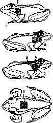
of the tail muscles), development of the lateral folds and the nictitating membrane. The second category includes the formation of the tympanic membrane (Fig. 1), particularly its elastic part, and the perforation of the opercular membrane, through the opening of which the forelimbs are released during metamorphosis, having previously been located in the branchial cavity. The latter phenomenon has been studied repeatedly; at present, it can no longer be admitted that the formation of a perforation in the wall of the branchial cavity is determined only by factors localized outside this wall, specifically in the gills, as Gelf believes, or in the skin glands of the forelimb, as Weber thinks. Cross transplantation experiments of the skin of the peribranchial area to the back and of the skin of the back to the site of future perforation with sufficiently prolonged observation showed the following: the skin that undergoes perforation during metamorphosis has local specificity, as it is perforated even when transplanted to the back; however, these specific properties of the skin itself are not sufficient for the typical perforation in size and form, for which the influence of some agents acting on this skin is necessary. It is highly probable that such an agent is the products of resorption of the gills, since the transplantation of a piece of resorbing gills under the skin of the back leads to the histolysis of the dorsal integuments, which has already been shown by Gelf.
The transplantation method in the study of the function of endocrine glands has been used for a very long time. One of the first experiments that laid the foundation for clarifying the significance of the sex glands for the development of sexual characteristics was the research of Berthold, showing that autotransplantation of the testis to a castrated rooster prevents the disappearance of the comb and sexual instinct in him. Further successes in the study of the development of sexual characteristics are also entirely connected with the transplantation method (see Sex). Likewise, the transplantation of other endocrine glands has provided substantial help in studying their physiological and formative significance (see Endocrinology). The application of the transplantation method encounters a whole series of difficulties, especially in higher vertebrates, since even homoplastic transplantations are not successful in all cases. A number of studies was therefore directed toward clarifying the causes of the existing failures, attempting to take into account the blood groups of the donor and recipient, and to determine the role of the possible formation of specific antibodies. Despite the encouraging results of some experiments, the question is by no means yet considered resolved. Figure 1. Formation of the tympanic membrane in Gelf's experiments: 1-tympanic membrane; 2-scar from the incision when removing the cartilage; 3-skin transplant from the area of the tympanic membrane, transferred to the back; 4-transplant of skin from the back to the site of the tympanic membrane.
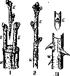
Figure 2. Various methods of grafting in plants: /- grafting proper; //- copulation; III- budding; V-scion; W-stock. (According to Strasburger.)
Transplantation in plants, called grafts, have been known since ancient times and are used to achieve rapid propagation of the shoot of a cultivated plant variety on a wild plant, which serves as the rootstock and provides the root system for the first. The transplanted part of the cultivated plant is called the scion, the wild plant that receives the graft is called the stock. The methods of transplantation in plants, namely proper grafting, copulation and budding, differ in the size of the scion and the method of its connection with the stock (Fig. 2).
Related articles
Mentioned in
Cite this page
“Transplantation.” Soviet Medical Encyclopedia. English translation of Bolshaya Meditsinskaya Entsiklopediya, 1st ed. (Moscow, 1928–1936), ed. N. A. Semashko. https://sovietmedicalencyclopedia.pages.dev/article/transplantation/