Fusion
Historical document, translated for reference. It reflects medical knowledge of the 1920s–30s and is not medical advice.
Summary
This article from the 1930s Soviet medical encyclopedia discusses the concept of fusion (conplantation) as a transplantation technique connecting significant sections of two organisms or entire organisms, with examples from various species including hydras, worms, and amphibians.
Encyclopedia article (1928–1936)
FUSION (conplantation), a term proposed by Isaev to denote such transplantations (see) when it is a matter of connecting together significant sections of two organisms or of connecting two entire organisms in pairs. Fusion of the latter type received the name parabiosis (see). Fusion in animals was first carried out by Trembley (Trembley) in 1774 in work with hydras. Trembley succeeded in fusing pieces of the same individual and pieces of different individuals of the same species, i.e., auto- and homoplastic conplantations.
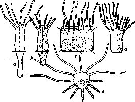
Figure 1. Sectoral chimeras in hydras: a-Pelmatohydra oligactis; b-Hydra vulgaris; c-operation of fusion; d-chimeras from the side; e-chimeras from above. (From Isaev.)
Subsequently, a number of authors succeeded in heteroplastic conplantations on hydras. In the experiments of Wetzel, attempts at fusion of the brown hydra (Hydra fusca) with the green hydra (Hydra viridis) remained unsuccessful. The connected pieces stuck together for some time, but then separated. On the contrary, fusion of Hydra fusca and H. grisea succeeds very well. The connected parts as a rule retained their characteristic pigmentation. Isaev performed two types of fusion between the common hydra (H. vulgaris) and the gray stalked hydra (Pelmatohydra oligactis). In one series of experiments, each hydra was cut lengthwise and unfolded into a flat piece; two such pieces from different hydras were placed together and kept in contact for some time, after which connection occurred and a single organism was formed, colored gray on one side and having long tentacles on that side, and colored reddish on the other side with shorter tentacles (fig. 1). Subsequently, certain regulatory changes occurred, and the number of tentacles decreased. In another series of experiments, Isaev passed an individual of one hydra species inside an individual of another species. For some time the coloration of the fused parts was preserved, but then the reddish parts were as if displaced by the gray ones. This replacement, however, does not mean the complete disappearance of the properties of the common hydra (Hydra vulgaris) in the fused individual. When such fused hydras budded, they gave rise to some offspring completely identical to the normal gray hydra and some that carried the characteristics of the fused parent. If we denote normal gray hydras by the letter P, and the intermediate ones, called by Isaev "oligactoids," by the letters PH, then the offspring of the fused hydra can be schematically denoted as: Generations I II III IV V P P P P P Average hydra 1 PH <^PH PH The results presented indicate that after fusion, with the apparent disappearance of the properties of one of the partners, cellular elements of both partners remain in the fused organism, which are revealed during budding. The presence of these elements was shown in a number of experiments by Goetsch with hydra fusion, and the cellular elements of one of the partners could migrate through the organism of the other partner, reaching areas sufficiently distant from the site of fusion. In addition to experiments on hydras, successful results were obtained from fusion of worms. For such experiments, different species of ciliated flatworms and oligochaete roundworms were used. T. Morgan, in experiments with the planaria Bipalium kewense, obtained fusion of large pieces of worms by compressing them between plates, which achieved contact of the wound surfaces. L. Morgan used for experiments the planaria Phagocata gracilis and Planaria maculata. Success in fusion was achieved by placing pieces between moist strips of very thin paper, the ends of which were fixed with pieces of glass and pins. L. Morgan discovered that with careful connection of parts, regeneration does not occur, and the fused organism remains unchanged. With unsuccessful connection of pieces, regeneration begins on the surface of the cut, and the fused individuals separate. The condition for maintaining fused individuals in connection with each other is the connection of the ends of their nervous system. If this condition is not met, then along the course of the nerve cords, regeneration begins with subsequent separation of the partners. Fusion experiments in annelid worms were carried out on different species of earthworms. Jost sutured pieces of the earthworm Lumbriculus rubellus and Allobophora terristris auto-, homo-, and heteroplastically. He obtained various combinations such as "short" worms, in which the middle part was cut out and the front and back ends were fused; "long" worms, composed of the front and back end of one worm and the middle of another; the front or back ends were fused with each other; T-shaped conplantations were performed, etc. Heteroplastic fusions succeed with great difficulty, however, in Jost's experiments, a worm obtained by fusion of pieces of Lumbriculus and Allobophora lived for more than 8 months, and the parts retained their characteristic species-specific features. The influence of one partner was not even observed during the regeneration of the other. Thus, during regeneration of a part of Lumbriculus, the regenerating part retained the typical pale coloration, and the darkly colored part of the fused individual, taken from Allobophora, had no effect on the regenerate of the part taken from Lumbriculus (fig. 2). Crampton fused pupae of butterflies, cutting them in different directions,
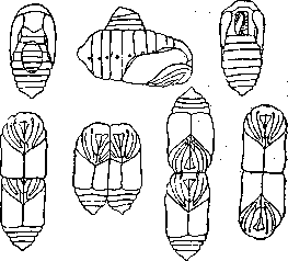
Figure 3. Fusion of butterfly pupae, cut in different directions, and their various parts. (From Crampton from Korschelt.)
folding together the cut surfaces and pouring the connection site with paraffin (fig. 3). From such fused pupae, it was sometimes possible to raise butterflies; mutual influences were manifested in the fact that the metamorphosis of the fused pieces occurred simultaneously, but1changes in coloration usually did not occur; each part in heteroplastic conplantations as a rule retained its own characteristics. However, in Crampton's experiments there are also such data: the posterior part of the abdomen of the pupa Callosamia promethea was fused with the pupa Samia cecropia and in the butterfly it took on the coloration of Samia; the same was observed when a piece of the pupa of the latter species was fused with the pupa Telea polyphemus. The circulation of the hemolymph of the larger partner may, in Morgan's opinion, be the cause of such a change in coloration. It should be noted that in Crampton's experiments, only the coverings were fused, while the internal organs remained separated. Fusion experiments were also carried out on vertebrate animals, and particularly demonstrative results were obtained on amphibian tadpoles. First, these studies were carried out by Born, extracting embryos from the egg membrane. He cut them into two parts and then, bringing the wound surfaces into contact, held the pieces of tadpoles together with silver wires. Homoplastic fusions in Born's experiments succeeded easily, however, fusion of Rana esculenta with R. fusca or R. arvalis is also quite possible. Connections of parts of R. esculenta and Bombina bombina are very short-lived, as is the connection of embryos of tailed and tailless amphibians (Triton taeniatus and Rana esculenta). In
cases of successful connections, internal organs fused—nervous, digestive, and circulatory systems, and the tadpole, normally
feeding by
swallowing. Completed the rear half of Rana syl
transformation into a frog vatica with the posterior half
skin. Effective het- of Rana palustris. (From Harri- eroplastic conpla- son from Korschelt.)
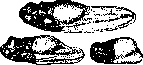
nations were carried out on amphibians by Harrison. By fusing the anterior part of the embryo Rana sylvatica with the posterior part of the embryo R. palustris, he observed the preservation of each part's characteristic coloration (fig. 4). In most fusion experiments, the authors came to the conclusion about the absence of influence of one partner on the other in terms of the development of morphological characteristics. At the same time, the developmental processes of the fused individuals proceeded synchronously (butterflies, tadpoles), which indicates the unquestionable presence of mutual influences. The same is indicated by the above-mentioned results of some of Crampton's experiments. It should be noted that many of the relevant works cited here are presented insufficiently detailed, without the necessary account of possible interactions and require re-investigation. The method of conplantations promises to yield much of value in solving a number of fundamental problems of biology concerning the properties of the organism as a whole, the connection of its parts, their relative autonomy, their local specific features, etc.
Related articles
Cite this page
“Fusion.” Soviet Medical Encyclopedia. English translation of Bolshaya Meditsinskaya Entsiklopediya, 1st ed. (Moscow, 1928–1936), ed. N. A. Semashko. https://sovietmedicalencyclopedia.pages.dev/article/fusion/