Parabiosis
Historical document, translated for reference. It reflects medical knowledge of the 1920s–30s and is not medical advice.
Summary
Parabiosis refers to the surgical connection of two organisms to study their mutual influences through shared circulatory systems. This experimental method was used to study humoral influences, including hormonal effects and toxin transmission between the connected organisms.
Encyclopedia article (1928–1936)
PARABIOSIS (from the Greek para- beside and bios- life), a term having two meanings. 1. The union of two organisms for the purpose of studying their mutual influences through the mediation of the circulatory and lymphatic systems. Parabiosis experiments were performed on mammals, birds, and amphibians through direct cross-connection of blood vessels
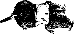
Figure 1. Rats immediately after the parabiosis operation.
by suturing the edges of skin incisions or establishing communication between the peritoneal cavities of the partners (figs. 1 and 2). In the latter case, the skin, muscles, and peritoneum were sutured layer by layer. In birds, parabiosis was performed by suturing the skin of the forearms, which had been plucked (fig. 3). The first acute parabiosis experiment was conducted by Frederick, who studied the effect of blood chemistry on the activity of the respiratory center. In two dogs, the circulatory systems were connected so that blood from the carotid artery of the first dog flowed to the head of the second and vice versa. If the trachea of one of the partners was clamped and thus the carbon dioxide content in the blood was increased, the other dog began to breathe more frequently. This form of acute physiological experiment, in which the connection between individuals is brief, does not make it possible to trace the influence of one individual on another in cases where indicators of the response do not react as quickly to humoral stimulation as the respiratory center in Frederick's experiments. For purposes of long-term observation, the joining of "29 partners by one of the above-mentioned methods was used. With all methods of parabiosis, the resulting connection of the blood vessels of the partners, whether through the fusion of capillaries or the connection of lymphatic vessels, often led to undesirable results for the experimenter. Many laboratory animals react to being joined by the death of one of the partners. The most sensitive in this respect are guinea pigs, the least sensitive are white rats. Experiments on rats are the most complete. The possibility of establishing a connection between the circulatory systems of animals joined for parabiosis is demonstrated both by direct macro- and microscopic examination of the point of union and by the fact that certain substances pass from the blood of one partner into the blood of the other. Thus, in 1909, Ranzi and Ehrlich, Friedberger and Nasetti showed that during immunization of ONE OF THE partners with bacterial toxins, the corresponding antitoxins could be detected in the other. Similar facts regarding the passage of antitoxins against cancer through the bloodstream were established by Rous and later against rat sarcoma by Matsuyama. Zappelioni (1910) tested the effect of tetanotoxin on parabiotic rats. In one series of experiments, tetanotoxin was injected into the muscles of a limb, in another - under the skin of the abdomen of one of the partners. In all cases, regardless of dosage, which ranged from maximum to minimum in a ratio of 10:1, the partner not directly exposed developed tetanus 2-3 days later and correspondingly died later. Morpurgo, studying the effect of kidney removal in one partner on the development of uremia in the other, established that the latter did not occur in all cases, i.e., for the most part, the kidneys of the unoperated partner were able to perform the function of serving both organisms. The presence of a common circulatory system led in a number of cases to circulatory disturbances, with one partner being in better conditions of blood supply than the other. Sauerbruch and Heide were the first to point out these relationships (Sauerbruch, Heide, 1908). In the experiments of these authors, such parasitism of one partner on the other was particularly noticeable when the paired rabbits were of different sexes, obviously due to the different power of the heart in the female and male. Morpurgo in his experiments on rats basically confirms the conclusions of Sauerbruch and Heide. The pumping of blood from one individual to another in parabiosis is particularly clearly shown in the experiments of M. Zavadovsky. In his work on parabiosis in roosters, in a number of joined pairs, a distinct pumping of blood from one rooster to the other occurred. This was associated with a significant increase in the size of the organs of the "full-blooded" and "bloodless" rooster. The latter died within 8-15 days (fig. 4). The parabiosis method was used by a number of authors to study hormonal influences and, in particular, to clarify the effect of sex hormones on the development of sexual characteristics. Pioneers in this field were Sauerbruch and Heide, but they failed to obtain clear results. In Morpurgo's experiments, 6 pairs of parabiotic rats of different sexes lived for more than 3 months. During this time, no noticeable changes in sexual characteristics could be noted. The testes and penis of parabiotic males were no smaller than those of the controls. The nipples of parabiotic females remained as well developed as those of the controls. Matsuyama (1921), who conducted parabiosis experiments on white rats, connected the skin, muscles, and peritoneum layer by layer, creating communication between the peritoneal cavities. The results of Matsuyama's work regarding changes in the sexual sphere of parabiotic rats are as follows. The gonads of opposite-sex pairs undergo degeneration. Degeneration of the testes occurs particularly early. Degeneration of the ovaries begins somewhat later and is expressed in their cystic transformation.
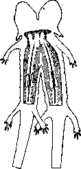
Figure 2. United Amblystoma larvae. On the
opened areas, the ovaries of both partners are visible. (From Björn.)
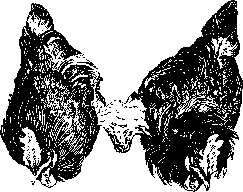
Figure 3. Roosters sutured along the forearm (according to Zavadovsky).
Figure 4. On the left - heart, spleen and liver of the "full-blooded" rooster, on the right - the "bloodless" rooster. In the result
of this, the organs of the "bloodless" rooster atrophied.
' as a result of this, the female in P. with the male becomes incapable of conception. Indeed, in one case fertilization of such a female occurred, but the pregnancy was interrupted before term, and defects in placenta formation were discovered. Autopsy revealed deep cystic degeneration of the ovaries. The male, joined with the pregnant female, showed extreme degeneration of the testes. One can therefore think that the humoral influences emanating from the body of the pregnant female have an even more depressing effect on the male sex gland than on the non-pregnant one. Very interesting materials were obtained by Matsuyama as a result of experiments on P. of normal and castrated animals. When a female was joined with a castrate, she remained infertile, and the ovaries showed a picture of cystic degeneration. The male, joined with a castrate, experienced degeneration of the testes. N. Goto (K. Goto, 1925) performed very extensive work on P. in rats. Of the 206 pairs he joined, 78, i.e. 38%, lived for more than a month; one pair lived for 2 years 4 months. The sex of the animals did not have a significant effect on survival. Thus, heterosexual pairs survived in 39% of cases, while homosexual pairs in 37%. Animals from the same litter gave somewhat better results (41% survived) than from different parents (37% survived). The operation is more successful on immature rats than on adults. Communication between the blood vessels begins on the 5th day of P. The study of P. of normal animals with castrates leads Goto to conclusions that differ substantially from those of Matsuyama. P. of a normal or semi-castrated female with a castrate leads to sharp changes in her sex organs. These changes consist in hypertrophy of the ovaries, proliferation of follicles, sometimes in the formation of cysts and a large number of yellow bodies. The changes occurring in the uterus, the author considers secondary and dependent on disorders in the ovaries. They reduce to hypertrophy of the mucous membrane, sometimes hydrometra or pyometra occur. In P. of a normal male with a castrate, hypertrophy of the seminal vesicles and prostate is noted in the first. Histological examination of the testes gave no indications of any deviations from normal. Based on the data presented, Goto asserts that in the blood of castrated rats there is a special substance, named by him 'castrahormone'. This 'castrahormone' directly or indirectly causes irritation of the ovaries and their hypertrophy. By joining a normal female with a normal or castrated male, Goto, in agreement with the data of Matsuyama, found atrophy and degeneration of the sex glands in both partners: namely, cessation of spermatogenesis, resp. oogenesis. A special study of changes in the active mesenchyme associated with the parabiotic state was undertaken by Sauerbruch (1923). He established that with prolonged P., the multiplication of lymphoid elements increases, and particularly clear changes are noted in the liver reticulo-endothelial elements. The author interprets these changes as a result of mutual influences of the organisms joined in P., in particular as a reaction to the constant introduction of foreign proteins and metabolic products into the bloodstream. This exchange of blood may have a non-specific depressing effect on the activity of the sex glands, which was noted in the interaction of individuals of different sexes and in P. of normal animals of both sexes with castrates. In experiments with P. of mammals, it was not possible to establish a specific influence of sex hormones on the development of sexual characteristics in a partner of the opposite sex or in a castrate partner. Such a change in sexual characteristics, particularly of the sex glands themselves, was observed under conditions that could be called natural P., namely in the so-called free-martin of heifers (see Hermaphroditism, - hermaphroditism in animals). An attempt to understand these phenomena is represented by experiments conducted on amphibians, in which fusions are easily performed on embryonic and larval stages. Already Harms (Harms, 1911) showed that in P. in amphibians, the sex hormone passes from one partner to another and affects its sexual characteristics. A castrated male in P. with a normal one retained his morphological secondary sexual characteristics (nuptial pads) and mating instinct. Burns (1925) conducted experiments on P. in embryos of amblystoma (Amblystoma punctatum). The operation was performed at the stage immediately following the closure of the medullary groove. Of the operated animals, 80 pairs survived, and all pairs turned out to be of the same sex (44 pairs of males and 36 pairs of females). The sex of the embryos being joined could not be determined in advance, so the distribution of sexes in such experiments should follow the law of chance. In other words, one would expect such a ratio: 1♂♂: 1♂♀: 1♀♂: 1♀♀, i.e. there should be as many heterosexual pairs as homosexual ones. The discrepancy between the experimental results and the theoretical calculation can be interpreted either as a consequence of selective mortality of heterosexual pairs or as a consequence of the transformation of one sex into another. Burns stops at the second explanation, believing that as a result of P. of heterosexual individuals, half of the females turned into males and half of the males into females. Since histological observations of Burns gave no indications of the phenomenon of transformation of the sex glands, his conclusions remained unconvincing. Very clear results in similar experiments were obtained by Witschi (Witschi, 1927), who studied the results of P. in the American tree frog (Hyla silvatica). He performed pairwise fusion of embryos at the stage following the closure of the medullary groove, i.e. 50-70 hours after the start of development at room temperature. The distribution of sexes in control animals was normal: 100♂ to 96♀; theoretically, 98♂ to 96♀ was expected (probable error ±4.7). Among the 56 surviving parabiotic twins, one could expect such a distribution of sexes: 14♂♂: 14♂♀: 14♀♂: 14♀♀ (probable error ±2.2). Microscopic examination of all 56 pairs gave results very close to expected: 16♂♂:17♂♀:10♀♂:13♀♀. However, microscopic examination of the gonads showed that of the 17 pairs where the left was male and the right female, 7 females showed a clear tendency to transform into males. The same picture was given by 4 females out of 10 pairs where the left was female and the right male. In no case was such a transformation of the male sex toward the female observed. Witschi believes that his results satisfactorily explain the phenomenon of free-martin, where also the transformation of sex goes only from ♀ to ♂, if one assumes that the male sex hormone in mammals and amphibians dominates over the female one. It is necessary, however, to note that in amphibians the transformation of one sex into another is accomplished with great ease under the influence of various influences. In mammals, the relationships are more complex, and such an extrapolation can hardly be accepted unconditionally.
Related articles
Mentioned in
Cite this page
“Parabiosis.” Soviet Medical Encyclopedia. English translation of Bolshaya Meditsinskaya Entsiklopediya, 1st ed. (Moscow, 1928–1936), ed. N. A. Semashko. https://sovietmedicalencyclopedia.pages.dev/article/parabiosis/