Decerebration (DECEREBRATE)
Historical document, translated for reference. It reflects medical knowledge of the 1920s–30s and is not medical advice.
Summary
This article from the 1928–1936 Great Medical Encyclopedia describes the surgical procedure of decerebration and the resulting decerebrate rigidity. It details the historical background by Sherrington, surgical techniques including tracheotomy and brain stem transection, and the physiological characteristics of extensor and flexor tonus.
Encyclopedia article (1928–1936)
DECEREBRATION, DECEREBRATE RIGIDITY (from Latin de-, negative particle, and cerebrum, brain). Decerebration is the surgical operation of transecting the brain stem. Decerebrate rigidity is the muscle rigidity that develops after the influence of the red nucleus on the lower divisions of the brain is eliminated. Decerebration of an animal was first performed by the English physiologist Sherrington. The operation
is performed as follows: after putting the animal to sleep with ether or chloroform, the trachea is exposed and transected halfway. A tracheal tube is inserted into the peripheral incision, the other end of which is connected to an artificial respiration apparatus. Air from this apparatus is passed through a narcotic substance and therefore enters the lungs together with the vapors of the latter, which subsequently maintains the animal's anesthesia. After tracheotomy, vagotomy is performed, i.e.,
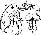
transection of both vagus nerves. In this way, cardiac arrest resulting from excitation of these nerves at the moment of brain transection is avoided. Then, the carotid arteries are ligated to prevent massive hemorrhage during skull opening and brain transection. After the operation on the neck, decerebration is begun. At the time of the operation, the animal must be in deep anesthesia; strong compression of the paws, ears, or touching the corneal reflex should not produce reflex movements. Having made sure of deep anesthesia, the skin is incised along the midline of the head and the temporalis muscle is removed on both sides. Then a hole is made with a trephine in the region of the temporal bone, and the upper part of the cranial vault covering the cerebral hemispheres is removed with bone forceps. The dura mater is removed from the exposed brain, and finally the entire brain stem is transected with a blunt spatula in front of the tentorium, in the midbrain region. The entire anterior part of the brain is removed from the cranial cavity, and a little cotton is placed inside. To avoid massive hemorrhages during the opening of the cranial cavity and brain transection, blood flow in the vertebral arteries must be temporarily stopped: the assistant compresses them by squeezing the neck with the thumb and index finger behind the first cervical vertebra (Fig. 1). When passing from the second vertebra to the first, the vertebral arteries lie superficially on the sides of the vertebral body, so they can be compressed at this place. After decerebration, they are no longer compressed, and anesthesia is stopped. To prevent blood from flowing out of the brain, the head is raised upward for 10-15 minutes. The animal must be artificially warmed with a warm cloth or other means, since it lacks normal thermoregulation. Artificial respiration is stopped only after good normal respiration is restored. A few minutes after decerebration, as the animal awakens from anesthesia, extensor tone develops, i.e., decerebrate rigidity. Figure 1. A - position of the cat's head during decerebration and level of brain transection (I-V); t - middle tooth of the tentorium cerebelli; h - hyoideum; 1, 2, 3 - 1st, 2nd, and 3rd cervical vertebrae. The course of the vertebral arteries is shown on the side of the 1st vertebra. The position of the ligature for their ligation is also shown there by dashed lines. B - the first three vertebrae from above. The position of the ligature for compressing the vertebral arteries is clearly visible. (After Sherrington.) In this form, the operation proceeds in rabbits, cats, dogs, and monkeys. Characteristics of decerebrate rigidity. The rigidity developing immediately after the operation is of an extensor character: all those muscles contract that must protect the body or parts of it from falling due to gravity. Their antagonists simultaneously experience inhibition. As a result, all limbs are extended, the head and neck are raised upward toward the dorsal side, the tail is bent in the same direction, and the spinal column is also arched in this direction. During this tonus, defensive flexion and scratching reflexes occur with lesser intensity. This happens because the flexor muscles are in a state of strong tonic inhibition. The less blood that flowed out during the operation and the less narcotic substance used, the stronger the extensor rigidity and the longer it lasts (Sherrington, Magnus). A few hours after the operation, extensor rigidity weakens and comes to naught. Then, instead of it, flexor tonus and flexor rigidity develop. The limbs are bent, the head, neck, and tail are lowered downward toward the ventral side (Beritov). In prolonged experiments, this shift occurs a few days later (Bazett, Penfield). Origin of decerebrate rigidity. All researchers unanimously state that decerebrate rigidity does not develop when the brain is transected in front of the optic thalami (incision I in Fig. 2), nor if the incision passes at the anterior boundary of the corpora quadrigemina (incision II in Fig. 2). It occurs only upon transection of the corpora quadrigemina and medulla oblongata behind the red nuclei (incisions III, IV, and V, Fig. 2). But for the development
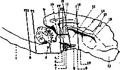
Figure 2. Brain of a cat indicating the sites for transection. Transection along lines I and II does not disturb normal tonus. Transection along lines III, IV, and V produces decerebrate rigidity. Transection along line VI or VII no longer produces decerebrate rigidity; 1 - III cervic.; 2 - II cervic.; 3 - n. VIII; 4 - pons; 5 - gangl. interpedunc.; 6 - nucl. ruber magnocellul.; 7 - n. oculomot.; 8 - nucl. ruber parvocellul.; 9 - corpus mamillare; 10 - tr. optic.; 11 - bulb. olfact.; 12 - corp. striatum; 13 - corp. callosum; 14 - ventric. III; 15 - thalamus; 16 - columna fornic.; 17 - corp. quadrig. ant.; 18 - corp. quadrig. post. (After Magnus.)
of decerebrate rigidity, it is not necessary to transect the entire brain stem; it is sufficient if the incision affects only those pathways that emerge from these nuclei to the spinal cord, i.e., the rubrospinal tract in the region of their decussation.
Related articles
Cite this page
“Decerebration (DECEREBRATE).” Soviet Medical Encyclopedia. English translation of Bolshaya Meditsinskaya Entsiklopediya, 1st ed. (Moscow, 1928–1936), ed. N. A. Semashko. https://sovietmedicalencyclopedia.pages.dev/article/decerebration/