Vagus Nerve
Historical document, translated for reference. It reflects medical knowledge of the 1920s–30s and is not medical advice.
Summary
The vagus nerve, the tenth cranial nerve, has diverse functions with corresponding anatomical substrates including sensory, motor, and parasympathetic nuclei. It innervates numerous organs in the head, neck, thorax, and abdomen, with parasympathetic fibers generally having effects opposite to those of the sympathetic nervous system.
Encyclopedia article (1928–1936)
VAGUS NERVE (wandering nerve), belongs to the X pair of cranial nerves. Varied in its functions, it has for each of them a corresponding anatomical substrate in the form of nuclei: there is a sensory nucleus, a motor nucleus, and a parasympathetic nucleus (parasympathetic division—see below; also Vegetative Nervous System). Sensory fibers of the V. n. originate in the cells of two ganglia: gangl. jugulare, located near the foramen jugulare, and gangl. nodosum s. plexiforme, located below. The ganglion jugulare is a mixed ganglion,
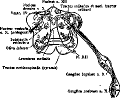
N. vagus and its nuclei in the medulla oblongata.
having relation to both sensitivity and the vegetative system; the second one—exclusively to sensitivity. The processes of the cells of these ganglia, as well as of the spinal cord, divide into peripheral and central; the latter, as part of the root of the V. n., are directed into the medulla oblongata and enter the fis. retro-olivaris; in the medulla oblongata the fibers are directed backward and, without reaching the floor of the fourth ventricle, enter the composition of fasc. solitarii, where they divide into short ascending fibers and long descending fibers, which on their way downward constantly send collaterals to the cells of nucl. terminalis (gelatinous substance located near fasc. solitarius behind and outside). Fascic. solitarius, gradually thinning, ceases to be distinguished as a separate bundle at the level of the pyramidal decussation and is lost in the upper cervical segments of the spinal cord. According to some authors, nucl. terminalis continues to the floor of the fourth ventricle, where it occupies the outer part of alae cinereae; its inner part is occupied by the parasympathetic nucleus of the V. n., near the cells of which the sensory fibers end—reflex. From the sensory nuclei originates the central sensory neuron of the V. n.—The motor, or anterior nucleus of the V. n. lies in the medulla oblongata, in the lateral column, in substantia reticularis; it consists of two parts—nucleus ambiguus (see Ambiguus nucleus) and a special lateral group of the anterior nucleus of the V. n. The motor nucleus appears in the lower parts of the medulla oblongata and extends to its upper parts. The cells of this nucleus are of large size, their axons are first directed backward, connect with the sensory fibers, then turn outward and forward and exit from the medulla oblongata through fis. retro-olivaris.—All fibers of the V. n.—centrifugal and centripetal—exit or enter the medulla oblongata through 10-15 roots, from which a common trunk is formed, exiting through for. jugulare from the cranial cavity; on its path there are two ganglia, about which was said above; when it exits for. jugulare, it receives from n. XI ramus internus, then lies between art. carotis interna and vena jugularis interna and in this position descends into the thoracic cavity; this part of it belongs entirely to the parasympathetic system. In accordance with the extensive area of distribution of the branches of the V. n., it is divided into sections: cervical, neck, thoracic, and abdominal.—The cervical section of the V. n. extends from the medulla oblongata to gangl. nodosum; at this level from it depart ramus meningeus, going into the cranial cavity to the meninges, to sinus occipitalis and transversus; ramus auricularis, innervating the external auditory canal and the posterior surface of the auricle. The boundary of the neck section is the exit of n. laryngei; from the neck section depart: anastomoses to n. XII; rami pharyngei, innervating the muscles of the pharynx, velum palatinum and m. uvulae (part of the fibers of these branches has a vegetative function); ram. lingualis n. vagi, going to the tongue; n. laryngeus superior, which divides into ramus externus and internus; the first is motor, innervates m. constrictor pharyngis inferior and m. crico-thyreoideus, the second is sensory, extends in the mucous membrane of the epiglottis and larynx, to the vocal fissure; n. laryngeus inferior s. recurrens (see), supplying with its branches the larger part of the laryngeal muscles, as well as giving fibers to the heart.—In the thoracic and abdominal sections of the V. n., in addition to motor fibers (smooth muscle), vascular and secretory, there are also sensory fibers for the respiratory tract, for the gastrointestinal tract and for the muscles innervated by the V. n., including cardiac. (See fig. 14-18 in the article Vegetative Nervous System, as well as figures in articles on individual organs.) The motor nuclei of the V. n., innervating the striated muscles of the pharynx, larynx, soft palate and upper part of the esophagus, represent simple motor centers of the medulla oblongata, muscle centers similar to those in the spinal cord; their function is reduced to the function of these muscles. Due to the termination near them of sensory fibers or their collaterals, they perform simple reflexes (e.g., contraction of the soft palate under the influence of irritation of the uvula); in addition, they participate in complex combined movements, such as swallowing (see), in the formation of voice, in the act of breathing (see), in the act of vomiting, etc. As a sensory nerve, the V. n. innervates the occipital parts of the dura mater, the external auditory canal, the posterior surface of the auricle, the soft palate, pharynx, larynx. As for the sensitivity of internal organs, the question still remains controversial, which fibers, sympathetic or cerebrospinal, innervate them.—Knowledge of the anatomy and physiology of the V. n. indicates the symptoms that will be observed when the V. n. or its nuclei are affected. More severe symptoms occur when the motor nuclei are affected; they lead to disturbance of swallowing, speech, articulation and breathing; the combination of these symptoms is highlighted under the name of bulbar symptoms, which occur in a number of diseases: bulbar paralysis, amyotrophic lateral sclerosis, Landry ascending paralysis, poliomyelitis (see), in progressive paralysis, in diseases of peripheral nerves with involvement of the V. n. (see Polyneuritis), in involvement of the root of the V. n. in various meningitides (see). The appearance of bulbar phenomena in the above-mentioned diseases makes the prognosis in most cases hopeless.
E. Kononova.
The parasympathetic part of the V. n. The vagus nerve, together with n. oculomotorius, n. glossopharyngeus and n-vi sacrales, forms, according to the terminology of Langley, the so-called parasympathetic system. The parasympathetic part has both centrifugal (to the larynx, pharynx, esophagus, stomach, heart, bronchi, lungs) and centripetal (n-vus depressor cordis, sensory fibers of the lungs and bronchi) fibers. The table on page 339 shows the functions of the centrifugal fibers of the V. n. and the effect of their sectioning on the corresponding organs. Centripetal fibers give a reflex decrease in the tone of vasoconstrictors and excitation of the centers of the V. n.: vasodilating (?), cardiac and respiratory. In all places of its distribution, the V. n. has on the innervated organs an action opposite to the action of the corresponding endings of the sympathetic nervous system. This antagonism is proved not only by the opposite physiological purpose of these nerves, but also by their different reaction to the poisons of the vegetative nervous system: the endings of the V. n. are excited by poisons of the choline group (choline, acetyl-choline, muscarine, physostigmine, pilocarpine) and paralyzed by atropine. The mentioned poisons have an elective action on parasympathetic nerve endings and, according to the creators of the classification of the vegetative nervous system, on sympathetic endings. Organ Heart. Bronchi. Esophagus. Gastrointestinal peristalsis. Stomach and pancreas glands. Liver. Kidney. Thermoregulation. Action of irritation V.n. Decrease in the number of sinus contractions per minute, worsening of conductivity and contractility, arrest in diastole, narrowing of coronary vessels. Predominantly constricting action. Spasm of the lower part. Gastric peristalsis is enhanced. Spasm of the middle part of the stomach, opening of pylori, enhancement of pendulum movements of the intestines and their tone. Enhancement of secretion, mainly for the pancreas. Enhancement of glycogen breakdown (?). Increase in the amount of urine and its solid constituents. Action of sectioning Tachycardia. Dilation. Dilation of the lower part, spasm. Intestinal peristalsis is preserved. Loss of reflex excitability of glands. secretion. Inconstant effects. Thermoregulation is disturbed (overheating). endings do not act. Recently, the opinion has been expressed that there are no specific vagotropic and specific sympathotropic poisons and that the action of any vegetative poison is amphotropic, and depending on pH and the ratio of ions in the tissue fluid, a given poison acts, predominantly, on one or another system. (See Vegetative neuroses.)
Related articles
Mentioned in
Cite this page
“Vagus Nerve.” Soviet Medical Encyclopedia. English translation of Bolshaya Meditsinskaya Entsiklopediya, 1st ed. (Moscow, 1928–1936), ed. N. A. Semashko. https://sovietmedicalencyclopedia.pages.dev/article/vagus-nerve/