Thoracic Cavity
Historical document, translated for reference. It reflects medical knowledge of the 1920s–30s and is not medical advice.
Summary
This article from the 1928–1936 Soviet Great Medical Encyclopedia details the anatomy, boundaries, and contents of the thoracic cavity. It covers the development of the coelom, pleura, mediastinum, and the arrangement of vital organs within the chest.
Encyclopedia article (1928–1936)
THORACIC CAVITY (cavum pectoris) is enclosed within the rib cage, the walls of which, lined by the endothoracic fascia (fascia endothoracica), bound it anteriorly, laterally, and posteriorly. Inferiorly, the thoracic cavity is separated from the abdominal cavity by the diaphragm, which projects into it in the form of a dome (see Diaphragm). Because of this, as well as the fact that a medially located ridge formed by the bodies of the thoracic vertebrae projects into the cavity, the shape of the thoracic cavity does not completely correspond to the external shape of the rib cage, and its overall dimensions are smaller than those of the latter. The thoracic and abdominal cavities develop from a single common cavity, the coelom, and in lower vertebrates are not yet separated. In
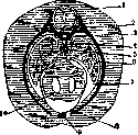
Figure 1. Cross section through a rabbit embryo: 1 - spinal cord; 2 - rib; 3 - notochord; 4 - aorta; 5 - esophagus; 6 - lungs; 7 - pleural cavity; 8 - pericardial cavity; 9 - sternum; 10 - heart. (From Hertwig.)
mammals, the separation of the coelom into cranial and trunk parts occurs with the development of the diaphragm (see figure 1). The pericardial sac, containing the heart primordium, develops from the cranial part. The descent of the latter entails the isolation of the cardiac sac and causes lateral protrusions that subsequently turn into pleural sacs with their cavities. The developing lungs project into them from both sides, pushing the medial layers of the pleural sacs ahead of them. Thus, the thoracic cavity contains two pleural cavities (cavum pleurae dextrum et sinistrum) housing the lungs, and the cavity of the cardiac sac (cavum pericardii), located between the previous two and containing the heart. Underdevelopment of the diaphragm, most commonly its left dome, results in a connection between the thoracic cavity and the peritoneal cavity; correspondingly, the pleural cavity in this case contains various parts of the abdominal viscera. (On other malformations see Thoracopagus, Thoracoschisis). The medial layers of the pleural cavities bound throughout the entire extent the space between the anterior surface of the vertebral bodies and the posterior surface of the sternum and costal cartilages (I-VI ribs
Figure 2. Cross section
through the mediastinum at
the level of the sixth thoracic
vertebra: 1 - azygous vein and right
sympathetic trunk; 2 -
esophagus and right vagus nerve;
3 - right bronchus; 4 - superior
vena cava; 5 - pericardial cavity;
6 - internal mammary vessels;
7 - retrosternal adipose
tissue; 8 - semilunar valves of
the pulmonary artery; 9 -
semilunar valves of the aorta;
10 - trunk of the pulmonary artery;
11 - left bronchus; 12 - descending
aorta and left vagus nerve;
13 - hemiazygous vein and left
sympathetic trunk. (After Corning.)
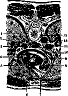
). This space bears the name of the mediastinum (mediastinum), and the parts of the pleura bounding it and embracing the root of the lung are the mediastinal pleura (pleura mediastinalis) (see figure 2). In it are located, besides the ventrally located heart and its sac, the aorta and vessels departing from it, the following organs situated outside the serous cavities, partly covered by the parietal layer of the pleura or pericardium and surrounded by loose connective tissue connecting with the endothoracic fascia: pulmonary arteries and veins, right and left brachiocephalic veins, venae cavae, azygous and hemiazygous veins, arteries departing from the aorta for the esophagus, bronchi, and lungs (see figure 3); thoracic duct, the trunks forming the right lymphatic trunk, and lymph nodes, phrenic nerve, vagus nerves, esophagus, trachea, thymus gland (in children) and fatty tissue; located on the lateral surfaces of the spine (on the costal heads), beneath the parietal pleura, the sympathetic trunk border cords are essentially already outside
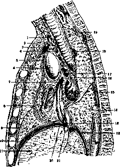
Figure 3. Sagittal section at the level of the entry of the venae cavae into the right atrium: 1 - brachiocephalic artery; 2 - manubrium of the sternum; 3 - left brachiocephalic vein; 4 - anterior mediastinal lymph nodes; 5 - reflection of the pericardium onto the anterior wall of the ascending aorta; 6 - severed convexity of the ascending aorta; 7 - superior vena cava; 8 - right wall of the right atrium; 9 - middle lobe of the lung; 10 - diaphragm; 11 - right lobe of the liver; 12 - peritoneal reflection on the posterior wall of the liver in the region of the caudate lobe; 13 - inferior vena cava; 14 - lower lobe of the lung; 15 - part of the trunk of the azygous vein; 16 - bronchial lymph nodes; 17 - right pulmonary artery and the site of its division into upper and lower branches; 18 - severed arch of the azygous vein; 19 - upper lobe of the right lung; 20 - anterior peritoneal reflection onto the liver; 21 - hepatic vein emptying into the inferior vena cava. (From Shevkunenko.)
the mediastinum. By a conventional frontal plane drawn through the trachea and bronchi, the mediastinum is divided into anterior and posterior parts. In the anterior part are embedded loose adipose and connective tissue, lymph nodes, and the thymus gland in children (or its remnants in adults) with its arteries (branches of the internal mammary artery) and veins (emptying into the brachiocephalic and internal mammary veins). The heart is located partly in the anterior and partly in the posterior mediastinum (see figure 4). The trunks departing from the aortic arch are covered at their origin by more superficially located brachiocephalic veins emptying into the superior vena cava. The latter and the aortic arch, located most superficially, are separated from the anterior wall of the thoracic cavity by means of the anterior costomediastinal recess, and at the level of the II-IV costal cartilages the layers of the pleura on both sides approach one another almost closely. More to the left and somewhat more superficial
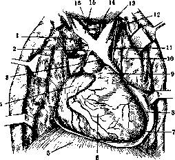
Figure 4. Thoracic cavity (anterior chest wall and anterior wall of the pericardium removed): 1 - trachea and right recurrent laryngeal nerve; 2 - aortic arch; 3 - transitional fold of the pericardium; 4 - right atrium; 5 - diaphragm; 6 - right ventricle; 7 - apex of the heart; 8 - left ventricle; 9 - pulmonary artery; 10 - left phrenic nerve; 11 - left vagus nerve; 12 - left subclavian artery; 13 - left common carotid artery; 14 - left recurrent laryngeal nerve; 15 - brachiocephalic artery; 16 - thyroid gland. (After Corning, with modifications.)
than the aortic arch lies the pulmonary artery. The right phrenic nerve, having entered the thoracic cavity between the subclavian artery and vein, then lies between the superior vena cava and the mediastinal pleura, and still lower between the pericardium and the pleura. The left phrenic nerve is located deeper than the right, but just like it, ventral to the root of the lung and lies between the pleura and the pericardium. Behind the aortic arch is the thoracic part of the trachea, the bifurcation of which falls somewhat to the right and lower than the aortic arch, so that the latter arches over the left bronchus. To the right and left of the trachea, in the region of the bifurcation and inferior to the latter's site, lymph nodes are embedded - right, left, and inferior tracheobronchial lymph nodes. Behind and somewhat to the left of the trachea, at the level of the superior thoracic aperture (see Rib cage), is the esophagus. In the thoracic part, up to the level of the sixth to ninth thoracic vertebrae, it lies against the anterior surface of the vertebral bodies, and at the level of the third to fourth thoracic vertebrae is located to the right of the thoracic aorta. At the level of the eighth thoracic vertebra, the esophagus departs from the spine anteriorly and, penetrating the opening of the diaphragm, lies anteriorly and somewhat to the left of the aorta. Below the tracheal bifurcation, the esophagus lies against the parietal layer of the pericardium. Passing between the main bronchi and the esophagus are the vagus nerves, with the left lying on its left and anterior surfaces, and the right on the posterior. Between the aorta (on the left), azygous vein (on the right), and esophagus (anteriorly), along the spine runs the thoracic duct (see Azygous vein and Thoracic duct). It, as well as the esophagus, is crossed in a transverse direction by the right posterior intercostal arteries departing from the aorta, passing along the anterior surface of the vertebral bodies. The mediastinal pleura only partially covers the esophagus (on the left - a small area above the diaphragm and above the aortic arch, on the right - below the root of the lung). The part of the pleura lining the diaphragm is named the diaphragmatic pleura; the part of the pleura covering the costal region and adjacent to the endothoracic fascia is the costal pleura. All these parts bear the general name of parietal pleura; while the part that is a continuation of the mediastinal pleura onto the lung itself is the visceral pleura. The diaphragmatic pleura passes into the costal pleura at the bottom of the cleft formed by the diaphragm and the costal wall. This cleft, lined thus by the layers of both pleurae, bears the name of the costodiaphragmatic recess or phrenicocostal sinus (see figure 5). During quiet breathing, the recess does not open, and the lung does not fully fill the pleural cavity. Only during a deep breath, when the diaphragm drops significantly, does the edge of the lung enter the recess, which thus turns from a cleft into a cavity. The lower boundaries of the recess are essentially simultaneously the lower boundaries of the pleural cavity and the lateral and partly anterior and posterior regions of the thoracic cavity and vary depending on the shape of the rib cage: with a narrow and long shape of the rib cage they run lower than with a short and wide one, while anteriorly they are higher. In 56% of cases the boundary of the recess on the left is higher than on the right (Melnikov), in 42% - lower, and in 2% - identical. The mediastinal pleura, passing
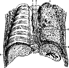
Figure 5. Frontal section along linea axillaris media (right lung removed): 1-v. azygos; 2-section of the esophagus; 3-aorta; 4-lymphoglandulae hili pulmonis; 5-lien; 6-sinus costo-diaphragmaticus. (From Shevkunenko.)
Anteriorly and posteriorly into the rib, it also forms two sinuses on each side-sinus costo-mediastinalis. Their boundaries anteriorly, as well as the position of the organs of the T. c. in general, vary depending on the shape of the thoracic cage (see Pleura, Thoracic Cavity).- The upper wall of the T. c. proper does not exist. In addition to the organs that pass through the superior aperture of the thoracic cavity, the upper part of the parietal pleura (cupula pleurae) also extends into it on each side, which is filled by the apex of the lung and in the middle protrudes above the clavicle by 3-5 cm (varies greatly). The pleural cavity under normal conditions is filled by the lungs, so that the visceral pleura is adjacent to the parietal, and on the surface of the lungs there remain indentations from the ribs, large vessels (aorta, a. subclavia, a. carotis communis sin., v. cava superior), heart and esophagus; however, the sinuses are filled, and not completely, only with forced deep inspiration (see Lungs). The pericardium represents, like the pleural sacs, a closed serous cavity, into which the heart is inserted. Thus, here too a distinction is made between the pericardial leaflet covering the heart itself (pericardium viscerale) and the leaflet forming the outer wall of the cavity, which anteriorly connects with the loose fatty and connective tissue to the posterior surface of the sternum, on the sides-with the mediastinal pleura, posteriorly-with the thoracic aorta and esophagus, and below fused with the superior surface of the diaphragm (see Heart).
Related articles
Mentioned in
Cite this page
“Thoracic Cavity.” Soviet Medical Encyclopedia. English translation of Bolshaya Meditsinskaya Entsiklopediya, 1st ed. (Moscow, 1928–1936), ed. N. A. Semashko. https://sovietmedicalencyclopedia.pages.dev/article/thoracic-cavity/