Diaphragm
Historical document, translated for reference. It reflects medical knowledge of the 1920s–30s and is not medical advice.
Summary
The diaphragm is a dome-shaped muscle separating the thoracic and abdominal cavities, with complex anatomy including tendinous and muscular parts, and crucial functions in respiration and circulation.
Encyclopedia article (1928–1936)
168 Anatomy and development of the D. Diaphragm, dia-phragma (from Greek diaphragma - partition), is the thoraco-abdominal barrier separating the thoracic cavity from the abdominal cavity. The diaphragm has an irregular shape of two domes (fig. 1 and 2) with a small saddle-shaped depression in the center for the heart - the cardiac fossa (planum cardiacum). The D. consists of a tendinous center (centrum tendineum) and a muscular part (pars muscularis s. carnosa). The tendinous center forms the entire surface of the domes and cardiac fossa, remotely resembles in outline a cloverleaf and is built from intertwining
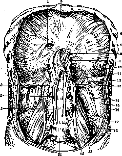
Figure 1. Diaphragm from below and in front: 1-medial crus of diaphragm; 2-intermediate crus; 3-lateral crus; 4-foramen of vena cava; 5-sternal part of diaphragm; 6-costal part of diaphragm; 7-tendinous center; 8-esophageal hiatus; 9-lumbar part of diaphragm; 10-aortic hiatus; 11 and 12-medial and lateral lumbo-costal arches; 13-lumbo-costal triangle (Bochdaleki); 14-m. quadratus lumborum; 15-m. transversus abdominis; 16-m. obliquus internus abdominis; 17-m. obliquus externus abdominis; 18-m. iliacus; 19-m. psoas major; 20-m. psoas minor; 21-body of V lumbar vertebra. (After Spalteholz.)
tendinous and elastic fibers. In the muscular part of the D. are distinguished: the sternal part (pars sternalis), the costal part (pars costalis) and the lumbar part (pars lumbalis). The sternal part is the weakest, individually variable, begins with bundles from the inner surface of the xiphoid process of the sternum (processus xiphoideus sterni). The costal part begins with bundles from the inner surface of the cartilages of VII - XII ribs between the attachment sites of the bundles of the transverse muscle of the abdomen. The bundles of the D. rise steeply upward; between them and the wall of the thoracic cavity there is a narrow costodiaphragmatic space (sinus costodiaphragmaticus); it is lined with parietal pleura and changes in shape and size during respiration. The lumbar part consists of three crura.-Of these, the medial crus (crus mediale) begins from the body of the last thoracic and lumbar vertebrae (up to III on the left and up to IV on the right) and weaves its tendon fibers into the anterior longitudinal ligament of the spine (lig. longitudinale vertebrarum anterius); the intermediate crus (crus intermedium), the weakest, begins
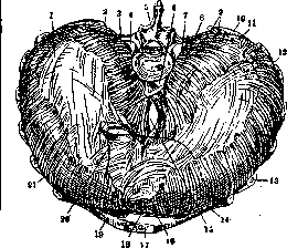
Figure 2. Diaphragm from the side of the thoracic cavity: 1-tendinous center; 2-lateral lumbo-costal arch; 3-II lumbar vertebra; 4-opening for m. psoas; 5-I lumbar vertebra; 6-aortic hiatus; 7-medial lumbo-costal arch; 8-intermediate crus; 9-lateral crus; 10-XII rib; 11-lumbo-costal triangle (Bochdaleki); 12 and 21-costal part of diaphragm; 13-VII rib; 14-pericardium border (marked with dots); 15-esophageal hiatus; 16-sterno-costal triangle (Larrey); 17-body of sternum; 18-sternal part of diaphragm; 19-VI rib; 20-foramen of vena cava. (After Braus.)
from the body of the II lumbar vertebra; finally the lateral crus (crus laterale) takes its origin from the so-called Gallus' tendinous arches; of these one (medial) is stretched from the body of I (or II) lumbar vertebra to its transverse process (arcus lumbo-costalis medialis s. arcus psoatis), and the second (lateral)-from the transverse process of I (or II) lumbar vertebra to the free edge of the XII rib (arcus lumbo-costalis lateralis s. arcus quadrati lumborum). From under the medial arch emerges the major psoas muscle (m. psoas major), and from under the lateral- the quadratus lumborum muscle (m. quadratus lumborum). In the D. there are a number of openings for vessels, nerves and some organs. Thus, between the tendon bundles of the medial crura and the spine - the opening (hiatus aorticus) for the aorta, sympathetic peri-aortic plexus (plexus periaortalis) and the lymphatic thoracic duct (ductus thoracicus); above it, between the intertwining medial crura - the opening for the esophagus (hiatus oesophageus), as well as both Ml vagus nerves. In the tendinous part of the D. - the opening for the inferior vena cava (foramen venae cavae), as well as for branches of the thoraco-abdominal nerve (rami phrenico-abdominales nervi phrenici). Between the medial and intermediate crura pass: veins - the azygos (v. azygos) on the right and the hemiazygos (v. hemiazygos) on the left, the greater splanchnic nerve (n. splanchnicus major) and the lesser splanchnic nerve (n. splanchnicus minor), which sometimes pierces the intermediate crus; between the intermediate and lateral crus lies the trunk of the sympathetic nervous system (truncus sympathicus). Further, in the D. are distinguished between the muscle fibers the tendinous fields: the sterno-costal triangle (trigonum sterno-costale, s. trigonum Morgagni, s. trigonum Larrey) and the lumbo-costal (trigonum lumbo-costale, s. trigonum Boehdaleki). The first of them are pierced by blood vessels (vasa epigastrica superiora).-The D. is covered on the side of the abdominal cavity over most of its extent by the parietal sheet of peritoneum, which is separated from the D. by retroperitoneal fat; peritoneum is absent only where the coronary ligament of the liver departs from the D. and where the duodenum, pancreas, kidneys and adrenal glands adjoin the D., as well as around the openings of the esophagus and vena cava; below the D. are also adjacent the liver, stomach and spleen. On the side of the thoracic cavity, the parietal sheet of pleura covers the D. only where the lungs adjoin it, and in the costodiaphragmatic sinuses. Between the D. and pleura is connective tissue (fascia endothoracica), and where the pericardial sac adjoins the D., only very dense connective tissue firmly fuses them together. The topographic position of the diaphragm is extremely variable (figure 3): in corpses - depending on age and pathological processes, and in the living, in addition, from respiratory displacements of the D., from the condition of the organs of both cavities and finally from constitution. On average, in the normal state, the following is accepted: the lower borders of the D. correspond to the attachment sites of the D. to the thoracic wall, the apex of the right dome is at the IV, and of the left - at the V intercostal space, the cardiac fossa - at the level of attachment of the fifth ribs to the sternum (figure 4). During respiration, the cardiac fossa and both domes move extremely slightly (no more than 2-3 cm), but the shape of the lateral parts of the diaphragm mainly changes: the chest expands, and the reserve costodiaphragmatic spaces increase in size, into which the expanding lungs rush. The D. develops quite complexly: first its connective tissue basis is formed, and then muscular elements grow into it. In turn, the basis of the D. develops in its anterior part from the transverse septum (septum transversum), separating from the ventral and lateral walls of the general body cavity of the embryo; the posterior part of the basis of the D. develops from the dorsal wall of the pleuro-peritoneal canals connecting the pleural and abdominal cavities of the body in the embryo. The muscular elements grow from the muscle primordia of the IV and V cervical muscle segments (myomeres), of which apparently the first gives the costal-sternal part of the D., and the second - the lumbar part. In its development, the D. descends from the cervical region along with the heart and lungs. Correspondingly to this, in case of underdevelopment of the D., slits remain in it, and through them diaphragmatic hernias can pass. Comparatively-morphologically, the D. is a new formation and is present in full development only in mammals. The diaphragm receives blood from branches of the subclavian artery (a. pericardiaco-phrenica and a. musculo-phrenica from a. mammaria int.), from the thoracic aorta (a. phrenica superior) and from the abdominal aorta (a. phrenica inferior).-The veins of the diaphragm run parallel to the arteries and drain blood in three directions: from the lower surface - into v. cava inferior, mostly with two trunks at the point where it pierces the D.; from the upper - mostly through vv. pericardiaco-phrenicae and musculo-phrenicae into vv. mammariae anteriores and in part - from the area of the medial crura into vv. azygos and hemiazygos. In addition to these drainage pathways, there are rich anastomoses with vv. intercostales of the lower six intercostal spaces, in part with vv. lumbales ascend
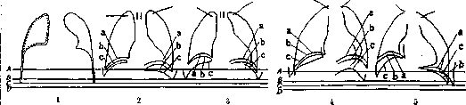
Figure 3. Diaphragm: 1-on a corpse; 2-in a living person in a standing position; 3-in a living person in a lying position; 4-when lying on the left side; 5-when lying on the right side; a-position of the diaphragm during expiration; b-during quiet inspiration; c-during deep inspiration. The horizontals A, B, C, D represent the position of the shadow of the diaphragm on the body surface in the living: A-during expiration (on the left side) during quiet breathing (beginning of the reserve space and costodiaphragmatic angle in position a); B-the lowest point reached by the lungs in a normal person (beginning of the reserve space and costodiaphragmatic angle in position c); C-the lowest point of pleurae parietalis (end of the reserve space); D-beginning of the diaphragm on the ribs. In fig. 1 pleura parietalis is shown as a solid line, pleura visceralis-as a dotted line. In fig. 2-5 the position of the clavicles is indicated. (From radiogram from Braus.)
B. M. E. vol. IX. dentes, in part with vv. oesophageae; and finally all three of the above venous systems anastomose with each other. The lymphatic vessels of the D. are connected with the lymphatic system of the thoracic and abdominal
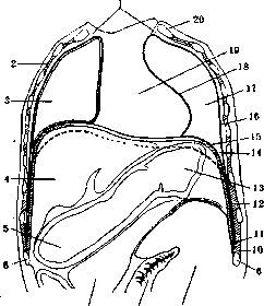
7 8 9
Figure 4. Frontal section through the trunk of an adult human, from the front (schematically): 1-apertura thorac. sup.; 2-intercostal muscles; 3-right lung; 4-liver; 5-duodenum; 6-10th rib; 7-pancreas; 8-small intestine; 9-colon descend.; 10-attachment of the diaphragm to the chest wall; 11-pleural reflection; 12-sinus phrenico-costalis; 13-stomach; 14-costodiaphragmatic angle; 15-pleura diaphragm.; 16-pleura costalis; 17-pleural cavity (lung removed); 18-pleura mediast.; 19-mediast. ant.; 20-1st rib. (After Braus.) cavities. From the upper surface of the diaphragm, lymph by means of supradiaphragmatic glands is directed to the sternal, anterior and posterior mediastinal and intercostal glands, from which it goes either into the right lymphatic duct or into the thoracic duct; from the lower intercostal glands into receptaculum chyli. From the lower surface of the diaphragm, lymph through the upper para-aortic, pre-aortic and esophageal lymphatic glands is directed into receptaculum chyli. Finally, owing to the presence of a third system of lymphatic ducts piercing the diaphragm, a close anatomical connection is established between the subpleural and subdiaphragmatic spaces. This connection explains the possibility of inflammatory processes passing from one to the other, in particular - the formation of pleural effusions in subdiaphragmatic abscesses and paranephritis in purulent pleurisy on the same side. The routes of infection transmission in these cases may be either the area of Boddal's triangle (see above) or the aforementioned 3rd system of piercing lymphatic currents. innervation. The innervation of the diaphragm has been studied only recently through the works of Felix, Goetze, and Kutomanov. This innervation is carried out by the phrenic nerve (n. phrenicus, fig. 5), intercostal and sympathetic nerves. The question of whether the n. phrenicus is the only motor nerve of the diaphragm, as found by Russel, Fuchs, and others, or whether other nerves also participate in the motor function, remains open. Felix gives the following scheme: the middle part, adjacent to the spine, is supplied by the phrenic nerve and the sympathetic; on both sides of this area a relatively small area is supplied with motor fibers of the XII intercostal nerve, the rest of the diaphragm is innervated only by the phrenic nerve (fig. 6). The operation of severing the phrenic nerve in humans (phrenicotomy) has recently made it possible to firmly establish the presence in it of centripetal sensory fibers. An increase in blood pressure, a change in the rhythm of respiration and finally irradiating pains in the shoulder are constant symptoms of acute irritation of the phrenic nerve. The sympathetic fibers of the n. phrenicus are received from the two lower cervical sympathetic ganglia and sometimes from
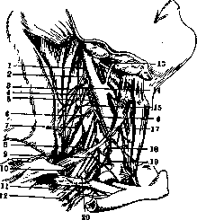
Figure 5. The phrenic nerve and its origin: 1-anastomosis between II and III cervical nerves; 2-external branch of n. accessorii; 3, 7 and 15-m. trapezii; 4-ramus auricularis magnus; 5-ramus cervicalis transversus; 6-ram. subclavicularis; 8-n. trapezius et rhomboideus; 9-n. subclavicularis; 10-n. scapul. sup.; 11-ram. inf. n. pectoralis; 12-n. pectoralis minor; 13-n. hypoglossus; 14-anastomosis between n. cervic. III and n. accessorius; 16-descending branch of plexus cervicalis; 17-n. phrenicus; 18-plexus sympathicus a. cervicalis; 19-plexus sympathicus a. scapul. sup.; 20-n. pectoralis sup. (After Hovelacque.)
the first thoracic; they form the 'suprapleural plexus' (plex. suprapleuralis), located on the dome of the diaphragm. In addition, from below the diaphragm receives sympathetic fibers from the solar plexus. Experiments by Ken Kure showed that severing these sympathetic fibers leads to degeneration of the diaphragmatic muscles, whereas severing the spinal nerves causes atrophy from inactivity. In addition to the phrenic nerve, the diaphragm is supplied with sensory fibers from the last six intercostal nerves (VII-XII).
D. Romashov. The function of the diaphragm can be divided into two parts: static and dynamic. The static function of the diaphragm has as its purpose to equalize the difference in intracavity pressures of the thoracic and abdominal cavities and thereby under normal conditions to prevent the pulling of abdominal organs into the thoracic cavity. This function of the diaphragm is in a close and direct dependence on the correct coordination of three factors: positive pressure in the abdominal cavity, negative pressure (force of retraction of the lungs) in the thoracic cavity and the tone of the diaphragm itself, which, according to experiments by Duchenne, is very significant and can resist great force. The dynamic function of the diaphragm is in close dependence on the tone of the diaphragm and includes three separate functions: respiratory, cardiovascular and motor-digestive. 1. The respiratory function of the diaphragm, considered in former times as the most important and significant function of the diaphragm, at present can no longer be recognized as such. Since the introduction of phrenicotomy as a method of excluding the function of the diaphragm, it could be ascertained that exclusion of the diaphragm does not threaten the respiratory function of the lungs. Even bilateral exclusion of the diaphragm does not affect the respiratory function of the lungs at all (Dunner). Persons with a pronounced asthenic constitution breathe almost entirely without the diaphragm, and according to the assertion of Mosso, in men during sleep there is exclusively thoracic (costal) type of breathing. 2. The cardiovascular function of the diaphragm is expressed in the fact that during inspiration both the pericardium and the lowest part of the superior vena cava lying in it expand. At the same time, the lowering of the diaphragm and the simultaneous increase in intra-abdominal pressure squeeze blood from the liver into the inferior vena cava, which promotes the constant outflow of venous blood into the right heart. 3. The motor-digestive function of the diaphragm is the most diverse. In relation to the esophagus, the diaphragm has great importance in that with every inspiration the esophageal opening of the diaphragm narrows almost to complete closure of the esophageal lumen, with every expiration it expands again and thus acts as an actual sphincter of the esophagus. On the stomach, the diaphragm exerts a significant influence in the sense of periodic movements of it, synchronous with the respiratory movements of the diaphragm. Great importance also has, on the one hand, the constant accumulation of air in the cardiac part of the stomach as a result of inspiratory sucking in of air ('aerospiration'), on the other hand - the removal of excessively accumulated air by means of belching (contraction of the abdominal press with simultaneous strong inspiratory lowering of the diaphragm and open cardia). The diaphragm acts similarly on the intestine. At the moment of expiration, gases rush into the splenic flexure, at the moment of inspiration - they move toward the exit opening. Similarly, the diaphragm aids defecation, significantly enhancing with its inspiratory lowering the action of the abdominal press. Finally, mention should be made of the massage action of the diaphragm on the bile ducts and especially on the gallbladder. This should also include the completely similar action of the diaphragm on the renal pelvis. Methods of studying the diaphragm. Anamnesis has almost no significance. At
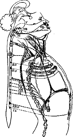
Figure 6. Schematic representation of respiratory innervation. The respiratory center in the substantia reticularis extends from the level of calamus scriptorius to the level of atlas. Excitations reach it from the intermediate brain, from the posterior quadrigemina, from trigeminus (V) (nasal mucosa) and from the pulmonary branches of the vagus. From the respiratory center, centrifugal impulses go through n. facialis (VII) to the musculature which raises the wings of the nose and opens the mouth, through n. vagus (X) (n. recurrens) to the musculature of the larynx, through n. phrenicus to the diaphragm, through intercostal nerves to the intercostal musculature and through the upper lumbar nerves to the musculature of the abdominal wall. To n. phrenicus are attached sympathetic fibers from ganglion stellatum, and therefore from gangl. suprapleurale, which together with it reach the diaphragm. The diaphragm, in addition, receives a nerve bundle and ganglion cells from n. splanchnicus, and through p. Solaris sympathetic fibers and groups of ganglion cells also reach the lower surface of the diaphragm. (From Müller.)
By palpation, the respiratory excursions of the diaphragm (D.) can be traced by examining the liver and spleen, which almost exactly repeat the movements of the D. Percussion makes it possible to determine the boundary between two adjacent organs separated by the D. Observation of the phenomenon of Gerhardt-Litten (a linear circular shadow in the area of the lower edge of the lungs, which with each inspiration moves downward) makes it possible to establish the mobility of the D. The most important method of examination is the X-ray, which allows easy study of the shape, position, mobility and tone of the D. On X-ray examination, the D. appears as two arched lines, the right one higher and the left one lower, separated from each other by the shadow of the heart. The right arch, and sometimes also the left, sometimes consists of two or three intersecting arcs, depending on the uneven contraction of individual muscle bundles of the D. and giving the D. a festoon-like shape (Maingot's phenomenon, see separate table, fig. 5). The right arch is visible throughout its entire length, while the left is visible only in the part lying over the stomach and the splenic flexure of the colon (or the spleen). The height of the position of both domes varies in different people, but generally decreases somewhat with age. During breathing, both halves descend and rise uniformly and synchronously depending on the respiratory phase. The size and direction of their excursions depend on the type of breathing, on the physique and on the development of the musculature. The dependence on musculature is expressed in the fact that in people with weak musculature, the movements of the D. generally have a much smaller range. The physique affects in the sense that in people with asthenic build and with general ptosis, the D. hardly participates in respiratory movements. With the abdominal type of breathing, the D. lowers during inspiration and rises during expiration, with the size of its excursions depending on the strength of breathing, but its anterior parts (the weak sternal part) make significantly smaller excursions than the stronger posterior parts (lumbar), which is especially well seen on lateral irradiation. With purely costal breathing, on irradiation in the posteroanterior direction, one can see how the dome of the D. rises during inspiration, although by a small amount, upward. However, on lateral irradiation it is seen that only the weak sternal part of the D. rises upward together with the chest wall, while the stronger lumbar part descends downward during inspiration. With the most common mixed type of breathing, the inspiratory phase begins with abdominal breathing, as a result of which the D. descends downward, but by the end of the inspiratory phase, costal breathing predominates, as a result of which the D. rises upward together with the chest wall. During expiration the opposite occurs, so that in general the D. makes 4 movements during a complete cycle: down-up-down-up. The functional capacity of each half of the D. can be determined by X-ray examination by Müller's test (inspiration with the glottis closed, in which the weakened half of the D. makes a larger upward excursion than the healthy one) or by the phenomenon of expiratory acceleration of the movement of the D. (Dillon), in which after maximum inspiration the weakened half of the D. rushes upward even before the start of expiration or during expiration moves upward faster than the healthy one. Pathology of the D. The pathological condition of the D. can be the result of a violation of the coordination of the above factors of its normal state: intra-abdominal pressure, retraction of the lungs and the tone of the D. itself. The result of changes in the first two factors will be one or another change in the position of the D. with the general preservation of its dynamic function, and this change can be both unilateral and bilateral. Thus, the D. stands higher than normal with any increase in intra-abdominal pressure (ascites, meteorism, general obesity, tumors of the abdominal cavity, pregnancy in the last weeks) or with a decrease in lung retraction (bronchostenosis, cirrhosis of the lungs, pleural adhesions). Conversely, the D. stands lower than normal in case of decreased abdominal pressure (enteroptosis) or in case of increased intrathoracic pressure (pneumothorax, emphysema). With a change in the tone of the D. itself, not only a change in the position of the D. occurs, but also a violation of its function. In rare cases, there may be an increase in the tone of the D. (tetany, hiccups), but in most cases there is a decrease. The latter may depend on weakening of the muscle in a disease of the D. or when inflammation spreads from the diaphragmatic pleura or from the peritoneal covering to the muscle itself - 'acute paralysis of the diaphragmatic muscle' by Ohm. Most often, however, the loss of tone of the diaphragm depends on paralysis or paresis of the phrenic nerve or can be artificially caused by its sectioning or tearing out for therapeutic purposes in diseases of the lungs (see separate table, fig. 4). An inevitable consequence of the loss of tone of the D. will be its displacement upward (the intra-abdominal pressure exceeding the intrathoracic). However, on the right side this will be prevented by the heavy liver with its ligaments, while on the left side, on the contrary, this will be facilitated by the gas-containing organs lying under the D. (stomach and splenic flexure of the colon), which seek to displace the D. upward. The result of such an upward displacement of the D. will be a displacement of organs of both the chest cavity (esophagus, lungs, heart) and the abdominal cavity (stomach, splenic flexure). The most pronounced form of such a disease, almost always left-sided, is the so-called diaphragmatic eventration (more correctly - unilateral persistent elevation of the diaphragm - see separate table, fig. 6). The main signs of this disease are a high-arching D., a large amount of gas in the stomach and splenic flexure, aortic heart shape, its displacement to the right, in far advanced cases deformation of the stomach and paradoxical movements of the affected half of the D. The displacement of the D. and neighboring organs in rare cases can occur without any sensations for the patient; for the most part, however, it gives a whole series of sometimes extremely severe phenomena, which can be reduced to three groups. 1. Group of pleuropulmonary phenomena: they are usually the least severe and are expressed by chest pain, shortness of breath and cough. 2. Group
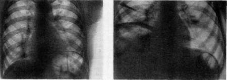
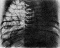
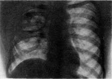
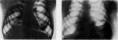
Figure 1. Paralysis of the left half of the diaphragm due to acute infectious inflammation of the phrenic nerve in a young woman. Figure 2. Subdiaphragmatic gas abscess. High position of the right half of the diaphragm. Figure 3. Congenital left-sided diaphragmatic hernia in a newborn. Figure 4. Paralysis of the right half of the diaphragm due to phrenicotomy. Figure 5. Festooned diaphragm (on the right) (Maingot's phenomenon). Figure 6. Unilateral persistent elevation of the diaphragm (so-called eventratio diaphragmatica).
See article Diaphragm. Cardiovascular phenomena, the most severe, sometimes reaching the force of true anginal attacks: heaviness, pressure, and pain in the heart area, shortness of breath, bradycardia, sometimes tachycardia, extrasystoles, and pulse arrhythmia, often disappearing immediately after belching. 3. Group of gastrointestinal phenomena (the most numerous). These include phenomena from the esophagus, expressed in dysphagia, periodic or constant, sometimes in the form of paradoxical dysphagia, as Leichtenstern named it. Phenomena from the stomach: belching, sometimes with a huge amount of air, pain in the stomach area, nausea, vomiting, sometimes ulcerous symptoms (bloody vomiting). Phenomena from the intestines: feeling of fullness, bloating, especially in the left epigastric area, sometimes persistent constipation as a result of the air lock that forms in the splenic flexure (Dillon). The described symptoms are not characteristic and therefore, before the introduction of X-ray research, were attributed to various diseases. Only since the time of Roentgen, the number of described cases of unilateral persistent elevation of the Diaphragm has increased so much that this elevation can be considered a not uncommon disease. Its origin should be attributed to paralysis or paresis of the phrenic nerve, which most often occurs as a result of some infection (see separate table, fig. 1). Sometimes this was also observed in newborns, in whom, as a result of some obstetric operation, an injury was inflicted on the phrenic nerve. A very similar "gastrocardiac symptom complex" was described in 1912 by Roemheld. However, the phenomena he described, also far from rare, although they depend on the abnormal position of the Diaphragm, are the result not of a lesion of the Diaphragm itself, but of pathological fermentation in the digestive tract in the presence of cardiac neurosis. Similar symptoms can also be caused by a subphrenic abscess, in which there is a decrease in the functional capacity of the Diaphragm, on the one hand due to the spread of the inflammatory process to the Diaphragm, on the other hand due to sometimes very significant displacement of the Diaphragm itself by the abscess (see separate table, figure 2).-Mention should be made of acquired or congenital defects of the Diaphragm, the so-called diaphragmatic hernia (see separate table, fig. 3). This disease can proceed completely asymptomatic or gives symptoms that cannot be distinguished from the symptoms of the above-described unilateral persistent elevation of the Diaphragm. Only careful and comprehensive X-ray research makes it possible to make a correct differential diagnosis. Among the most reliable differential diagnostic signs are the paradoxical movement of the Diaphragm during the Mueller test and the expiratory acceleration of the movement of the Diaphragm in the Dillon phenomenon. As for diaphragmatic hernia, both acquired and congenital, the only correct method of treatment is surgical intervention, which gives complete success. Surgical intervention is also equally successful in the case of subphrenic abscess.-As for other diseases of the Diaphragm, which are the result of loss of its tone and causing its displacement upward, in such cases treatment should pursue 3 tasks. 1. It should develop and strengthen the diaphragmatic muscle, since it has not yet completely lost its tone and has not undergone atrophy; for this, it should be systematically exercised and the patient should be persistently trained in the sense of correct breathing with the help of the Diaphragm. 2. It should contribute to the reduction of gas accumulation under the Diaphragm. For this, one should take care of proper gastric and intestinal digestion by prescribing an appropriate diet. Especially, one should avoid the consumption of any nutritional substances containing a large amount of fiber that easily causes fermentation. 3. Finally, with reasonable hygienic measures, the patient should be accustomed to the least possible burden on the digestive tract and especially avoid drinking hot liquids, with which not only is air swallowed, but also a large amount of air is sucked into the stomach. All these measures give the patient considerable relief. However, where the Diaphragm has completely lost its tone and, due to complete atrophy, has turned into a thin, inelastic membrane,-the mentioned measures can hardly help the patient, and then one should also resort to surgical intervention, which has been successfully performed on more than one occasion.
я. dilatation. Injuries to the D. are either penetrating (open) or subcutaneous (closed). The most common causes of penetrating injuries are stab, incised, and gunshot wounds. Stab and incised wounds are most frequently found on the left side, as they are inflicted by the attacker's right hand (most often with a knife or dagger). It is interesting to note that in most cases the wound is inflicted from top to bottom, while in the statistics of Italian surgeons (Rossi) a direction from bottom to top is noted. The skin wound is located mostly in the VII, VIII, and IX intercostal spaces on the lateral surface of the body. The size of the wound in the D. varies from a few mm to a huge opening 15 cm long. Gunshot wounds to the D., which were particularly common during the imperialist war, also vary greatly in size. Especially severe are wounds inflicted in the lying or standing position (projectile dropped from an airplane). In this case, the wounding projectile, passing through the length of the body, may carry with it fragments of bones shattered by it, causing huge lacerated wounds. The clinical picture of wounds to the D. depends on the size of the wound and the nature of associated injuries. An isolated uncomplicated injury to the D. may proceed with almost no symptoms pathognomonic for this type of injury, or may give only minor symptoms depending on irritation of the n. phrenici: pain on deep inspiration, radiating to the shoulder. For this reason, such injuries to the D. are often not diagnosed, and treatment is limited to suturing the skin. Such errors often result in the development of diaphragmatic hernias. An injury to the D. accompanied by prolapse of internal organs (most often the omentum) is diagnosed much more easily. A completely different clinical picture must be observed with injuries to the D. of larger size, often complicated by prolapse of abdominal organs (stomach, liver, spleen, intestines) and kidneys into the thoracic cavity and their injury. In this case, there is lag of the corresponding half of the D. during breathing, tympany on the corresponding half of the chest, increased participation of the intercostal muscles in breathing, pain and a feeling of pressure in the chest, retention of gas and feces in the absence of peritonitis symptoms, shock, displacement of the heart (in left-sided injuries). Injury to the liver, spleen, lung, or kidney, causing profuse internal bleeding, usually quickly leads to the death of the patient brought to a medical facility in extremis. According to the statistics of Iselin (iselin), 63.9% of cases of complicated injuries to the D. are fatal, while in uncomplicated injuries to the D. a fatal outcome was observed in only 16.9% of cases. Subcutaneous (closed) injuries - ruptures of the D. - occur under the influence of external violence (compression of the chest or upper abdomen by car buffers, cartwheels or automobile wheels, falling from a great height), more rarely - from a sudden increase in intra-abdominal pressure (labor contractions, vomiting movements). The symptoms are similar to those in penetrating injuries, but the absence of a wound makes diagnosis even more difficult and thus worsens the prognosis. Here again, the nature of the clinical picture depends on the degree of damage to the diaphragm and the greater or lesser involvement of abdominal organs in the process. The difficulty of accurate diagnosis, dangerous complications both immediate (death from bleeding, peritonitis) and delayed make surgical intervention mandatory at the slightest suspicion of injury to the D. The best approach is provided by transpleural laparotomy. - The operation for an uncomplicated injury to the D. consists of suturing the opening in the D.; the prolapsed internal organs require inspection and reduction into the abdominal cavity. Injury to organs of the abdominal and thoracic cavities significantly complicates the operation and sometimes forces one to abandon the transpleural route and switch to ordinary laparotomy through the anterior abdominal wall. Damaged organs are treated according to the general rules of abdominal surgery depending on the nature of the organ and the degree of injury. Sauerbruch, who has extensive material on injuries to the D. both in peacetime and in wartime, notes a decrease in mortality depending on the expansion of indications for surgical intervention at the slightest suspicion of injury to the D. - Inflammatory processes of the D., being almost always secondary, depend on the spread of the process from a neighboring organ of the thoracic or abdominal cavity and the kidney; only rarely do transfers occur via lymphatic pathways from distant sites; these include acute inflammatory processes (pleurisy, subdiaphragmatic abscess) and chronic (tuberculosis, actinomycosis, syphilis), as well as echinococcus. Being only secondary complications of diseases of neighboring organs, these diseases, lacking their own specific symptomatology, do not require special description. What has been said now applies in full to new growths of the diaphragm, which are always secondary, originating from neighboring organs and tissues.
Related articles
Mentioned in
Cite this page
“Diaphragm.” Soviet Medical Encyclopedia. English translation of Bolshaya Meditsinskaya Entsiklopediya, 1st ed. (Moscow, 1928–1936), ed. N. A. Semashko. https://sovietmedicalencyclopedia.pages.dev/article/diaphragm/