Epigastrium
Historical document, translated for reference. It reflects medical knowledge of the 1920s–30s and is not medical advice.
Summary
The epigastrium is the upper region of the abdominal cavity, corresponding to the anterior abdominal wall. This article describes its anatomical boundaries, musculoskeletal structure, blood supply, innervation, clinical significance, and relationship to underlying organs.
Encyclopedia article (1928–1936)
EPIGASTRIUM, epigastric region, represents the upper portion of the abdominal cavity, in projection onto its anterior-lateral wall. More precisely, the boundaries of the epigastrium are defined: inferiorly by a horizontal line connecting the anterior ends of the X ribs, superiorly by the projection onto the anterior wall of the diaphragm (fig. 1). By two vertical lines bisecting the Poupart ligaments, the epigastrium is usually divided into 3 parts: 2 lateral-regions hypochondriacae and 1 median-regio epigastrica (epigastric region). In relation to the spine, the entire region as a whole corresponds to the level of vertebrae DVIII-XII and LI. The shape of the epigastrium on one hand depends on the type and form of the thoracic cage (see), in particular on the size of the infrasternal angle (angulus infrasternalis), on the other hand it changes in connection with fat deposition, muscle tension, and changes in the organs contained within it. Excessive obesity, enlargement of organs (liver, spleen, etc.), distension of the stomach and intestines and other causes that increase intra-abdominal pressure contribute to the protrusion of the regio epigastrica above the level of the costal arch and the elevation of the costal part of the epigastrium. Reverse phenomena create its recession. With age, the relief of the region also changes in the direction of recession.-The foundation of the wall of the epigastrium is (fig. 2) the anterior bony
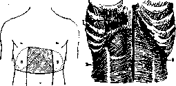
Figure 1. Boundaries of the epigastrium: I-regio epigastrica; z-regio hypochondriaca.
Figure 2. The bony-muscular foundation of the epigastrium: 1-m. rectus abdominis; 2-m. obliquus abdominis internus; 3-m. transversus abdominis; cartilaginous portion of the ribs (on the right V-X, on the left VI-X), xiphoid process, musculature of the intercostal spaces (see Thoracic cage), upper portion of the rectus abdominis muscles, right and left external, internal, oblique, transverse, and finally the linea alba composed of the crossing fibers of the last three muscles (see Abdominal wall). The bony-muscular layer in front has skin and subcutaneous fatty tissue with fascia superficialis, without interruption and not strengthened by the transition from the thoracic region to the abdomen (which allows the unhindered movement of subcutaneous emphysema, hematoma, abscesses, etc., from the chest to the abdomen and back). Behind the muscles and ribs lies fascia transversalis and peritoneum. - In the layer of fasciae superficialis are located the terminal branches of the VI-IX intercostal cutaneous nerves, small subcutaneous arteries from the intercostal and art. epigastrica superior, and a dense network of subcutaneous veins, with anastomoses draining blood upward into v. epigastricae superficiales superiores and through vv. medianae tegmentosae xiphoideae and its branches into vv. mammariae internae, downward-into vv. paraumbilicales and partly into vv. epigastricae superficiales inferiores, to the sides-into vv. thoracicae longae tegmentosae Braun's and through them into vv. axillares (see Caput medusae). The lymphatic outflow is predominantly directed to the axillary lymph glands. The bony-muscular layer is supplied with arterial blood from the system art. mammariae internae by two of its branches: in the isolated bed of the rectus muscles (see Abdominal wall) art. epigastrica superior, passing behind the muscle, in the regio hypochondriaca-the art. musculophrenica running along and behind the costal arch (fig. 3), with its branches art. intercostales anteriores (see Thoracic cage). The venous outflow goes along the same paths in the reverse order, therefore predominantly into the system vv. mammariae internae and from them into vv. apopu-tae. The lymphatic outflow from the bony-muscular portion of the epigastrium is directed into the system of the sternal current and further into lgl. parasternales (along v. and a. mammariae int.); the first stage on this path is the group of lymph glands behind the angle of convergence of the cartilages of VI and VII ribs and 2-3 lymph glands behind the xiphoid process.--The innervation of the muscles of the epigastrium occurs from the intercostal nerves of V-VIII segments (fig. 3). In the region of the epigastrium are contained primarily 4 organs: liver with gallbladder, cardiac portion of the esophagus, stomach, and spleen. Additionally within the boundaries of the epigastrium lie: duodenum, part of the transverse colon, bursa omentalis, pancreas, both adrenal glands (fig. 4), upper
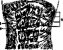
Figure 3. Diagram of the arterial supply and innervation of the epigastrium: 1-a. epigastrica superior; 2-a. musculophrenica; 3-nn. intercostales.
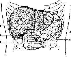
Figure 4. Projection of organs onto the anterior wall
in the region of the epigastrium: 1-liver; 2-gallbladder; 3-duodenum; 4-colon transversum; 5-stomach. pole of the kidneys (mostly left), v. cava inferior, legs of the diaphragm and between them at the level of LI hiatus aorticus and the abdominal aorta emerging from beneath it with its first branches, in particular arteria coeliaca. On either side of the latter are located the nodes of the solar plexus (which explains the irradiating pains "under the xiphoid process"), lgl. coeliacae-nodal lymphatic glands for truncus lymphaticus intestinalis, and finally the beginning of ductus thoracicus with its cisterna chyli (to the right of the aorta in hiatus aorticus).-The clinical significance of the epigastric region corresponds to the importance in pathology of the anatomically related organs. However, access to them for palpation is hindered by the nature of the structure. In cases of pain, the muscular contraction (defense musculaire) of the rectus muscles of the regio epigastrica and the short transverse fibers of the transverse muscle and its aponeurosis create an insurmountable obstacle for palpation and often complicate conditions for differential diagnosis. In a surgical aspect, the transverse direction of the fibers of m. transversi and its aponeurosis complicates suturing in longitudinal median incisions. The narrowness of the regio epigastrica and the low compliance of the ribs sometimes force to supplement the abdominal wall incision with the temporary severing of the cartilages of the costal arch in order to improve access to the deeply located organs in the dome of the diaphragm. Finally, the linea alba regionis epigastricae is often the site of hernia formation (hernia epigastrica), with pains that usually simulate gastric diseases, (see Hernias).
Related articles
Mentioned in
Cite this page
“Epigastrium.” Soviet Medical Encyclopedia. English translation of Bolshaya Meditsinskaya Entsiklopediya, 1st ed. (Moscow, 1928–1936), ed. N. A. Semashko. https://sovietmedicalencyclopedia.pages.dev/article/epigastrium/