Thorax
Historical document, translated for reference. It reflects medical knowledge of the 1920s–30s and is not medical advice.
Summary
The thorax is composed of the thoracic spine, twelve pairs of ribs and their cartilages, and the sternum. It forms a truncated cone shape, with variations in aperture size and shape that affect the structures passing through it.
Encyclopedia article (1928–1936)
THORAX, is composed of the thoracic spine at the back, twelve pairs of ribs and their cartilages at the sides, and the sternum at the front. Usually only the first seven pairs of ribs reach the sternus, more rarely eight; the VIII, IX, and usually X ribs connect with the rib above them by their cartilages and form the so-called costal arch (arcus costarum); the X (sometimes), XI, and XII ribs end freely. The general shape of the thorax can be compared with a truncated cone, flattened in the anterior-posterior direction. The anterior
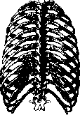
Figure 1. Thorax from behind.
wall (planum sternale), formed by the sternum and adjacent costal cartilages, is shorter than the posterior wall and is inclined to the vertical at an angle of about 20°; the size of the angle varies individually and depending on the phase of respiration (see Sternum). On the posterior wall, a row of spinous processes forms a crest (crista spinalis), on either side of which are two ridges formed by the tips of the transverse processes of the vertebrae (see figure 1). The grooved depression between these ridges is called sulcus dorsalis, between the crest of the transverse processes and the angles of the ribs-sulcus costo-vertebralis minor, between the crest of the spinous processes and the angles of the ribs-sulcus costo-vertebralis major. Each pair of ribs has its own shape and direction, but in general, due to the varying degree of their curvature along the surface, edge, and twisting, all of them are directed (especially starting from angulus costae) from top to bottom and forward in the form of weakly twisted ribbon-like strips, which in the anterior part and in the cartilaginous part rise obliquely upward. The intercostal spaces in the upper and lower parts of the thorax are shorter and wider than in the middle; their widest portion is at the border of the bony and cartilaginous parts of the ribs, the narrowest-in the anterior part. The thorax has two openings-upper and lower. The first-apertura thoracis superior-is formed behind by the first thoracic vertebra, on the sides by the first ribs, and in front by the sternum. Its plane is inclined (forward and downward), at an angle to the horizontal, open backward. Thus the upper edge of the anterior wall is lower than the posterior. The magnitude of this depression equals the height of the entrance to the upper aperture, which in newborns and embryos is almost zero, while in adults it varies considerably (Melnikov). According
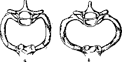
Figure 2. Forms of the upper aperture: b-wide.
a-narrow; by form, two types of aperture are distinguished (see figure 2): 1) elongated in the frontal direction and flattened in the anterior-posterior; 2) elongated in the sagittal direction and compressed from the sides. To determine the form, the ratio of the transverse dimension to the sagittal, multiplied by 100 (thorax index), is used. The first dimension is determined by the distance between the Lisfranc tubercles, the second-between the incisura jugularis and the spinous process of the VII cervical vertebra. If the index is greater than 95-the aperture is wide, if less than 95-narrow. With a wide aperture, a wide thorax with a wide manubrium sterni is noted, whose incision and suprasternal fossa are smoothed; with a narrow one-a narrow thorax and sternum with sharply expressed incisura and suprasternal fossa (Lisitsyn). Through the upper aperture into the neck region project the apices of the lungs and pass a. carotis communis, a. subclavia, a. mammaria interna, v. jugularis communis, v. subclavia, ductus thoracicus, ductus lymphaticus dexter, n. vagus and its ganglion recurrens, n. phrenicus, n. sympathicus, trachea and esophagus. The lower opening of the thorax-apertura thoracis inferior-is formed behind by the XII thoracic vertebra, on the sides by the lower edge of the XII rib, the end of the XI rib, the costal arch (cartilages of the VII, VIII, IX, and X ribs), and the sternum (xiphoid process)-in front. It is closed by a muscular-aponeurotic diaphragm (see Diaphragm) and is significantly larger in size than the upper one. Its edge is sharply arched, and in form it also
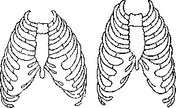
Figure 3. Short and wide thorax.
Figure 4. Narrow and long thorax. varies, corresponding to the general form of the thorax. Both costal arches form an angle (angulus infrasternalis thoracis), the size of which varies depending on the form of the thorax from 90° to 120° (see figures 3 and 4). In the latter case, the costal margin forms not an angle but an arc of large radius (Melnikov). Into the cavity of the thorax projects a centrally located ridge, composed of the bodies of the thoracic vertebrae (prominentia vertebralis), on either side of which are pulmonary grooves-sulci pulmonales. The intercostal spaces are filled with intercostal muscles-mm. intercostales externi and interni, with the first filling the space from the head of the rib to its cartilaginous part, being replaced further by strong ligaments (lig. costochondralia), while the second-from the sternum to the costal angles; the space between them is filled with loose connective tissue. On the inner surface of the ribs and muscles is lined (see figure 5) with the endothoracic fascia (fascia endothoracica), to which adheres the parietal pleura. On the posterior surface of the sternum is attached m. triangularis sterni and m. transversus thoracis, also lined with a fascial sheet, to which adheres loose connective tissue filling the anterior mediastinum.-On the outside, the bony wall of the thorax is covered with muscles taking origin or attaching here. In front are located: m. subclavius, originating from the I rib and attaching to the clavicle, m. pectoralis major, originating from the sternal end of the clavicle, from the anterior surface of the sternum almost
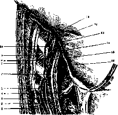
Figure 5. Layers of the thorax: 1-m. transversus abdominis; 2-f. transversalis, which directly above passes to the diaphragm; 3-retroperitoneal tissue with excised f. retroperitonealis; 4-peritoneum; 5-space between thorax and m. transversus abdominis; 6-f. transversalis (place of transition to diaphragm); 7 and 8-f. endothoracica; 9-space between m. obliquus abdominis externus and thorax; 10-ribs, cartilages, and intercostal muscles; 11-pleura parietalis lining the thorax; 12-pleura parietalis covering the diaphragm; 13-diaphragm; 14-l. diaphragmatica; 15-space prae-diaphragmaticum; 16-liver. (From Shevkunenko.)
along its entire extent and from the sheath of the rectus abdominis and attaching to the crest of the tubercle major of the humerus; beneath this muscle is the m. pectoralis minor, originating from the bony part of ribs III-V and attaching to the coracoid process. Both muscles have sheaths formed by the laminae of the fascia pectoralis and f. coraco-clavi-pectoralis; superficial to the muscles is the superficial fascia, subcutaneous fatty tissue, and skin. In the lower part of the Thorax beneath these three layers is the m. obliquus abdominis externus, covered on its outer and inner surfaces by fascial laminae, and deeper to it the m. obliquus abdominis internus, also enclosed in a fascial sheath. In the lateral parts are the m. serratus ant. (originating from the first eight ribs and attaching to the medial border of the scapula) and posteriorly and laterally the m. latissimus dorsi, the origin of which is covered by the trapezius muscle. Beneath these last muscles are the mm. rhomboideus major and minor and still deeper the m. serratus posterior superior and inferior and the long and short muscles of the back. The lower wall of the thoracic cavity, as stated above, is the diaphragm. Vessels and nerves of the Thorax. The anterior part of the thoracic region is supplied mainly by the a. mammaria int. and its branches-the aa. intercostales ant., which anastomose widely with the aa. intercostales post. (from the aorta thoracica), which supply the posterior parts. The lateral regions are supplied by branches of the a. axillaris (a. thoracica suprema, a. thoracica acromialis, a. thoracica longa) and from the a. subscapularis (a. thoracica dorsalis). The veins of the anterior part collect into two trunks accompanying the a. mammaria int. on each side and merging at the level of the 2nd-3rd intercostal space into one trunk located medially to the artery and emptying into the v. subclavia. The veins of the lateral and posterior regions form two networks-superficial and deep. The first is subcutaneous, anastomosing with the veins of the neck, abdominal wall, as well as with the veins of the axillary fossa. The deep network is formed by the posterior intercostal veins, which on the right empty into the v. azygos, with the exception of the 2nd-3rd upper ones, which empty into the v. anonyma dextra or into the v. cava superior or form a special trunk-the v. intercostalis suprema dextra, emptying into the v. azygos; the posterior intercostal veins on the left empty into the v. hemiazygos (the first 5-7 branches into the upper part, the lower ones into the lower part).-The lymphatic vessels from the external intercostal muscles collect into trunks directed posteriorly and emptying into the lymphoglandulae intercostales post., and then into the ductus thoracicus; the lymph vessels from the internal intercostal muscles are directed forward to the lymphogl. intercostales anter. and empty on the left into the thoracic duct, and on the right into the truncus lymphaticus dexter (see Thoracic duct). The vessels of the major pectoral muscles and the muscles of the lateral parts, as well as the subcutaneous tissue and the area of the mammary gland, partially empty into the lymphogl. intercostales anter., into the supra- and infraclavicular glands and mainly into the glands of the axillary fossa. Poirier and Cuvier also indicate another pathway-intrathoracic: several small trunks together with the ram. perforans a. mammar. int. penetrate into the thoracic cavity and empty into the glands accompanying this artery (see article Mammary gland, figure 6).-The nerves of the anterior part are nn. thoracici anteriores and nn. intercostales; of the lateral parts are nn. intercostales, nn. thoracici longi and partially the rami posteriores nn. thoracicum, which supply mainly the posterior part. Additionally, the m. cucullaris is innervated by the XI cranial nerve pair (n. accessorius Willisii). For practical purposes, the Thorax is divided into regions delimited by the following vertically drawn lines (see figure 6): 1) linea mediana anterior-drawn along the middle of the sternum; 2) lin. sternalis-along the edge of the sternum; 3) lin. mamillaris-through the nipple (more precisely-through the middle of the clavicle); 4) lin. parasternalis-in the middle between lin. mediana ant. and lin. mamillaris; 5) lin. axillaris ant.-along the anterior edge of the axillary fossa; 6) lin. axillaris post.-along its posterior edge; 7) lin. axillaris media-in the middle between the two preceding; 8) lin. scapularis-through the inferior angle of the scapula; 9) lin. mediana post.-along the spinous processes of the vertebrae. A detailed topographic-anatomical division into regions of practical value has no significance. The shape of the Thorax is determined by the ratio of three dimensions: 1) anteroposterior-from the level of attachment of the VII rib to the sternum to the corresponding (lying in this horizontal plane) spinous process; 2) transverse-at the level of the most distant points of the VII ribs and 3) the height of the thorax, measured from the incisura jugularis to the planum subcostale (distantia jugulo-pubica- J. p.). The latter is drawn horizontally through the lower points of the X ribs (Cunningham). The corresponding indices are calculated (the ratio of the transverse to the sagittal dimension or to J. p. x 100).-The greatest practical significance is the width index (the ratio of the transverse to the anteroposterior x 100), which ranges from 110 to 178. A Thorax with an index less than 130 belongs to the narrow group, more than 140 to the wide group; from 130 to 140-transitional forms (Melnikov). The absolute dimensions of the Thorax also vary considerably: the length of the anterior wall from 15 to 20 cm, the posterior 25-32 cm; the transverse diameter of the apert. thoracis sup.-from 8 to 12 cm, sagittal-6-10 cm; apert. thoracis inf.: transverse-from 20 to 30 cm, sagittal-16-25 cm.-The general shape of the Thorax is influenced by race, age, sex, and profession. The latter is manifested depending on the development of the musculature of the shoulder girdle and chest, conditioned by the nature of the occupation. Age differences and sexual differences are particularly pronounced. The Thorax of a newborn still has all the features of the embryonic type: due to the relatively large size of the liver, the lower part is disproportionately large, mainly due to the anteroposterior dimension, while the transverse is small, especially in the upper part; the direction of the ribs is almost horizontal, as a result of which the length of the Thorax is reduced. With age, these ratios change, and the Thorax of the elderly is characterized by a pronounced inclination of the ribs (decrease in muscle tone) and a small infrasternal angle, greater length and reduced sagittal and transverse diameters. The Thorax of women is smaller in all dimensions and in general it is relatively short and wide. Systematic lacing of the Thorax in a corset gives it a completely special shape (the so-called 'corset chest,' which should actually be classified in the group of deformations); in this case, the Thorax, which is a cone with the base downward, is transformed into a cylinder or even a cone with the base upward; the lower ribs are displaced downward, sometimes to such an extent that the infrasternal angle almost disappears and the costal arches lie against each other (Jossel). The shape and general appearance of the Thorax have essential significance in determining the type of body build ('constitution'). The numerous classifications proposed on this subject and based on the morphological features of the individual are largely determined by the shape of the Thorax (first of all), as well as the abdomen and head. The respiratory, cerebral, and asthenic types (Sigaud, Kretschmer, and Chernorutsky) are characterized by a narrow, long Thorax, low rib position, and an acute infrasternal angle (index less than 130); the digestive, picnic, and hypersthenic types-by a wide and short Thorax, expanding downward, and a large rib angle (index more than 140); the muscular, athletic, and normasthenic types-by a proportionally developed Thorax (index 130-140). Kretschmer gives the following figures for chest circumference (average value at inspiration and expiration). For asthenics: men-84.1, women-77.7; for athletes: men-91.7, women-86.0; for picnics: men-94.5, women-86.0. There are numerous attempts to express the relative chest circumference in indices. Of these, practically acceptable are: 1) Pignet's index: -L-(P+T), where L-height in cm, P-weight in kg, T-chest circumference in cm. An index less than 10 denotes a 'strong' constitution, more than 26-'weak'; 2) Brugsch's index: T X 100 -£- , where T-chest circumference, L-height. Index less than 50-narrow-chested, more than 55-broad-chested; 3) Martin's index, in combination with Brugsch's index= J.p. X100 , T X 100 ,
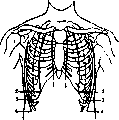
Figure 6. Guiding lines on the thorax: 1-lineae para-stemales; 2-lineae ma-millares; 3-lineae sca-pulares; 4-lineae axil-lares. (According to Corning.)
diameter of the clavicle); 4) lin. parasternalis-in the middle between lin. mediana ant. and lin. mamillaris; 5) lin. axillaris ant.-along the anterior edge of the axillary fossa; 6) lin. axillaris post.-along its posterior edge; 7) lin. axillaris media-in the middle between the two preceding; 8) lin. scapularis-through the inferior angle of the scapula; 9) lin. mediana post.-along the spinous processes of the vertebrae. A detailed topographic-anatomical division into regions of practical value has no significance. The shape of the Thorax is determined by the ratio of three dimensions: 1) anteroposterior-from the level of attachment of the VII rib to the sternum to the corresponding (lying in this horizontal plane) spinous process; 2) transverse-at the level of the most distant points of the VII ribs and 3) the height of the thorax, measured from the incisura jugularis to the planum subcostale (distantia jugulo-pubica- J. p.). The latter is drawn horizontally through the lower points of the X ribs (Cunningham). The corresponding indices are calculated (the ratio of the transverse to the sagittal dimension or to J. p. x 100).-The greatest practical significance is the width index (the ratio of the transverse to the anteroposterior x 100), which ranges from 110 to 178. A Thorax with an index less than 130 belongs to the narrow group, more than 140 to the wide group; from 130 to 140-transitional forms (Melnikov). The absolute dimensions of the Thorax also vary considerably: the length of the anterior wall from 15 to 20 cm, the posterior 25-32 cm; the transverse diameter of the apert. thoracis sup.-from 8 to 12 cm, sagittal-6-10 cm; apert. thoracis inf.: transverse-from 20 to 30 cm, sagittal-16-25 cm.-The general shape of the Thorax is influenced by race, age, sex, and profession. The latter is manifested depending on the development of the musculature of the shoulder girdle and chest, conditioned by the nature of the occupation. Age differences and sexual differences are particularly pronounced. The Thorax of a newborn still has all the features of the embryonic type: due to the relatively large size of the liver, the lower part is disproportionately large, mainly due to the anteroposterior dimension, while the transverse is small, especially in the upper part; the direction of the ribs is almost horizontal, as a result of which the length of the Thorax is reduced. With age, these ratios change, and the Thorax of the elderly is characterized by a pronounced inclination of the ribs (decrease in muscle tone) and a small infrasternal angle, greater length and reduced sagittal and transverse diameters. The Thorax of women is smaller in all dimensions and in general it is relatively short and wide. Systematic lacing of the Thorax in a corset gives it a completely special shape (the so-called 'corset chest,' which should actually be classified in the group of deformations); in this case, the Thorax, which is a cone with the base downward, is transformed into a cylinder or even a cone with the base upward; the lower ribs are displaced downward, sometimes to such an extent that the infrasternal angle almost disappears and the costal arches lie against each other (Jossel). The shape and general appearance of the Thorax have essential significance in determining the type of body build ('constitution'). The numerous classifications proposed on this subject and based on the morphological features of the individual are largely determined by the shape of the Thorax (first of all), as well as the abdomen and head. The respiratory, cerebral, and asthenic types (Sigaud, Kretschmer, and Chernorutsky) are characterized by a narrow, long Thorax, low rib position, and an acute infrasternal angle (index less than 130); the digestive, picnic, and hypersthenic types-by a wide and short Thorax, expanding downward, and a large rib angle (index more than 140); the muscular, athletic, and normasthenic types-by a proportionally developed Thorax (index 130-140). Kretschmer gives the following figures for chest circumference (average value at inspiration and expiration). For asthenics: men-84.1, women-77.7; for athletes: men-91.7, women-86.0; for picnics: men-94.5, women-86.0. There are numerous attempts to express the relative chest circumference in indices. Of these, practically acceptable are: 1) Pignet's index: -L-(P+T), where L-height in cm, P-weight in kg, T-chest circumference in cm. An index less than 10 denotes a 'strong' constitution, more than 26-'weak'; 2) Brugsch's index: T X 100 -£- , where T-chest circumference, L-height. Index less than 50-narrow-chested, more than 55-broad-chested; 3) Martin's index, in combination with Brugsch's index= J.p. X100 , T X 100 ,
t distantia jugulo-pubic, T - circumferentia thoraco-pubica, L), gives an idea of dolicho- and brachymorphy of the subject, with the index 30.5 + + 50.0 determining the boundary between types (Shevkunenko). Depending on the form of the thorax, the position and ratio of the organs contained in it differ. In a wide thorax with an upper aperture elongated in the frontal direction, the aortic arch is located high [up to the level of the upper edge of the first thoracic vertebra (D1)], tending to take a direction in the frontal plane (Lisitsyn); the heart is positioned transversely and such that its left border may be located outside of lin. mamillaris sin. (Nedrigailova). In a narrow and long thorax with an aperture compressed from the sides, the aortic arch is located low (up to the lower edge of D12) and tends to take a sagittal direction (Lisitsyn); the heart is positioned vertically, in extreme forms barely protruding beyond the left edge of the sternum-"drop heart" (Nedrigailova). The boundaries of the mediastinal pleura in a narrow and long thorax shift more to the right, and the boundaries of the sinus costo-diaphragmatici in the posterior parts are higher than in a wide and short thorax, and lower in the front (Melnikov). The diaphragm in a narrow thorax is located higher, and its openings are closer together than in a wide one (Moskalenko). The embryonic development of the thoracic cage is determined by the embryogenesis of its elements- the spine, ribs, and sternum. The mesenchyme surrounding the chorda is the basis, from which through two successive transformations the membranous vertebral column develops into a cartilaginous and then a bony spine. In humans in the second month of intrauterine life, independently of the spine, ribs develop by the transformation of the so-called "intermuscular ligaments"-lig. intermuscularia into cartilage. Their primordia are present near the vertebrae along the entire length of the spine. In humans and mammals, only the ribs of the thoracic section, growing in the ventral direction, reach considerable length, and in a 3-month-old embryo the length of the first 7 pairs of ribs reaches the ventral surface of the thorax, where they form on each side "sternal ridges", from which the sternum develops. In lower vertebrates (fish, reptiles) ribs develop (approximately according to one type) in all sections of the spine, which in selachians (sharks) still consists of cartilaginous vertebrae, while in ganoid and bony fishes it is already bony. In some fish (Crossopterygii) each vertebra has two pairs of ribs-upper and lower, but in most there are either upper or lower ribs. The latter are laid in the connective tissue layers and are located near the wall of the secondary body cavity. In amphibians, the thoracic section of the spine has ribs in the form of short appendages of the transverse processes (not reaching the sternum), corresponding to the upper ribs of fish. In snakes, the ventral ends of the ribs do not reach the midline and end freely, while in lizards, remaining cartilaginous, they connect directly with the sternum or with the rib (cranially) lying in front. A feature of the avian thorax, besides the structure of the sternum (see), is the ossification of the ribs and the fusion of the vertebrae. In mammals, as well as in humans, the distal ends of the ribs either connect with the sternum or articulate with the rib lying in front, forming a costal cartilaginous arch, or end freely, being embedded in the muscles of the abdominal wall. The number of thoracic vertebrae in them varies from 9 to 24 (Choloepus Hoffmanni), but most have 13. The general form of the mammalian thorax approximates that of the human embryo: compressed from the sides and elongated in the anteroposterior (dorsoventral) direction. (For deformities of development of the thorax see Thoraco-schisis, Thoracopagus.) Deformities of the thorax are either a particular manifestation of diseases of the skeletal system in general or are caused by a violation of normal static conditions, with the presence of the first circumstance inevitably also manifesting the second; the cause of deformities can also be both intra- and extrauterine diseases of the organs of the thoracic cavity, mainly the lungs and pleura. Changes in the shape of any part of the ribs, sternum or spine, on the one hand, lead to a change in the shape of the thorax, and on the other-they may be associated with a change in the shape of the thorax in general; moreover, diseases of the spine often also lead to a violation of static conditions. Extensive tumors of the thoracic wall (both of soft tissues and of the skeleton) may not
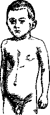
Figure 7. Pectus carinatum. (After Mütller.)
not only cause deformation of the thorax at the site of the tumor, but also lead to displacement of the organs of the thoracic cavity and compensatory deformation of the corresponding sections. Deformations of the thorax can also be caused by underdevelopment of one or another muscle or a group of them (most often the m. pectoralis major); in this case, the essence of the deformation lies not only in the absence of the muscle as such, but also in the absence of muscular traction, and consequently in the underdevelopment of the corresponding section of the thorax. Essentially, this is not a deformation, but only an asymmetry, a phenomenon common in humans, merely expressed to a more severe degree. With insufficient growth of bones in general, caused by abnormalities of endochondral development, the essence of the process lies in slowed, insufficient, or prematurely discontinued multiplication of cartilage cells at the border of cartilage and bones (achondroplasia), to which can be added the softening of the main tissue of the growth lines (chondrodystrophia malacia) or overgrowth of cartilage (chondrodystrophia hyperplastica). In these cases, the thorax takes on an asymmetrically ugly form depending on the intensity of the process in its various sections. The most diverse curvatures of the spine and ribs are observed in the same way as a result of past osteomalacia. It should be noted that in this disease in men, the process is more often localized in the ribs and spine, whereas in women (mainly during pregnancy) it is localized in the pelvic bones. The changes in the thorax in osteomalacia, in form, approach those observed in rickets. The latter sometimes appear as early as the third month of life and initially consist of a slight thickening of the sternal ends of the ribs, as well as thickening at the border of their cartilaginous and bony parts, subsequently reaching considerable sizes. These nodular thickenings, in appearance, gave rise to compare them with rosary beads ('rachitic rosary'). Subsequently, bends, flattenings, and even indentations of the costal cartilages are observed, thus forming two grooves on either side of the sternum. In severe degrees of these deformations, multiple fractures of the ribs are often observed, and an arched concavity forms in the lateral sections of the thorax; the sternum at this time protrudes forward considerably, so that the entire thorax, appearing unevenly compressed from the sides, on a horizontal section has the shape of a triangle and, by similarity, bears the name 'chicken breast' (pectus carinatum, see figure 7). In even more severe cases, the sternum protrudes so much that the cartilages of the lower ribs, which articulate with it, attach behind it, are sharply concave, the xiphoid process is very mobile, depressed backward, the costal arches are approximated; due to the muscular traction of the diaphragm, the lower opening of the thorax is disproportionately widened, and the edges of the ribs bounding it are turned forward. There are indications that so-called funnel chest is also a consequence of rickets, however, in this case, various developmental defects in the form of cleft lip, syndactyly, etc., are often observed, which speaks in favor of a developmental defect. The essence of this deformation consists in a funnel-shaped depression of the anterior part of the thorax, reaching such a degree that it can extend to the spine itself, without unpleasant sensations for the patient (Tikhov).-The above-mentioned changes are sometimes accompanied by deformations of the spine, expressed more often in curvature and protrusion backward of the lower thoracic vertebrae, less often in the form of lateral curvatures, mainly in the upper section. In Barlow's disease (see Barlow's disease), as well as in severe adult scurvy, sometimes the separation of the cartilaginous and bony parts of the ribs and the depression of the sternum with the cartilages inward into the thorax occurs. Lateral curvature of the spine is called scoliosis (scoliosis). Often, lateral curvature of one section is accompanied by compensatory curvatures in another (see figure 8). In these latter cases, scoliosis is called complex and can involve various sections of the spine. Curvature to the side is usually combined with rotation of the vertebrae in the affected section. Due to all these causes, changes in the form of the thorax occur to a greater extent, the more pronounced the scoliosis, and consisting in the formation of the so-called 'rib hump': a convex posterior rib hump on the side of the curvature and a concave one on the other half of the thorax. With complex, sharply expressed scoliosis, the deformation of the thorax reaches degrees that impair the function of the organs of the thoracic cavity. In rare cases, scoliosis is the result of a disease in embryonic life, causing improper development of bones (according to Bohm's data, congenital scoliosis occurs as often as rachitic scoliosis). According to Drachmann's statistics, it is observed in 1.3% of school-age children due to improper posture during studies, etc. (see Scoliosis). To the same extent, curvatures of the spine backward (kyphosis), more often being a consequence of the tuberculous process of the vertebral bodies (gibbus), also have a deforming effect on the thorax, and sometimes reach such degrees that the lower ribs can touch the iliac crests.
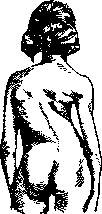
Figure 8. Left-sided scoliosis. Severe deformation of the thorax. (According to Muller.)
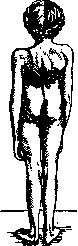
Figure 9. Gibbus in the lower part of the thoracic spine. (According to Muller.)
In this case, the sternum protrudes forward considerably and is shortened, with its lower end far from the spine; the curvature of the ribs is flattened, the costal arches are approximated, and the entire thorax is compressed from the sides and elongated in the anteroposterior direction (see figure 9). Similar, but less pronounced deformations are also observed in Bechterew's disease - Strümpell-Marie. (Deformations of the thorax caused by developmental abnormalities of the sternum-see Sternum; deformations in aplasia of ribs and in cervical ribs-see Rib.) Chronic progressive expansion of the lungs in emphysema leads to expansion of the thorax, taking on a barrel-shaped form. The supraclavicular spaces gradually become smoothed, the intercostal spaces bulge, the ribs lose their normal inclination, the epigastric angle is obtuse, almost smoothed.- A special group of thorax deformations consists of changes in its shape due to chronic pathological processes in the pleura and lung, when, due to the wrinkling of the proliferating connective tissue in them, depression of the corresponding side of the thorax and subsequent curvature of the spine (scoliosis) occur, as well as due to surgical intervention involving the removal of a considerable number of ribs (thoracotomy).
Related articles
Mentioned in
Cite this page
“Thorax.” Soviet Medical Encyclopedia. English translation of Bolshaya Meditsinskaya Entsiklopediya, 1st ed. (Moscow, 1928–1936), ed. N. A. Semashko. https://sovietmedicalencyclopedia.pages.dev/article/thorax/