Pneumothorax
Historical document, translated for reference. It reflects medical knowledge of the 1920s–30s and is not medical advice.
Summary
This article from the 1928–1936 Great Medical Encyclopedia examines the etiology, pathogenesis, and clinical occurrence of spontaneous pneumothorax. It details how pulmonary tuberculosis, destructive lung diseases, and ruptures of subpleural emphysematous bullae lead to the accumulation of air or gas in the pleural cavity.
Encyclopedia article (1928–1936)
PNEUMOTHORAX (from the Greek pneuma — air and thorax — chest), the accumulation of air or gas in the pleural cavity. Spontaneous pneumothorax, unlike artificial pneumothorax (see below), occurs spontaneously in connection with: 1) damage to the lung when the integrity of the chest wall is violated (see Lungs — surgical treatment of lung diseases), 2) diseases of the chest organs. The most frequent cause of pneumothorax is destructive processes in the lungs leading to a violation of the integrity of the visceral pleura. Up to 90% of all pneumothoraces are attributed to pulmonary tuberculosis. Comparatively rarely, pneumothorax occurs in abscesses, gangrene (see Lungs), malignant tumors, and upon the breakthrough of an echinococcus into the pleura. Even more rarely, it occurs upon ulceration of the esophageal wall (cancer), the breakthrough of a subdiaphragmatic abscess containing gases, and stomach cancer (destruction of the diaphragm). To the present time, the question of the possibility of the onset of pneumothorax in pulmonary emphysema remains controversial. Most authors, based on anatomical findings, adhere to the following view: the onset of pneumothorax is possible only upon the rupture of a subpleural emphysematous bleb, which may form as a result of cicatricial changes in the lungs in various pathological processes (most often tuberculous). This same cause should explain the occurrence of pneumothorax in individuals who previously had no complaints regarding this disease and considered themselves healthy (the so-called "idiopathic" pneumothorax). It must be assumed that isolated cases of pneumothorax in whooping cough, influenzal pneumonia, bronchial asthma, and typhoid fever occur in the exact same manner as a result of the rupture of subpleural emphysematous blebs or "apical cicatricial blebs" (Fischer). In addition to the just-mentioned causes of the occurrence of pneumothorax on the path from the lung to the pleura, one must also take into account the reverse path — from the pleura to the lung: 1) the breakthrough of pus (empyema) from the pleural cavity into a bronchus; 2) the rupture of pleural adhesions with a violation of the integrity of the visceral pleura and alveoli. In this latter case, air begins to flow into the pleura comparatively slowly, in small portions, without causing severe disorders in individual cases. With such an occurrence of pneumothorax, patients do not experience suffering (pains, shortness of breath, etc.), and the pneumothorax is an accidental finding during the examination of the patient. In pulmonary tuberculosis, pneumothorax occurs most often in cases of acutely and subacutely proceeding processes. In chronic forms of tuberculosis with a tendency toward the development of fibrosis, there are extensive pleural adhesions that prevent the flow of air into the pleural cavity. Statistical data on the frequency of pneumothorax among tuberculous patients range from 0.1% to 10%. Such a discrepancy in figures is explained by the fact that observations were conducted on different materials. Sanatorium patients, for example, yield a lower percentage of pneumothorax than hospital patients. In addition to this, with the use of X-rays, the diagnosis of pneumothorax has been significantly facilitated, especially in cases of partial pneumothorax. In men, pneumothorax is observed more often than in women. By localization, it is more frequent on the left side. The reason for the frequency of left-sided pneumothorax is seen in the fact that the movements of the left atrium and ventricle prevent the formation of pleural adhesions in the adjacent areas of the lung.
One distinguishes complete and partial pneumothorax. In complete pneumothorax, the air that has broken into the pleural cavity compresses the lung completely against the spine; in partial pneumothorax, the air is located above one or another section of the lung, depending on the presence of pleural adhesions. Both complete and partial pneumothorax may have a free communication with the bronchus, with air entering and leaving the pleural cavity freely during inspiration and expiration—open pneumothorax. Open pneumothorax can form both in cases of wounds or injuries to the chest wall (open externally) and in cases of tearing of the lung (open internally). In those cases where air enters the pleural cavity only during inspiration or expiration until a certain pressure is formed in the pleural cavity, and then communication between the pleural cavity and the external air ceases, we have a closed pneumothorax. The entry of air into the pleural cavity ceases definitively from the moment the perforation opening is closed by a reparative process; air begins to be absorbed from the pleural cavity, and the severity of functional disorders subsides. The pressure in the pleural cavity can be either positive or negative, depending on the anatomical conditions of the formation of the perforation opening and the conditions of lung compression. In the case where air enters only during inspiration with a progressive increase in pressure in the pleural cavity, we have the so-called valvular pneumothorax, which reproduces the mechanism of a check valve (see Lungs, Figure 26). The mechanism of valvular pneumothorax is explained by most authors in the following way: a small piece of lung tissue or pleura closes the opening upon an increase in pressure in the pleural cavity, and upon a decrease in pressure, this flap moves away from the opening and opens it; thus, air accumulates in the pleural cavity with each inspiration. In the majority of cases of valvular pneumothorax, the mechanism of air entry into the pleural cavity is nevertheless somewhat different: "the fistulous canal connecting the cavity with the pleural cavity can have two directions. Either it runs parallel to the direction of the elastic forces of the lung, or it approaches them at an angle. In the first case, upon the collapse of the lung, the canal decreases in length, but remains open; in the second, the outer wall of the canal, due to the increased pressure in the pleural cavity, will closely adhere to the inner wall, and the opening will be closed. With fluctuations in intrapleural pressure corresponding to deep inspiration and expiration, the canal will alternately close and open, i.e., it will have the property of a valve" (M. Bock). In pneumothorax resulting from the rupture of pleural adhesions with disruption of the integrity of the alveoli or from the rupture of emphysematous subpleural vesicles, there will likewise be a compression of the perforated area during expiration and free passage of air during inspiration. A similar mechanism of "valve" functioning can be observed for a long time. As a result of the absorption of air from the pleural cavity, the intrapleural pressure drops below the elastic force of the lung, the perforation opening opens, and air enters the pleural cavity again (V. Chukanov). Pathological anatomy. To determine the presence of air in the pleural cavity on a corpse, the following method is used: a skin flap in the form of a pocket is separated in front on the affected side of the chest, into which water is poured, and an puncture of the intercostal space is made under water—air escapes from the pleural cavity in the form of bubbles. Upon opening the chest cavity, the lung is found compressed against the spine or pushed aside—depending on adhesions—in one direction or another. To find the perforation opening on the pleura, it is recommended to fill the pleural cavity with water and pump air into the lungs through the trachea with a pump; the appearing bubbles will approximately indicate the site of perforation. In ruptures of a cavity, the perforation opening is sometimes widely gaping. In some cases of pneumothorax, the perforation opening cannot be found, especially in those cases where it is insignificant and the pleura is covered with fibrinous deposits. If the patient dies rapidly from pneumothorax, an autopsy reveals a pleura unaffected by the inflammatory process. In those cases where the patient survives pneumothorax for a day or more, an autopsy—as a rule—also reveals an exudate of one character or another. An exception to this rule is pneumothorax formed as a result of the rupture of subpleural emphysematous vesicles or "apical scar vesicles". Upon the rupture of a cavity, pyogenic cocci (streptococcus, staphylococcus, diplococcus, etc.) almost always enter the pleural cavity, causing the subsequent formation of purulent exudate (pyopneumothorax). In open pneumothorax, the conditions for pyogenic cocci to enter the pleural cavity are most favorable. In cases of rapid closure of the perforation opening, especially if it was formed as a result of the rupture of pleural adhesions, a sero-fibrinous exudate appears (seropneumothorax). Statistics show that cases of pneumothorax with serous exudate are observed much more frequently than with purulent exudate. Upon the rupture of a caseous subpleural focus, upon the rupture of pleural adhesions containing tubercle bacilli, and in some cases also upon the rupture of a cavity with rapid closure of the perforation opening, Koch's bacilli enter the pleural cavity, causing inflammation of the pleura with the accumulation of sero-fibrinous fluid. In individual cases, pure tuberculous pus is found without the presence of pyogenic infection. The amount of fluid varies—from several cubic centimeters to 2–3 liters or more. Symptomatology. The onset of spontaneous pneumothorax is most often observed during physical exertion, sometimes quite insignificant: sneezing, coughing, putting on boots, jumping, dancing, etc.; in some cases, pneumothorax forms during a resting position in bed; the destructive process in the lungs destroys the visceral pleura, and air begins to enter the pleural cavity sometimes in such a large amount that it immediately creates a severe picture. The patient complains of sharp pains in the side, severe shortness of breath. For the most part, cyanosis, frequent and small pulse, cold sweat, phenomena of severe collapse, and sometimes even the death of the patient within a few minutes or hours are observed. The severity of functional disorders depends on the anatomical conditions of the formation of pneumothorax, the degree of compression of the lung and mediastinal organs, and on the functional capacity of the other lung. In individual cases, the onset of pneumothorax passes almost unnoticed by the patient, and pneumothorax is discovered accidentally during an X-ray examination of the patient. In those cases where air, depending on the small size of the perforation opening, enters the pleura slowly, in small portions, the body gets the opportunity to adapt to the disruption of the functional activity of the respiratory and circulatory organs. Within the next few days, a rise in temperature occurs in connection with the inflammatory process in the pleural cavity, and subsequently the fate of the patient most often depends already on the character of the inflammatory process and the degree of tuberculosis damage to the other lung. With the addition of exudative purulent pleurisy to open pneumothorax, the patient, a relatively short time after the formation of pneumothorax, begins to expectorate pus, and the expectoration occurs episodically, immediately in a large amount ("full mouth"). Sometimes expectoration is associated with a change in the patient's position, when the level of pus becomes higher than the cardiopulmonary fistula and pus begins to flow into the bronchi. The total amount of expectorated pus per day ranges from 100–200 cm3 to 1 liter. Upon examination of the patient with phenomena of increasing pressure in the pleural cavity, the characteristic position of the patient is noted: mostly he is in a semi-recumbent position on his back, leaning slightly to the affected side. The intercostal spaces on the affected side are smoothed out, and in some cases tense and protruded. The half of the chest on the side of pneumothorax is protruded, almost does not participate in respiration, the other half produces enhanced respiratory movements. The heart beat is sharply displaced, especially in right-sided pneumothorax. Upon palpation, tenderness of the intercostal spaces is noted; in right-sided pneumothorax, the liver is displaced downward. Vocal fremitus is absent or sharply weakened and is noted only over those areas of the lung where there are extensive pleural adhesions preventing compression. Upon percussion on the side of pneumothorax, a loud and low sound is noted; in some cases, the sound is high and tympanitic, depending on the tension of the air bubble in the pleura. Similar changes in the percussion sound can spread significantly further than the usual boundaries of the lung, sometimes extending to the other half of the chest, occupying, for example, the place of absolute cardiac dullness (displacement of the mediastinum and heart). The lower border of the altered percussion sound reaches the edge of the costal arch; in right-sided pneumothorax, hepatic dullness disappears. The liver is displaced downward, and its lower edge reaches the navel. Mobility of the percussion sound boundary over the pneumothorax area is absent. If an effusion joins the pneumothorax, then upon percussion of the lower parts of the chest, a dulled or dull sound is found.
When the patient's position changes, the upper boundary of this dull sound changes due to the displacement of fluid, always maintaining a horizontal level. The displacement of the boundaries of dullness upon changing the patient's position (bending the patient forward or backward) indicates quite definitely the presence of fluid and air in the pleural cavity. The free displacement of fluid makes it possible to detect just as accurately the peculiar "succussion splash" (succussio Hippocratis), obtained by rapidly changing the patient's position or by shaking the chest. The "succussion splash" is sometimes audible to the patient himself, as well as to those around him, and in many cases is the first symptom of which patients complain. Auscultation in closed pneumothorax reveals sharply weakened vesicular breathing or, which is observed more often, the complete absence of respiratory noise. If there is an open pneumothorax (communication with atmospheric air through a bronchus), respiratory noises with a metallic tint are heard. The intensity of this phenomenon varies depending on the increase in respiration. In weak patients, it may not be heard at all, in which case the method of auscultatory percussion is used. Simultaneously with listening to the patient from behind directly with the ear, one taps from the front on the pleximeter with the end of a hammer, a pencil, or a fingernail; in this case, a sound with a metallic tint is clearly heard (Heubner's sign). It is necessary to close the free ear while listening to this phenomenon. Heubner's sign is observed in both open and closed pneumothoraces, as well as in large smooth-walled cavities. In hydropneumothorax, the falling drop sound phenomenon—tintement métallique, first described by Laennec—is sometimes heard. This sound phenomenon most likely depends on a single rale in the lung, acquiring a metallic tint under resonance conditions, rather than on a drop falling into the pleural fluid from the walls of the pleura. If one listens to a patient with hydropneumothorax in the process of evacuating fluid or immediately after the patient has expectorated a sufficient amount of fluid (in open pneumothorax), one can catch during inspiration a peculiar sound first described by Dance and Beau and called by Unverricht the "water-pipe murmur" (Wasserpfeifengeräusch). The origin of this sound depends on the entry of air into the pleura through a pleuro-pulmonary fistula lying below the upper boundary of the exudate; air, passing from the lung through the layer of fluid, rises to the surface in the form of bubbles. The entry of air is associated with the opening of the pulmonary fistula due to a decrease in intrapleural pressure and the transformation of a mechanically closed pneumothorax into a "valvular" one. The described metallic phenomena depend on the presence of a smooth-walled cavity and a certain degree of air tension in it. Under these conditions, high overtones arise, causing the metallic tint of the sound. Manometric determination of gas pressure in the pleural cavity is of great importance in resolving the question of whether there is a closed or open pneumothorax (see below, artificial pneumothorax). In closed pneumothoraces, the pressure is usually below zero, in open ones it is equal to atmospheric pressure, in valvular ones during inspiration it is about zero, and during expiration +5-6 cm of water column. If air is introduced into the pleural cavity, in closed and "valvular" pneumothoraces the pressure rises, while in open ones it remains unchanged, since the gas escapes through the pleuro-pulmonary fistula. The diagnosis of complete spontaneous pneumothorax, with proper consideration of the described symptoms and a thorough analysis of the patient's condition, does not present great difficulties. In doubtful cases, the question is resolved by X-ray examination. The extraordinary transparency in the place where the lung is replaced by air, the absence of the pulmonary pattern, the displacement of the mediastinal organs and the heart to the healthy side are a characteristic picture for pneumothorax. The collapsed lung in the form of a hemisphere, a small lump with irregular contours, is pressed against the hilus. The contours of the ribs and their cartilaginous ends clearly protrude. The diaphragm exhibits paradoxical movement: during inspiration it rises upwards, during expiration it lowers (Kienböck). The costo-diaphragmatic angle is enlarged. The pulmonary pattern on the healthy side stands out sharper than normal due to an increase in the amount of blood in the vessels. In hydropneumothorax, there is a regular horizontal level of the upper boundary of the fluid (see separate table to the article Heart defects, Fig. 7). The fluid level oscillates in a wave-like manner upon shaking a patient with pneumothorax and upon the contraction of the heart. X-ray examination reveals the most insignificant accumulation of fluid, usually located in the costo-diaphragmatic angle and escaping ordinary clinical examination. With a significant accumulation of fluid in the pleura, a small air bubble is pushed upward and is not detected by ordinary physical research methods. Quite often, fluid fills the entire pleural cavity, resembling the picture of ordinary exudative pleurisy, and only in the dome of the apex is a horizontal level of fluid barely noticeable, definitely speaking in favor of hydropneumothorax. In isolated cases, this horizontal level is detected only after evacuation of the fluid (A. Ya. Sternberg). Great difficulties for a correct diagnosis exist in the presence of partial spontaneous pneumothorax. At the same time, from the point of view of differential diagnosis, one must keep in mind large cavities sometimes occupying an entire lobe of the lung or both lobes of the left lung. Careful consideration of clinical data, the results of physical research methods, anamnesis, and finally X-ray examination make it possible to make a correct diagnosis. An essential sign in large cavities is the absence of displacement of the heart and mediastinum to the opposite side. In addition to large cavities, in differential diagnosis one must take into account 1) a subdiaphragmatic abscess containing air, 2) a diaphragmatic hernia, 3) unilateral paralysis of the diaphragm. In subdiaphragmatic abscesses, one must take into account 1) the development of the disease and its course, indicating a process in the abdominal cavity, 2) the absence of cough and sputum, 3) insignificant displacement of the heart, 4) displacement of the liver downward, 5) during fluoroscopy, high standing of the diaphragm. The course of pneumothorax depends on the underlying disease. Upon the opening of an empyema into a bronchus, upon the rupture of a lung abscess or the opening of a localized subpleural gangrenous focus into the pleura, recovery can occur in a relatively short time if surgical intervention is performed in a timely manner. Pneumothoraces formed as a result of traumatic rupture of the lung, as well as as a result of rupture of subpleural emphysematous vesicles or apical cicatricial vesicles, most often end favorably, the air is slowly absorbed, and the compressed lung gradually expands. If a serous exudate joins the pneumothorax, which most often happens in tuberculous patients, the course of the pneumothorax will depend on the course of pleurisy. With an insignificant accumulation of serous fluid that does not exert pressure on the heart and mediastinum, provided the perforation hole is closed by a reparative process, the air and then the fluid can gradually resolve. In some cases of rapidly increasing exudate, death from asphyxia can occur due to compression of the mediastinal organs. One must be especially careful with right-sided hydropneumothorax, in which the fluid presses on the relatively weak muscle of the right heart and atrium; in these cases, death can occur unexpectedly from cardiac arrest. The course of spontaneous hydropneumothorax in tuberculous patients largely depends on the general state of the patient's strength and the degree of spread of the process, and mainly on the condition of the other lung. A pneumothorax formed during a unilateral tuberculous process can lead to an improvement of the latter: due to compression of the lung, the amount of sputum decreases, the temperature drops, the general condition of the patient improves; therefore, in some cases it is advisable to maintain compression of the lung by converting spontaneous pneumothorax into artificial pneumothorax. However, cases of a favorable effect of spontaneous pneumothorax in tuberculous patients are relatively rare. Most often, spontaneous pneumothorax leads to a sharp deterioration in the patient's condition, especially with damage to the tuberculosis of the other lung. The prognosis is definitely unfavorable in tuberculous patients if pneumothorax is formed as a result of the rupture of a cavity and a purulent exudate joins the pneumothorax. A severe process in the pleura quickly leads to exhaustion of the patient's strength, and, if surgical intervention (rib resection with opening of the pleural cavity) is not undertaken in a timely manner, amyloid of internal organs develops over time. Amyloid degeneration of internal organs is also observed in those cases where a long-term non-closing thoracic fistula remains after the operation. For the treatment of surgical pneumothorax associated with chest damage, see Lungs - surgical treatment of lung diseases. In pneumothorax formed as a result of the opening of an empyema, the rupture of a gangrenous focus or abscess into the pleura, it is necessary to immediately perform a thoracotomy operation to ensure the free outflow of pus outward. In pneumothorax in tuberculosis patients, as well as in the rupture of subpleural emphysematous vesicles, it is necessary first of all to take measures to eliminate severe symptoms.
For severe pain and shortness of breath, subcutaneous injection of morphine. A decline in cardiac activity requires the use of stimulants (camphor, digitalis preparations, caffeine, etc.). In cases of severe compression of the lung, displacement of the heart and mediastinum, and phenomena of severe collapse, which is most often observed in the so-called "valvular" pneumothorax, when the air pressure in the pleural cavity reaches high figures, there is a danger of the patient dying from asphyxia. In these cases, besides stimulants, one has to take measures to lower the pressure in the pleural cavity. For this purpose, air is evacuated from the pleura using an apparatus for artificial pneumothorax, and in the absence of such an apparatus, the air is simply released through a needle or trocar. However, this measure gives only temporary relief: with the lowering of pressure, the perforation opening opens, and air begins to flow into the pleural cavity again. In addition, there is a danger of purulent infection penetrating through the bronchopleural fistula; therefore, air is evacuated only under vital indications. In the most severe cases, when air evacuation gives only temporary relief and the patient's condition continues to remain threatening, recourse is made to the method of converting a closed or valvular pneumothorax into one open to the outside. For this purpose, a needle inserted into the pleural cavity is secured with adhesive plaster and bandages and left in this position until the formidable phenomena of asphyxia subside. With the formation of a closed pneumothorax, without severe symptoms of compression of the mediastinal organs, air evacuation is not recommended. In these cases, spontaneous cure may occur with the closure of the perforation opening. If exudative pleurisy joins the pneumothorax, it is first of all necessary to determine the character of the exudate (exploratory puncture). In serous and seroso-fibrinous exudates, one should adhere to an expectant posture and resort to fluid evacuation only in cases where the accumulating exudate hinders blood circulation and causes severe shortness of breath. When evacuating, one must bear in mind the possibility of the perforation opening opening, and therefore it is recommended to replace the removed exudate with air, maintaining the pressure in the pleural cavity slightly above zero. Usually it is necessary to introduce air in an amount equal to half of the removed exudate. The evacuation of fluid and introduction of air are carried out simultaneously using Potain's apparatus and the apparatus for artificial pneumothorax. The needle from the apparatus for artificial pneumothorax is punctured above the level of the exudate. Air is introduced into the pleural cavity after evacuating a certain amount of fluid, depending on the manometer readings. The formation of exudate in spontaneous pneumothorax in tuberculous patients is an almost constant phenomenon. In some cases, the serous exudate, after some time, turns into purulent (tuberculous empyema without pyogenic infection). With such cold purulent exudates, it is recommended to refrain from opening the pleural cavity by thoracotomy operation or introducing Bülau's siphon drainage, and instead, pus should be evacuated with simultaneous washing of the pleural cavity with either warmed physiological solution or rivanol solution (1:5,000-10,000). Jessen advises washing with a solution of iodine and potassium iodide: Jodi puri 1.0, Kali jodati 2.0, Aq. dest. 20.0 - 5.0 per 1,000 cm3 of water. The operation of pus evacuation with washing of the pleural cavity has to be repeated as the exudate accumulates. Washing technique: a needle or trocar with two cannulas is inserted into the pleural cavity. One cannula is connected to Potain's apparatus for evacuating pus and washing fluid, the other is connected by a rubber tube to a vessel containing the washing fluid. An ordinary enema mug can serve this purpose. It is better to have a graduated jar. In the absence of a trocar with two cannulas, the operation can also be performed with a single simple hollow needle. First, the pus is aspirated as completely as possible. If this cannot be done due to displacement of the mediastinum (the patient complains of severe chest pain and cough), air is introduced using the apparatus for artificial pneumothorax to equalize the pressure in the pleural cavity. In the absence of such an apparatus, air can also be let in through the needle used to aspirate the pus. For this purpose, a sterile gauze tampon is prepared, with which the outer opening of the needle is quickly closed after disconnecting it from the cannula of Potain's apparatus. Upon inhalation, air enters through the gauze tampon via the needle into the pleural cavity with the characteristic sound of a "water pipe". After two or three deep breaths, the patient states that they feel better (the pressure in the pleural cavity has equalized), then further evacuation can be continued. One should not fear the penetration of infection into the pleural cavity with the air. Sometimes the patient has to be tilted slightly toward the apparatus (the operation is performed with the patient sitting) in order to evacuate the pus more fully. Upon completion of aspiration, washing fluid warmed to body temperature in the amount of 200-300 cm3 is introduced and the washing fluid mixed with pus residues is aspirated again. This is repeated until almost clean washing fluid flows into Potain's apparatus. For a more accurate determination of the pressure in the pleural cavity during the pus aspiration process, a manometer is adapted to Potain's apparatus. For rapidly accumulating purulent tuberculous exudates, oleothorax is used. If spontaneous pneumothorax is complicated by purulent exudate due to the entry of pyogenic infection into the pleural cavity (streptococcus, staphylococcus, diplococcus, etc.), then pus evacuation and pleural washing give only a temporary effect. Bülau's drainage in tuberculous patients is used only in cases where the patient's condition does not allow hoping for recovery due to extensive damage to other organs (kidneys, intestines) or damage to the second lung. In the absence of such complications, surgical intervention should be performed immediately, and the choice of intervention method will depend on whether there is a constantly operating bronchopleural fistula or whether it is closed by the increased pressure of the air bubble and exudate. In the first case, it is necessary to strive to close the bronchopleural fistula by bringing the parietal pleura closer to the visceral one. Since the bronchopleural fistula in tuberculous patients most often forms in the upper lobes of the lungs, upper thoracoplasty is performed. Upon resection of the upper 5-6 ribs according to Sauerbruch, we can count on compressing the perforation opening. Before the thoracoplasty operation, it is necessary to evacuate the pus and wash the pleura. If it is impossible to determine the location of the fistula, complete thoracoplasty is performed. Some time after the plastic surgery, rib resection is done for the outflow of pus to the outside. The thoracoplasty operation can be performed only in strong young people provided there is no tuberculous lesion of other organs, especially the second lung. Sometimes, even in the presence of these conditions, one has to refrain from thoracoplasty due to the severe septic condition of the patient, especially in neglected cases. Then, first of all, it is necessary to perform a thoracotomy with resection of 1-2 ribs. In some cases, this intervention alone creates the condition for the closure of the bronchopleural fistula. The temperature drops, the patient's strength returns, and general well-being improves. Upon restoration of the patient's strength, the thoracoplasty operation is undertaken in cases where the lung cannot expand due to the loss of elasticity of the visceral pleura as a result of abundant fibrous layers, and an air pocket remains in the pleura. If the air pocket is not completely destroyed by Sauerbruch's thoracoplasty operation, an extrapleural plomb is added, or complete rib removal (decostatio unilateralis totalis) is performed, or the remnants of the air cavity are filled by transplantation of the pectoral muscles. V. Chukanov. Artificial pneumothorax (unilateral, bilateral) is a method of treating lung disease, mainly destructive forms of pulmonary tuberculosis, by repeated introduction of indifferent gases (nitrogen, oxygen, carbon dioxide) or atmospheric air into the pleural cavity for the purpose of compressing the lung or greater or lesser reduction of the elastic tension of the lung tissue with a simultaneous significant reduction in the functional activity of the corresponding lung.
History. The idea of the expediency of treating localized focal diseases of the lungs (tuberculous abscesses) by compression of the lung tissue with air introduced through an opening in the pleura was expressed repeatedly in the medical literature of the late 18th and first half of the 19th centuries (Gilchrist, Carson, Ramedge), but the state of thoracic surgery at that time and the lack of asepsis were insurmountable obstacles to the introduction of this method of treatment into medical practice. The accumulation of observations on the favorable effect of spontaneous pneumothorax and pleural exudates on the course of pulmonary tuberculosis created the prerequisites for the theoretical development of the artificial pneumothorax technique. Artificial pneumothorax was first introduced into clinical practice by Carlo Forlanini (1882-95). Independently of him, Copphy (England, 1885) and Murphy (America, 1898) also made the first attempts to use artificial pneumothorax for therapeutic purposes. Artificial pneumothorax began to gain wider dissemination from the beginning of the 20th century (1905-06) after the detailed development of the technique by Brauer and Saugmann. Artificial pneumothorax received final recognition after the international congress in Rome (1912) and since then has begun to spread rapidly and widely. In Russia, the first popularizer of artificial pneumothorax was A. N. Rubel (1912); of great importance is the research and educational work on artificial pneumothorax carried out by A. Ya. Shternberg in Leningrad (1907) and V. E. Vainshtein (Yalta, Moscow). In the USSR, artificial pneumothorax became widespread from 1920-23 after the discussion of the issue at the tuberculosis congress (Holzman) and at broad conferences in Moscow. From 1912, under the influence of Forlanini's own observations (successive induction of artificial pneumothorax on the other lung in the same patient), his pupils Ascoli and Fagioli, and Abbott, bilateral artificial pneumothorax also began to be used; from 1924-27, bilateral artificial pneumothorax began to spread in the USSR as well (Vainshtein, Kertsman, Ravich-Shcherbo, Holzman). In Germany, a negative attitude toward bilateral artificial pneumothorax reigned for a long time, and only in the very last years, under the influence of the works of Wiese, Frischbier, Mendel, and Liebermeister, has the resistance of the medical mass begun to give way to active work on introducing the method into everyday practice. Apparatus and technique of the operation. Gas is introduced at present everywhere according to the method proposed by Forlanini of puncturing the pleura with a needle or trocar connected by a rubber tube to an apparatus containing gas. The method of incising the soft tissues and passing the pleura 703
PNEUMOTHORAX
704 by a blunt path, proposed by Brauer for the formation of a primary bubble (prophylaxis of gas embolism, overcoming adhesion of the pleural layers), has not received distribution at all. Anesthesia of the site intended for the introduction of the needle or trocar is performed only here and there abroad when using trocars and needles with a sealed end. The puncture of a sharply sharpened needle, with a caliber no thicker than 1-1.5 mm, is so little painful that local
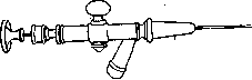
Fig. 1.
anesthesia becomes completely superfluous. With correct accounting of the manometer readings and careful passage of the needle through all tissues down to the pleural cavity, the danger of puncturing the lung and, all the more, significant trauma to it is negligible. In view of this, any devices proposed abroad and reduced to four main variations are completely superfluous: a) a rider on the needle, set further or closer to the end of the needle, corresponding to the assumed thickness of the traversed tissues; b) the use of cannulas or taps onto which the needle with a lateral outlet is placed Figure 2. for a syringe (for control aspiration to check the location of the end of the needle) (Saugmann's needle, Fig. 1); c) the use of trocars with stylets removable after penetration into the cavity (or, conversely, with a needle with a sharp end, the blunt hollow trocar is led out of the needle channel beyond the limits of its end) - Solomon's cannula (Fig. 2), Kovacs's needle, Zorraquin's needle (Fig. 3); d) the use of needles with a sealed end and a lateral slot of the channel (Fig. 4). The best needle is a platinum-iridium or nickel needle 6-7 cm long, 1-1.5 mm thick 1 with a short-cut, sharply sharpened end. When using steel needles ^^ ^ГЕШ^=^~~3^ Figure 3. they must be sterilized by dry heat (not higher than 100° to avoid melting the solder of the needle to the cannula), having previously wrapped each needle in cotton or gauze and inserted a mandrel into it. Platinum needles are sterilized by calcining on the flame of an alcohol lamp before each use (do not heat the solder spot). To clean the channel of the needle, which is sometimes clogged by tissue particles through which the needle passes, or by a drop of pleural fluid or blood, a mandrel is necessary (Fig. 5). Its length must exceed the length of the needle channel by 2-3 mm. Thanks to the mandrel, in the absence of manometer fluctuations, it is possible to orient oneself in the location of its end (the sensation of resistance indicates that the needle is in the tissue of the pleura or lung; traces of fluid or blood remaining on the cotton ball against which the mandrel withdrawn from the needle is wiped indicate blockage of the needle channel by exudate or blood from a vein). The mandrel in caliber must correspond to the needle. When using needles with a caliber of 1-1.5 mm, mandolin strings are very suitable for making mandrels. Before each use, the mandrel is passed through the flame of an alcohol burner. To avoid touching the cannula of the needle or the outer opening of the tap with a finger, the mandrel must be bent in a bayonet shape, and at the end intended for holding, it is advisable to make a loop for convenience. The moment of passing through the pleura is accompanied by the operator's hand feeling greater or less resistance of the membrane, sometimes - with thickening of the pleura due to prolonged inflammation - very significant: "the needle passes as if through thick leather, a sole"; with an unaltered pleura, this moment is vaguely perceptible. Apparatuses from which the gas introduced into the pleural cavity is obtained are proposed and are being proposed in a very large number of variations.
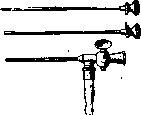
Figure 4. Kuss's needle.
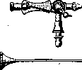

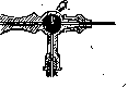
Figure 5. Carpi's needle with a mandrel.
All modifications can be divided into 3 groups: a) apparatuses consisting of two communicating, fixedly fastened vessels, of which one serves as a gasometer and the other as a hydraulic press, displacing gas through the needle into the pleura at a difference in fluid levels in both vessels. At the same time, when the cylinders are located on the same horizontal line, upon equalization of the fluid level in them, further displacement of gas can occur by means of air injection above the fluid of the press vessel with a Richardson bulb (lungs, not allowing sharp pressure increases, by pressures on the bulb) [type of apparatus of Forlanini (Fig. 6), Muralt (Fig. 7), Shternberg, Kondorsky, etc.]. When the vessel with liquid is located above the gasometer, this need for injection disappears (type of apparatus of Slobodyanik, Epstein, etc.), b) The same construction, but with the possibility of moving the vessel with liquid, raising or lowering it on a rack 70S and pinion using a system of pulleys (apparatuses of Kuss, Grass-Ulrici, Kachkachev, Oksyuzov); thanks to this detail, which allows finely regulating the pressure of the liquid on the gas and thereby controlling the speed and force of the flow of the gas jet into the pleural cavity, the construction is very expedient and deserves the widest
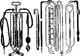
Figure 6. Forlanini's apparatus.
application, c) In the third group of apparatuses, in pursuit of portability, the cylinder-press is replaced either by a Richardson bulb (Morelli) or a large syringe (Leschke, Jousset, Carpi, Ravich-Shcherbo). A common drawback of this group is the difficulty of regulating the force and speed of introduction of gas into the pleura, and the danger of injuring the pleura if the gas is introduced too quickly. Apparatuses with communicating vessels for the sake of portability can be brought down to negligible sizes (micro-model of Slobodyan
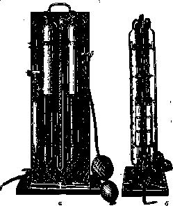
Figure 7. Muralt's apparatus. New model with two gasometers (a - front view, b - side view).
ka), and their assembly for transport can be permitted without difficulty. While permitting the use of any apparatus provided that the rules of asepsis and manometry are scrupulously observed, one should nevertheless definitely pronounce in favor of the necessity of standardizing the type of apparatus with a movable gasometer in the USSR. For the purpose of purifying the introduced gas from dust and microbes suspended in it, it is most convenient to use frequently replaced sterile cotton filters located along the path of the gas stream. Oil solutions of camphor, menthol, etc., proposed by Slobodyanik, Oksyuzov, and others, have no advantages over cotton filters, the use of which is much simpler. Nitrogen, which was initially introduced into the pleura and required the use of special cylinders with compressed gas and pressure-reducing valves to weaken the pressure of the stream when filling the gasometer, has long been replaced in the USSR and in some places abroad by atmospheric air. The use of oxygen (Muralt's new model of apparatus with two pairs of cylinders) or carbon dioxide (Ulrici) during primary insufflation for the purpose of preventing gas embolism is a completely unnecessary complication of technique, since with the correct performance of the operation the danger of gas embolism is reduced almost to zero, and with gross errors in methodology neither oxygen nor carbon dioxide guarantees against embolism. An indispensable condition for the correct design of the apparatus for artificial pneumothorax is the presence of a manometer (oscillometer) recording fluctuations in intrapleural pressure. In this case, the manometer must be in constant communication with the pleura, regardless of whether the gasometer is turned on or off. Besides spring oscillometers (Brauer), a U-shaped water manometer is most frequently used, recording the intrapleural pressure in centimeters of water column by the difference in the levels of both limbs. Additional mercury manometers recommended by some designers (e.g., Kachkachev) are unnecessary and even harmful, as they can lead to the temptation of using very high pressures (above 25-30 cm of water column), which are completely unnecessary in collapse therapy. The inclusion of an additional oil manometer (Berthier), which allows (thanks to the greater viscosity of the oil) more accurate reading of the intrapleural pressure at any moment without calculating the average readings of the oscillometer, is also not an absolute necessity. The scale of the manometer for the convenience of immediate calculation of the height of the column, consisting of the sum of equal segments of the liquid column above and below the zero point (law of communicating vessels), can have designations noting doubled readings [at the division of 1/2 cm-1 (1/4+1/4); at the division of 1 cm-2, etc.]. The liquid in the manometer should be tinted with a dye that does not precipitate from an aqueous solution and does not settle on the glass (eosin, methylene blue in a weak solution); for the convenience of setting the zero point, it is better to make the scale slightly movable. For more convenient filling of the manometer with liquid and to prevent the ejection of liquid during a sudden increase in pressure (abrupt pumping with the bulb, unexpected coughing of the patient), a bulbous expansion with a capacity of about 1/2 the volume of the liquid filling the manometer should be made on the open limb of the manometer. The pressure bulb is filled with a 3% solution of boric acid in distilled water. Tinting the liquid is unnecessary. Despite the great insensitivity of the pleura to infection by microbes entering from the outside, noted by surgeons and pathologists, and despite the low probability of a massive virulent infection from the air introduced into the pleura, the requirement of the strictest asepsis of all equipment for artificial pneumothorax remains in force. Needles are calcined or sterilized by dry heat and kept sterile. When special stopcocks are used for the needles, the latter are kept in alcohol and flamed before use. The same is done with cannulas onto which the needle is mounted, if stopcocks are not used. The operator's hands are washed (according to Spasokukotsky) with a 1/2% solution of ammonia (official solution) and lubricated with a 3-5% solution of iodine or bromferron. The site intended for the injection is lubricated in the same manner. The entire system of the apparatus is washed at least once a month with a 3-5% solution of carbolic acid (filled for a day). Cotton filters are changed at least once a decade. The room set aside for artificial pneumothorax should be considered as an operating room and used only for its intended purpose. Examination of patients and any other medical manipulations should not be performed in it. Aside from the personnel engaged in the operation and the patient on whom the operation is performed, there should be no one else in it. For primary patients, who should spend some time after insufflation in complete rest, and in case of complications, it is very advisable to have another room next to the artificial pneumothorax office, communicating with it and having a separate exit. A steady temperature not lower than 17° must be ensured for the artificial pneumothorax office (if necessary, additional heating with electric heaters). Special devices for warming the air introduced into the pleura, as experience of the majority of authors shows when introducing gas at room temperature, are unnecessary. The operation is usually performed with the patient lying on the side opposite to the side on which the insufflation is performed. A bolster should be placed under the patient's side to widen the intercostal spaces of the opposite side. The head should be placed straight on the table (or couch) without a pillow. The arm of the side to be collapsed is bent at the elbow and rests with the palm on the patient's ear. The physician pulls it back to obtain maximum separation of the ribs. When inserting the needle from the back, the patient is slightly turned with the shoulder toward the table (or couch) and the arm is pulled to the side and down. When operating on a surgical table, the surgeon stands; when operating on a special couch, he sits down to the side of the patient and works sitting. The latter method is more expedient, as it does not violate the asepsis of the operation at all, but saves the physician a lot of energy. The operation is mostly performed with an assistant who hands the iodine tampon, mandrin, switches the stopcocks, and keeps records, but with modern equipment, correct organization of the workplace, and proper preparation of patients, it is quite possible to work without an assistant. Records are kept either in the patient's medical history or on a special chart. The following entries are mandatory: date (day, month, year), side where the gas is introduced (r.-right, l.-left), initial pressure - before insufflation, when reading by the oscillometer, two figures are noted (limits of fluctuations during inspiration and expiration), e.g., -12----8; -4-0; then the amount of introduced gas in cubic centimeters is noted, and the final pressure, e.g., -6----2; -4----1-3; +6----1-12, etc. Often in partial pneumothoraces, the gas, entering a small cavity communicating by a narrow channel with other chambers of the bulla, creates a transient positive [pressure].
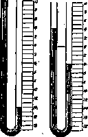
Figure 8. Position of the manometer when the needle enters the pleural cleft: a-during deep inspiration; b-during calm breathing. (In Figs. 8 and 9, the left limb communicates with the pleural cavity.)
pressure, which subsequently, after the gas has more or less significantly distributed itself, decreases—in such cases one should wait until the pressure stabilizes and record the final readings. Reading the manometer is the most critical moment of the operation, and without mastering it, one cannot undertake the induction and management of artificial pneumothorax. At the moment when the needle with its tip enters between the pleural layers (Fig. 8), the elastic traction of the lung, which tends to collapse, aspirates a small bubble of air located in the needle and in the end of the tube supplying gas to it. A continuous communication is established between the air in this primary bubble and the air above the manometer limb communicating with the needle, and thus the aspiration pull from the pleural cleft is transmitted to the liquid column of the manometer, which begins to rise and fall depending on the strength of the aspiration, the amplitude of inspiration and expiration, the presence of pleural adhesions, etc. As long as the pleural layers are not yet separated by gas, the manometer fluctuations are insignificant (amplitude of 2–4 cm); as the layers unstick, the amplitude of fluctuations becomes larger, retaining the character of "negative" fluctuations indicating the aspiratory pull of the collapsing lung. As the pleural cavity fills with gas and the lung collapses, the negative fluctuations become smaller and smaller, and a gradual equalization of atmospheric and intrapleural pressures is established (the amplitude of fluctuations is small, the readings are close to zero: -2 to -1; -2 to -1; -3 to 0, etc.). This indicates the achievement of optimal collapse in the absence of interpleural cords or filling of the cavity in the presence of adhesions. In cases where the mediastinum is very compliant and, upon gas introduction, shifts under the influence of its pressure and the aspiration pull of the other lung in the opposite direction, such pressure equalization cannot be obtained. In the presence of adhesions or diffuse fusions of the visceral and parietal pleura, the cavity fills rapidly, and the introduced gas is distributed under increasing pressure, which, if necessary, can be brought up to +20, +30 cm of water column without fear of disturbing the blood circulation in the lung (the pressure in the branches of the pulmonary artery is higher than 40 cm3 of water column). If the needle communicates with a bronchus or alveoli of the lung, oscillations around the zero point with equal fluctuations toward + or - are established (depending on the caliber of the bronchus) (Fig. 9). If the needle is stuck in airless tissues or clogged with fluid or a solid particle, no fluctuations are noted. Finally, if the needle has entered a vessel, the pressure becomes positive and the filter adjacent to the needle is stained with blood. When entering a vein, the pressure rises slowly and insignificantly; upon wounding an artery (observed extremely rarely), the pressure rise is rapid, a column of blood quickly penetrates the filter, and its pulsating fluctuations are noted. If the manometer readings are insufficiently distinct and arouse doubt, several verification procedures should be performed after instructing the patient: a) ask the patient to take a breath and hold it without closing the glottis. If the needle is in the pleural cavity, the pressure settles in the aspiration phase ("negative") and does not change while the patient holds their breath. If the needle is in a bronchus or alveoli, the pressure, after giving a negative swing upon inspiration, settles more or less rapidly at zero. If the needle is outside the air-bearing layers, no fluctuation occurs. This verification method is the most convenient and indicative; b) to check whether the needle is in a bronchus, a drop of an aromatic substance can be placed on the filter adjacent to the needle. When the needle enters a bronchus, the patient begins to smell the control substance in the exhaled air; c) sometimes, when the needle is in the subpleural fascia, clear pleural fluctuations can be transmitted to the manometer. However, negligible amounts of introduced gas (0.5–1 cm3) rapidly yield high positive pressures (the patient usually feels pain). In such cases, immediately stopping the gas inflow, the needle is carefully advanced until the appearance of even clearer and sweeping manometer fluctuations. It is absolutely mandatory to observe the basic rule: do not let in gas in the absence of manometer readings certifying that the needle tip is in the pleural cavity; during initial induction, initially give gas in small portions of 1/2–5 cm3 until a sufficient cavity is formed. Sometimes, with a thin pleura or very rapid insertion of the needle, or with fusion of the pleural layers, the needle enters very deeply beyond the bounds of the pleural cleft. In such cases, one should slowly withdraw the needle back while constantly observing the manometer. Sometimes, upon withdrawing the needle, it is possible to hit the cleft and gain the ability to introduce gas. In such cases, a puncture wound of the lung occurs, sometimes of some pulmonary capillary. This can lead: a) to minor hemoptysis (a few spittings) directly after the intervention or on the same day (the patient must be warned about this to avoid unnecessary anxiety); b) to the formation of a traumatic spontaneous pneumothorax of larger or smaller sizes. With unilateral artificial pneumothorax, this complication is mostly harmless and can pass without a trace for the patient; with bilateral artificial pneumothorax, it can assume formidable dimensions. Therefore, all such incidents must be noted in the records, even if by a conventional sign (for example, 4-), and such patients must be carefully monitored...
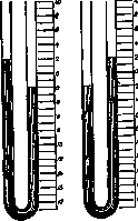
of the manometer upon entry
needles into the lung or bronchus: a - inspiration; b - expiration. should be carried out within the next few hours after the intervention. Outpatients are best detained for some time in the waiting room. When initially inducing an artificial pneumothorax, the patient must be instructed that he should lie without changing the position indicated by the doctor, breathe calmly, without holding his breath without the doctor's instructions, and not cough excessively; for those coughing, it is better to make an injection of 0.5-1.0 morphine or give 20 drops of a 1% solution of dionin 10 minutes before the insufflation, and if he feels the urge to cough, the patient should give a signal to the doctor (who must withdraw the needle from the pleura in advance). After the insufflation is completed, the patient should remain motionless for some time, then, under the supervision of a doctor, carefully sit up and move (if there are no severe pains) to another couch, on which he should stay quietly for several hours. After this, if there are no severe pains, the patient can move to the ward, or, in the case of outpatient induction, return home. With repeated insufflations in a hospital setting, patients can immediately return to the ward, while outpatients should rest in the waiting room for 10-20 minutes. The choice of site for needle insertion is determined by the nature of the process and the possible localization of pleural adhesions (the latter is far from always possible to determine due to the absence of precise signs of the presence of adhesions). Most often, one of the lower intercostal spaces between the axillary lines (e.g., between the 6th and 7th ribs) or along the scapular line (between the 8th and 9th ribs) is chosen. But sometimes, in the presence of adhesions at the bottom and the need to collapse the upper sections, it is necessary to attempt insertion of the needle in the upper spaces and even anteriorly below the clavicle. The question of the possibility of inducing an artificial pneumothorax can only be resolved by trial manometry. In cases of unsuccessful attempts to find the cavity during the first punctures (no more than 2 per session), they should be repeated after some time. The dosage of insufflations and the intervals between them depend on the patient's condition, his reaction to the insufflation, and the rate of gas absorption by the pleura (on average, at the beginning of treatment about 200 cm3 per day, subsequently 100 cm3 and less). In case of repeated failure of attempts, a course of electro-vibratory massage according to Sobelmann and Grinchar should be carried out, thanks to which in half of the cases where artificial pneumothorax cannot be induced, it is possible to loosen the adhesions, separate the adhered pleural leaves and still obtain a good collapse. Usually, with the first insufflations, moderate amounts of gas are introduced—300-500 cm3, with repetition of insufflations every other day until a good bubble is formed. Liebermeister (1931) recommends, guided by spirometry, from the very beginning to conduct insufflations in such a way as to approach the equalization of atmospheric and pleural pressures during the very first insufflations. He is not afraid to give up to 1,000 cm3 in suitable cases during primary induction. In the Moscow Regional Tuberculosis Institute, in a number of cases when artificial pneumothorax had to be induced in robust patients with a large vital capacity, large amounts of gas (700-1,000 cm3) were also allowed without any unpleasant sensations in the patients. Of course, in each individual case it is necessary to take into account the state of the patient's cardiac activity, his vital capacity of the lungs and the respiratory pause (the ability to hold the breath for a more or less prolonged time); Holzmann uses the Stange method. In the future, the regulation of intervals and the dosage of insufflations are determined by the indications of the spirometer (it can be brought up to 1/4 of the normal vital capacity of the given patient) and X-ray examination. Whenever possible, it is necessary to check the state of the artificial pneumothorax with X-rays after each insufflation, in any case initially at least once a decade, and then at least once a month. In the further course of treatment, when the pleura begins to absorb gas more slowly, the intervals between insufflations are gradually lengthened (1 time in 5, 7, 10, 15 days; towards the end of treatment, it is possible to reach 20 and 30 days in individual cases), but 1) too long intervals should be avoided to prevent untimely expansion of the lung (threat of adhesions, gaping of the cavity), 2) regular X-ray control [see separate table (articles 367-368), figures 7 and 8] over the state of the collapse should be maintained (for the purpose of timely intervention in case of impending deviations from the correct course of treatment). Thus, no schemes and templates can be allowed when carrying out treatment with artificial pneumothorax; the doctor must maneuver all the time depending on the dynamics of the given case. In cases where a complete collapse of the lung or selective compression of the affected areas is obtained, treatment can be carried out at low negative or close to zero pressures. In the presence of adhesions, an attempt should be made to achieve compression of the cavities by carrying out pneumothorax at positive pressures. However, if within 2-3 months it is not possible to obtain a firm collapse of the cavities, the question of additional surgical interventions (cauterization of cords, Jacobaeus operation, plombage, thoracoplasty, phrenicoexeresis) should be raised and the artificial pneumothorax, useless in such cases, should not be delayed, in order to avoid the formation of a pulmonary fistula or seeding of the uncollapsed lung. In cases of pulmonary hemorrhage, when artificial pneumothorax is induced for vital indications, a simultaneous introduction of large amounts of gas (up to 1,000 cm3) within the endurance of the cardiovascular system of the patient is permissible. Thanks to this, a rapid cessation of bleeding is achieved and the development of aspiration pneumonias is prevented. The methodology for performing bilateral artificial pneumothorax does not differ in principle from the described one. The question of simultaneous or two-stage induction of bilateral artificial pneumothorax is decided based on the patient's condition. Lunkevich and Willer speak out for simultaneous induction as a rule; Holzmann and his co-workers were convinced that in most cases patients tolerate two-stage induction of artificial pneumothorax more easily. The decision on which side to start the artificial pneumothorax is determined by the nature of the process. In cases where there is a suspicion that due to the duration of the process one can encounter adhesions on one side, one should try to induce artificial pneumothorax on this side first. In cases where the most active side with the freshest rashes is unmistakable, one should start with it. In the future, it is advisable to perform insufflation on both sides in one session. In the absence of adhesions on both sides, one must adhere to equally small negative pressures; in the presence of adhesions on either side and immobility of the mediastinum, one can try to achieve a better collapse on this side by increasing the pressure. The criteria for dosage in bilateral pneumothorax are the indications of spirometry and respiratory pause. If they are not lower than 50% of the norm for the given patient, insufflations can be performed calmly until the desired collapse is obtained. The physiopathology and therapeutic effect of artificial pneumothorax are based on the following main points: a) due to the separation of the visceral and parietal pleura by air, the elastic tension of the lung tissue decreases and the lung tissue collapses sometimes to the size of a complete collapse. Particularly rapidly, the affected tuberculous areas collapse (with the exception of continuous pseudolobular exudative pneumonias—coined by French authors)—a selective pneumothorax is obtained (Morgan). The decrease in volume and tension of one lung leads to the same changes in the other lung, which explains the sometimes observed therapeutic effect of artificial pneumothorax on foci in the other, uncollapsed lung.
Due to the great compensatory capacities of the lung, the exclusion of a certain part of it from breathing is compensated by healthy areas remaining outside the collapse or in slight collapse (the increase in vital capacity after insufflations above the limits at which it should have stopped according to the amount of gas in the pleural cavity); b) Due to the creation of an air interpleural barrier, the deceleration of lymph circulation in the collapsed lung, the compression and emptying of cavities, the intoxication of the patient is sharply and rapidly reduced. c) The mechanical compression of cavities creates favorable conditions for the adhesion and fusion of walls and the obliteration thus of cavities serving as sources of the spread of the process; d) Around the foci in the collapsed lung and around the cavities under the influence of the combination of all factors acting on the lung during artificial pneumothorax, including functional rest, more or less powerful fibrous capsules develop, which also inhibits the further progression of the process; e) The healthy tissue of the compressed lung during treatment is in a state of atelectasis (depending on the degree of collapse); upon the liquidation of the artificial pneumothorax and the expansion of the lung, these areas again enter into work. f) Artificial pneumothorax, proceeding at negative or weakly positive pressures with a patient's vital capacity of at least 70% of normal according to spirometry, does not cause any significant changes in gas exchange and circulation and, with a healthy heart and vascular system, is impeccably compensated; g) Bacilli located in the foci of the collapsed lung do not perish and do not lose virulence for a very long time even after the end of artificial pneumothorax (2 or more years later) and under the influence of usual causes provoking the tuberculous process (see Pulmonary tuberculosis) can give a new flare-up of the process amidst apparent complete health with the formation of new cavities and metastases (about 8% of all cases of completed artificial pneumothorax). Semeiotics of artificial pneumothorax. After the very first introduction of 250-300 cm3 or more of gas with a free pleural cavity, the auscultatory and percussive picture on the part of the collapsed lung changes sharply: a low loud percussion sound appears, Pneumothorax respiratory sounds weaken, rales are either not heard at all or their audibility is significantly weakened. X-ray control (preferably the next day after insufflation) makes it possible to note a light gap formed by the gas layer, either in the basal and lateral parts of the pulmonary field or enveloping the entire lung like a cloak. Subsequently, as the gas bubble develops and forms, another X-ray picture of artificial pneumothorax is established - depending on the completeness of the lung collapse and the lability of the mediastinum and diaphragm. In the absence of lateral, basal, and apical adhesions, the lung collapses along its entire extent with a selective predominance of the collapse of the affected area. Healthy lobes continue to breathe, although significantly less than usual; the affected area with a good collapse is almost motionless. The diaphragm, free from adhesions, makes fairly large excursions. The mediastinal organs are not displaced if the latter is stable. Respiratory sounds are not conducted or are sharply weakened. Sometimes a soft metallic sound is attached to them, like the vibration of a string (thin cords). Percussion gives a low box sound, sometimes with a tympanic tint. The outlines of pneumothorax on the X-ray vary depending on the presence of mediastinal adhesions and the degree of collapse of diseased and healthy areas. The next type is pneumothorax in the presence of apical and diaphragmatic adhesions, but with the possibility of creating a rational collapse of the affected areas by pressure on the remaining free surface of the lung. Next, partial pneumothorax should be noted, when the cavity is held by more or less thin cords with a good general collapse and, despite prolonged and energetic compression, the lung cannot collapse. Finally, the most unfavorable form for artificial pneumothorax are cases where broad planar adhesions do not allow the affected area to be compressed and only healthy tissue is compressed (Karpilovsky's negatively selective pneumothorax). In the latter cases of partial pneumothorax, in addition to the characteristic X-ray picture, the physical symptom complex also changes sharply: respiratory sounds and rales are heard; during oral auscultation, metallic rales of the uncompressed cavity are often heard (patients often indicate them themselves); in the area of the unstripped lung, percussion gives changes in the percussion sound usual for the affected lung tissue (muffling, tympanites). With an unstable mediastinum and the presence of adhesions prompting a forced collapse of the lung, further changes develop caused by the displacement of blood vessels and the heart (displacement of tones and cardiac dullness). Fluoroscopy establishes the degree of displacement of the mediastinum and its organs in the opposite direction and the displacement of the diaphragm, as well as all details of the location of the pulmonary bubble and adhesions. However, thoracoscopy (see) makes it possible to establish this location of adhesions immeasurably more accurately, since in planar projection they often take on outlines that do not correspond at all to their true dimensions. Partial displacements of the mediastinum (upper and lower weak spots) leading to the formation of mediastinal "hernias" are not uncommon. In addition to the displacement of the diaphragm under the pressure of gas and in connection with the processes of muscle degeneration with a significant loss of the normal function of the diaphragm in cases of pneumothorax, especially often in exudative pneumopleuritis, the phenomenon of balancing of the diaphragm with its paradoxical movement on the side of the collapsed lung is observed (Kienböck). During inspiration, the dome of the diaphragm on the side of the applied pneumothorax rises. The mechanism of this phenomenon is complex: partly the compliance of the diaphragm under the influence of the increase in abdominal pressure plays a role here, and partly in the case of exudates, the visibility of the elevation of the diaphragm is created due to pressure on the exudate from the mediastinum, which moves during inspiration towards the pneumothorax.
- The direct effect of artificial pneumothorax on the patient's condition becomes apparent with the correct placement of the bubble and lung collapse in the very first days: a) by a rapid lytic drop in temperature to normal, b) by an increase in the amount of sputum followed by its rapid decrease and disappearance, and further c) by a change in the hemogram and the erythrocyte sedimentation rate toward normal values, d) by weight gain, e) by a sharp improvement in the patient's general well-being, the appearance of appetite and vigor, and the rapid restoration of working capacity. The absence of these phenomena, in particular the failure of the temperature to level off and the sputum to disappear, indicates an unsatisfactory collapse. If, with a good collapse, the amount of sputum first drops sharply and then increases in the intervals between insufflations, this indicates an excessive lung collapse. The same is indicated by the absence of weight gain and even weight loss with a radiologically good lung collapse. It can be considered a rule that a well-localized, timely induced pneumothorax that has led to a good, stable collapse of the cavity gives the entire symptom complex of positive phenomena quickly (according to Holzmann's observations, within 4–6 weeks in 70% and within the first 2½–3 months in 90%), and this symptom complex is maintained until the end of treatment. Any deviation should be regarded as an indicator of insufficient collapse or an added complication. The temperature curve must be watched especially closely. Any temperature rises associated with insufflations, especially those regularly repeating after each insufflation, should make one ponder the condition of the other lung or the pleura of the collapsed side. A stepwise increase in temperature after insufflation with the appearance of pain in the collapsed side should be regarded as a signal of an emerging pneumopleuritis or, in the absence of pleural phenomena, as a result of metastasis of the process. Dyspnea during artificial pneumothorax performed at an optimal collapse with a sufficient reserve of vital capacity (at least ⅓ of normal) bothers patients little (only during fast walking, physical exertion), and its appearance should prompt the physician to carefully evaluate the condition of the heart, the other lung, and the degree of collapse. Spirometry and control of the respiratory pause provide great assistance in this. The described immediate results are observed in the vast majority of cases of significant lung collapse, even in those cases where pneumothorax gives a temporary and palliative effect; therefore, until the end of treatment, they do not yet give the right to judge the stable long-term results, since numerous complications during the course of treatment can nullify the very best results obtained initially. Complications during artificial pneumothorax can occur both during the initial induction and during any subsequent insufflation; therefore, the vigilance of the attending physician must not weaken, no matter how «easy» and «simple» the case may seem, and the meticulous execution of all technical details of the operation is mandatory from the first to the last insufflation. 1. Air embolism is currently extremely rare, from 0.03% to 0.1% in relation to the number of insufflations. This complication is not always fatal, but if all phenomena do not pass very quickly, persistent paresis, paralysis, partial anopsia, etc., may develop. Prophylaxis of embolism is achieved by continuous monitoring of the manometer readings (see above). 2. Subcutaneous, subfascial, and mediastinal emphysema develops either during the injection of air into the tissues (manometry error) or upon the penetration of air from the pleura into tissue spaces through the puncture channel during excessive air injection under high positive pressure (methodological errors), or upon the rupture of interpleural adhesions. Most of the emphysema passes almost asymptomatically (crepitus of the subcutaneous tissue, radiologically air layers in soft tissues), but it can also develop into a formidable and agonizing complication for the patient. 3. Injury to the intercostal nerve (neuralgia). Prophylaxis: introduction of the needle along the upper edge of the underlying rib. 4. Injury to subcutaneous vessels (embolism: rarely, hematomas: frequently). Prophylaxis: the same as in relation to the nerve. Pressure bandage for hematomas. 5. Injury to the lung. Most often, the matter is limited to the discharge of 2–3 spits of blood (warn the patient). Sometimes an asymptomatic spontaneous pneumothorax is observed (described by Khmelnitsky, Belyaeva), but in cases of sharply reduced lung elasticity and with bilateral pneumothorax, traumatic pneumothorax can turn into a severe complication (spontaneous pneumothorax). With ruptures of adhesions and perforation of the cavity wall, a valved pneumothorax or pleurocutaneous fistula may form (see above), which is one of the most severe complications of artificial pneumothorax. Prophylaxis of these complications: pressures close to atmospheric, and regulation of intervals between insufflations that does not allow pressure fluctuations. 6. Incomplete pneumothorax due to adhesions interfering with the collapse of the cavity (hanging cavity) and encapsulation of active foci usually leads sooner or later to metastasis of the process both in the collapsed and in the opposite lung. Thick-walled gaping cavities and massive exudative-pneumonic processes (lobitis) lead to the same. In such cases, it is necessary as soon as possible (no later than 3 months from the start of treatment) to raise the question of additional surgical interventions. If it is impossible to use other surgical methods of treatment, the question of continuing artificial pneumothorax must be decided in each individual case separately. Sometimes, by careful management of such a pneumothorax at positive pressures, it is possible to obtain a satisfactory effect. 7. Metastases and the appearance of fresh foci in the other lung (sometimes developing completely asymptomatically and detected only by X-ray and the appearance of bacilli in the sputum in cases where abacillary state has already set in). Prophylaxis: constant monitoring of sputum and X-ray, maintaining the collapse at the optimum. Therapy: in the absence of contraindications (heart, limitation of vital lung capacity, adhesions), induction of pneumothorax on the other lung. 8. The insufficiency of the respiratory apparatus (dyspnea) and cardiovascular system (dyspnea, tachycardia) developing in a number of cases, and reflex reactions from the stomach (vomiting) force the cessation of artificial pneumothorax. 9. Intercurrent pneumonias (rare in patients undergoing artificial pneumothorax treatment) can sharply worsen the patient's condition by an acute reduction of the respiratory surface of the lung. 10. Pneumopleuritis is the most frequent complication of artificial pneumothorax. If all cases are taken into account when an accumulation of fluid is detected in the pleural cavity (radiologically detectable exudates) without subjective sensations of the patient, then on average about 50% of all cases of artificial pneumothorax are complicated by pneumopleuritis; higher figures (70%) are noted by some authors (Dumarest, Murat). The percentage of pneumopleuritis proceeding with elevated temperature, pains (Sternberg's intercostal symptom), and the formation of a large serous exudate fluctuates, reaching up to 30%. Severe pneumopleuritis that does not resolve for a long time and turns into cold or hot empyemas is encountered significantly less frequently (according to the material of a number of authors, about 7%). The frequency and severity of pneumopleuritis are largely determined by the nature and extent of the process. The fresher the process, the earlier artificial pneumothorax is induced, and the better the collapse, the lower the chances for the occurrence of severe pneumopleuritis. Most pneumopleuritis cases appear 4–6–8 months after the start of treatment; in a number of cases, pneumopleuritis developed during the period of completion of artificial pneumothorax. Benign, "asymptomatic" pneumopleuritis in the sinuses (200–300 cm3) usually arises completely unnoticeably (often 24–48 hours after insufflation, during menstruation) and proceeds almost imperceptibly (splashing sound) for the patient. They often spontaneously disappear just as imperceptibly as they appeared, only to reappear from time to time. Typical tuberculous pneumopleuritis with serofibrinous exudate usually proceeds with an increase in temperature—a gradual rise over several days to moderate figures (2–3 weeks) and a gradual drop to subfebrile figures, sometimes dragging on for a long time. Pneumopleuritis is always accompanied by pain in the side. Sometimes these pains are very severe. In such cases, X-ray control reveals only minor traces of exudate and makes it possible to avoid insufflations during the period of acute development of pneumopleuritis, which can exacerbate and worsen the course of pneumopleuritis. As the pneumopleuritis subsides, the level of intrapleural pressure should be established by a manometer check, and if an increase is present, excess air should be withdrawn, while in the case of significant negative pressure indicators, it should be equalized by the introduction of gas. If the fluid stopping at a certain level is not absorbed within 4–6 weeks after the drop in temperature, it needs to be pumped out, simultaneously introducing a needle from the artificial pneumothorax apparatus to supplement gas and maintain the pressure at figures close to zero.
If this rule is not observed, the pleural hyperemia that develops during evacuation may lead to a new flare-up of pneumopleurisy, not to mention unpleasant subjective sensations for the patient. Evacuation should be carried out as dry as possible. Before withdrawing the needle, a few drops of a weak (2-3%) iodine tincture should be injected with a syringe to remove exudate from the needle channel and disinfect the puncture channel. A pressure bandage is applied to the puncture wound. The patient should, if possible, assume a position in which the puncture site will not be directed downward. Non-interference in serofibrinous pneumopleurisy, practiced by many, is irrational: the pleura becomes macerated and sclerosed, and the fibrin threads, like cables, pull the lung toward the diaphragm and costal pleura, and thus can lead to the premature termination of artificial pneumothorax and the cancellation of its effect. The next form—recurrent large pneumopleurisy with transition to tuberculous empyema—presents an even more severe complication than the previous one. Pleural sclerosis, intoxication in the presence of tuberculous pus (amyloid), abscesses at puncture sites, and pleuropulmonary fistulas followed by septic infection of the exudate (see) create a very serious situation in such cases and require special therapeutic measures (pleural lavage, siphon drainage, thoracoplasty). Usually, such pneumopleurisy occurs in cases of partial pneumothorax with the presence of subpleurally located caseous foci. Septic pleurisies are distinguished by a stormy course, high fever, and sepsis pathogens in the pus. Treatment consists of the earliest possible wide resection. In the majority of cases, obliteration of the pleural cavity develops more or less rapidly after pneumopleurisy. If this process develops even before the active foci in the lung have scarred, it can lead to new flare-ups and further progression of the process. In rare cases, pneumopleurisy is followed by a turning point in the course of the process, particularly striking when gaping cavities collapse and Koch's bacilli disappear from the sputum. As a rule, pronounced pneumopleurisy sharply reduces the effectiveness of artificial pneumothorax. Among the etiological factors causing pneumopleurisy, the following should be distinguished: a) septic infection (hematogenous-angina and similar diseases, perforation of lung foci during chest punctures during treatment); b) tuberculous infection—hematogenous, lymphogenous, and upon perforation of tuberculous foci; c) chest injuries, pleural injuries by a needle and trocar, and d) cooling of the patient, chilling of their extremities (during swimming, in the cold season, during work in the air, in a strong wind). The latter circumstance requires special training of the patient and physical-therapeutic preparation of their vasoregulators (rubbing down, rubbing of the legs and arms, alternating cold and hot foot baths or showers). Cytological examination of pneumopleurisy does not provide particularly characteristic reference points for their differentiation, but their bacteriological examination (for Koch's bacilli and sepsis pathogens) is very important and must be performed without fail. In connection with pneumopleurisy, after their resorption, fibrin clots form in the pleural cavity, located in the form of oval or spherical bodies of various sizes (sometimes the size of a chicken egg) in the sinuses and moving when the patient changes position. They are completely asymptomatic and require no treatment. No intervention is required either if a needle breaks off during insufflation or pleural puncture and the broken end enters the pleural cavity. Documented cases of long-term presence of needle parts (steel and platinum) in the pleural cavity indicate their complete harmlessness. In cases of adhesions in the lower parts of the chest, the needle can pass through the diaphragm and enter the subdiaphragmatic areas of the abdominal cavity. In this case, the manometer gives paradoxical fluctuations, opposite to what we have when the needle is introduced into the pleural cavity. Sometimes this paradoxical nature of fluctuations is not given due attention and gas is injected, believing that it is introduced into the pleural cavity. The pneumoperitoneum obtained in this way is tolerated by patients without subjective disorders and is detected by X-ray examination. Puncture of cavities has been described in the literature (the manometer gives fluctuations as when entering a bronchus). One of the frequent complications of artificial pneumothorax is mediastinal shift and mediastinal hernias. In cases where this shift leads to unpleasant subjective sensations, it can prove to be a strong obstacle to the proper management of artificial pneumothorax. In such cases, it is advisable to induce an artificial pneumothorax on the other side and equalize the intrapleural pressure on both sides. The majority of the described complications arise in connection with errors in the technique of performing artificial pneumothorax. The most frequent errors are: a) irregular X-ray control, infrequent control of sputum and erythrocyte sedimentation rate; thus, exudates and dissemination are not recognized in time; b) too long a wait in the case of uncollapsible cavities, hanging cavities, partial pneumothorax; untimely measures lead either to metastasis of the process or rupture of cavities; c) too long a wait in pneumopleurisy (see above); d) too infrequent refills: the lung expands, a stable compression of the cavities is not created; in cases of old pneumothorax, when the pleura becomes less pliable, too low a pressure develops and the threat of spontaneous pneumothorax is created; e) too frequent insufflations of large amounts of gas under high pressure, creating favorable conditions for the development of exudates, the possibility of spontaneous pneumothorax, circulatory disorders, dissemination; f) a change of physicians managing the treatment, without unity of the treatment plan and unity of methodology, creates the possibility of the above-mentioned technical errors and is a frequent cause of treatment failure; when transferring a patient to another physician, it is necessary to guarantee the correctness of the treatment. For indications and contraindications, see Pulmonary tuberculosis. Final results of artificial pneumothorax. The favorable immediate effect of artificial pneumothorax was mentioned above. It is observed in the overwhelming majority of cases. Much more important are the final results with long-term, multi-year observation of the patient in the usual environment after the cessation of artificial pneumothorax. The completeness and stability of the effect are influenced by the timeliness of inducing artificial pneumothorax. While the average percentage of restoration of working capacity is about 50%, and the disappearance of bacilli is about 70%, and mortality is about 40%, in cases of artificial pneumothorax in fresh infiltrates, more than 80% stable working capacity and disappearance of bacilli are obtained (similar data by Lunkevich and Biller, Altshullor and Johanson). Cases of fibrocavernous consumption give a significantly smaller percentage of positive outcomes and a larger percentage of relapses. As a result of these data, artificial pneumothorax acquires great social and prophylactic significance, since it can be applied in approximately 30% of all bacillus carriers, and when applied in fresh forms of pulmonary tuberculosis, it provides a rapid and stable elimination of the process. Bilateral pneumothorax is naturally less effective and gives about 40% positive outcomes with restoration of working capacity; but if we take into account that until now it has mostly been induced in far-advanced bilateral processes, this effect must also be recognized as very significant. Pneumothorax in children is used in individual cases both abroad and in our country (Markova, Markuzon); Holzmann has performed artificial pneumothorax in children from 5 to 12 years old. With the exception of smaller amounts of introduced gas (corresponding to the size of the respiratory surface), the management of artificial pneumothorax in children presents no special features. With a skillful approach, it is quickly possible to win the trust of children and ensure their ideal behavior (easier than in adults). It is much harder to discipline their parents. The question of where to start artificial pneumothorax—in a dispensary or a hospital (sanatorium)—is resolved by life itself. If it is possible to immediately send the patient to a hospital, it is of course better to induce artificial pneumothorax there. But if referral to a hospital threatens to be delayed, and the case is fresh and active, threatening dissemination every day, it is unacceptable to wait, and artificial pneumothorax must be induced immediately in the dispensary office. Even bilateral pneumothorax, despite the high frequency of complications, should not be delayed if one has to wait a long time for the possibility of placing the patient in a hospital or sanatorium. One of the most responsible moments is deciding the question of terminating artificial pneumothorax. At this point, along with extremists insisting on multi-year artificial pneumothorax, there are proponents of the opposite opinion, striving to shorten artificial pneumothorax treatment as much as possible. In fresh cases, these authors (Rubinstein) allow a 6-8 month duration of treatment. It is more correct to adhere to the criterion proposed at the time by V. E. Weinstein: artificial pneumothorax is discontinued 1-1.5 years after the moment of the disappearance of bacilli from the sputum, provided that monthly sputum checks are carried out. It is recommended to make a sputum culture (according to Hohn) before deciding the question.
Indicators of the erythrocyte sedimentation reaction, temperature, X-ray, and general well-being must certainly be taken into account. The termination of pneumothorax should be timed to a favorable season of the year (summer in the middle zone, winter-autumn in the south). It is best for the patient to be given an extended leave (or sanatorium treatment) for this period. During the reexpansion, one must closely monitor the X-ray picture of the lung, sputum, and other symptoms. Upon the appearance of signs of deterioration, artificial pneumothorax should be resumed. The process of termination itself is best carried out not in the form of a sudden complete cessation of insufflations, but by expanding the lung gradually through the lengthening of intervals and the reduction of the amounts of gas refilled. The opinion, widely held among phthisiatrians, that a terminated pneumothorax usually cannot be resumed is incorrect, since it has been possible to resume artificial pneumothorax long after its liquidation, even in cases of pneumopleuritis. As with the initial induction, the question is resolved by trial manometry. V. Kholyschansky.
Related articles
Mentioned in
Cite this page
“Pneumothorax.” Soviet Medical Encyclopedia. English translation of Bolshaya Meditsinskaya Entsiklopediya, 1st ed. (Moscow, 1928–1936), ed. N. A. Semashko. https://sovietmedicalencyclopedia.pages.dev/article/pneumothorax/