Respiration
Historical document, translated for reference. It reflects medical knowledge of the 1920s–30s and is not medical advice.
Summary
This article defines respiration as the fundamental vital process of gas exchange between an organism and its environment, involving the intake of oxygen and the expulsion of carbon dioxide. It details the comparative physiology of respiration across species, the structure of the human respiratory apparatus, and the mechanics of pulmonary ventilation.
Encyclopedia article (1928–1936)
Frequency of respiration, strength of respiratory muscles, and depth of respiration.
Classification and composition of alveolar air.
Respiration, the fundamental vital process, the task of which is to support gas exchange (see) between a living organism and the external environment surrounding it, whereby the organism introduces O2 from the outside and expels CO2, and also in some cases a number of other gaseous products of the decomposition of substances in the body, for example H, CH4, water vapor, etc. Comparative physiology of respiration. Respiration of unicellular organisms or those multicellular ones, all cells of which have direct contact with the external environment, occurs directly through the surface of the cells by way of diffusion. In more complex organisms, but with low metabolic intensity and thin external coverings, for example in worms (except annelids), respiration of cellular elements is provided through the skin and tissue fluid. With further complication of the organization of living forms, special organs of respiration arise; thus, in some arthropods—tracheae, i.e., numerous air-bearing tubules distributed throughout the body, communicating through closable openings (stigmata) on the external surface of the body with atmospheric air. The air of the tracheae serves for gas exchange with the tissue fluid (blood) of the insect. Respiration of fish is carried out by means of gills—numerous lamellar outgrowths from the wall of the foregut, abundantly supplied with blood vessels, whereby gas exchange takes place between the water washing the gills and the blood of the branchial vessels. Reptiles and amphibians already have lungs, but respiration in them occurs to a significant degree also through the skin. In birds, mammals, and humans, respiration is carried out almost entirely through the lungs, which in birds have a special structure and communicate with air sacs. Respiration of a human externally consists of alternating expansions and collapses of the chest cavity, parallel to which occurs the entry and exit of air (into the lungs and out of them) through the nose or mouth. This air exchange between alveolar and external air is only one link in the chain of processes ensuring, on the one hand, the delivery of O2 from the outside to the cellular elements of the body, and on the other, the removal of CO2 from the places of its formation to the outside. The cellular elements of the body, being, as Pflüger showed, places of O2 consumption and CO2 formation, can support gas exchange with the external environment only through the internal environment of the organism (blood and lymph). In the capillaries of the systemic circulation, internal or tissue respiration is carried out, whereby O2 partially passes from the blood into the tissues, and CO2 from the tissues into the blood. Venous blood flowing away from the tissues undergoes arterialization in the capillaries of the pulmonary circulation, entering into gas exchange with alveolar air, from which it draws O2 and into which it gives off part of the CO2—pulmonary respiration. Finally, the last link in the chain of respiratory processes accessible to direct observation is the ventilation of alveolar air (see), carried out with the help of respiratory movements—external respiration proper. Many authors extend the concept of respiration beyond the described processes of gas transport, and include in it also those metabolic processes that take place in cellular elements and are connected with the consumption of O2 and the formation of CO2. However, as Bethe rightly remarks, such an understanding of respiration would make the concept itself extremely vague and almost coinciding with the concept of 'life'. Respiratory apparatus. The respiratory apparatus proper in humans is the walls of the thoracic cavity with the muscles that bring them into motion, and the lungs with the air-bearing passages. The thoracic cavity represents a kind of cone, the base of which is concave corresponding to the convexity of the diaphragm (see), and the apex is rounded. The side walls of this cone-shaped space are formed by the skeleton of the chest cage and intercostal muscles, the base—by the diaphragm, and the apex—by the neck muscles, blood vessels, and nerves, whereby all the spaces between these formations are filled with connective tissue. Inside this hermetically sealed cavity are located the lungs (see), filling it almost entirely. Each lung is covered by the pleura (see), the visceral layer of which, at the place of the lung hilum, i.e., where the main bronchi (see), accompanied by vessels, enter the lungs, passes into the parietal layer. The parietal layers of both sides, having turned from the lung hilum forward and backward, pass inside the thoracic cavity partly completely freely (posteriorly), partly adhering to the pericardium (anteriorly), thus forming between them the mediastinal cavity (see). Having reached the walls of the thoracic cavity, the parietal layers grow to them, whereby between the layers of the pleura around each lung remains a capillary slit, containing in a normal state only an insignificant amount of fluid. This capillary slit has received the name of the pleural cavity, or cavity of the pleura, although in reality there is only a capillary space, and a cavity is formed only under certain pathological conditions, e.g., in pleurisy (see) or pneumothorax (see). The entire thoracic cavity is thus subdivided into a part occupied by the lungs with their cavity, containing air and communicating by means of air-bearing passages with the atmosphere, and into an extra-pulmonary part, the mediastinum, in which are located the heart and large vessels, esophagus, thoracic duct, trachea, main bronchi, etc., and both pleural cavities. Air-bearing passages are called the uniquely constructed system of tubes through which the connection of the lung cavity with the external atmosphere is carried out. One distinguishes upper air-bearing passages, from the openings of the nose and mouth to the vocal cords, and lower—from the latter to the alveoli (see) of the lungs. The normal path of air should be considered the nasal passages. Their significant surface, thanks to the conchae, and the abundant vascularization of their mucous membrane ensure the heating of the inhaled air almost to body temperature and its saturation with water vapor, as well as its liberation from the greater part of dust before the air reaches the larynx. During respiration through the mouth, all this is carried out to a significantly lesser degree, and therefore training oneself to breathe through the nose even during physical work has essential prophylactic significance. The lower section of the air-bearing passages begins with the trachea (see), which divides at the level of the IV-V thoracic vertebra into two primary bronchi, each of which gives in the corresponding lung by way of multiple branching an extensive system of increasingly thinning small bronchi, which, however, retain cartilaginous inclusions in their walls, which makes these tubes non-collapsible. The small bronchi finally pass into bronchioles, the thinnest tubules, already devoid of cartilage in the walls, and the bronchioles—into alveolar ducts, or alveoli, blind vesicles with cellular walls, abundantly permeated, also in their protrusions, by a network of capillaries of the pulmonary artery. In the alveoli, gas exchange (pulmonary) takes place between the blood and alveolar air. The thickness of the epithelial layer through which gases must diffuse during this is equal, according to Zuntz and Loewy, to 0.004 mm, and the entire surface through which diffusion occurs, i.e., the internal surface of the entire sum of the alveoli of the lungs, according to Aeby, is equal to about 100 m2, and according to the latest measurements by Wilson (1922)—approximately half as much. At birth, the lungs fill the thoracic cavity, being themselves in a collapsed (atelectatic) state. Such correspondence between the dimensions of the thoracic cavity and the lungs is preserved still in the first week of life; in the future, however, the growth of the dimensions of the thoracic cavity proceeds faster than the growth of the lungs, and since in the latter after the first breath there is always air under atmospheric or close to it pressure, then by this pressure the elastic lungs are stretched as the thoracic cavity expands so that the layers of the pleura all the time remain in close contact. Such gradual stretching of the lungs ends by the end of the formation of the organism, whereby the lungs fill the thoracic cavity, being stretched far beyond the limits of their natural volume. As a result, 'elastic recoil of the lungs' develops (i.e., their tendency to collapse), counteracting atmospheric pressure and preventing it from being transmitted entirely to everything that is located inside the thoracic cavity outside the lungs. Donders measured the magnitude of the elastic recoil by tying a manometer tightly into the trachea of a corpse and then producing a pneumothorax (see) by opening the thoracic cavity. Starting from the position of exhalation, he found the magnitude of the recoil to be equal to 6 mm Hg, and in the position of inhalation—up to 30 mm. The latter figure is undoubtedly exaggerated, since the position of inhalation on a corpse was achieved by inflating the lungs. Aron measured the pressure inside the pleural space on a living human by pricking a hollow needle connected to a manometer into it obliquely through the intercostal space. The pressure turned out to be always less than atmospheric and, moreover, during quiet inhalation by 4.64 mm Hg, and during quiet exhalation by 3.02 mm Hg.
The existence of constant negative pressure in the extra-pulmonary space and its increase with each inhalation reflect significantly on blood and lymph circulation, lying at the basis of the so-called suction action of the chest. Mechanism of lung ventilation. In the process of respiration, the chest cavity undergoes alternating expansions and contractions due to corresponding movements of the chest wall. During expansion of the chest cavity, the lungs expand passively due to the pressure of alveolar air; this latter becomes less than atmospheric, because the air is distributed in a larger volume and consequently conditions are created for the flow of air from the outside, from the place of higher pressure, into the alveoli, to the place of lower pressure—inhalation occurs. During contraction of the chest wall, the lungs collapse by virtue of their elastic tension, the air pressure in them becomes higher than atmospheric, and thus conditions are created for the flow of air from the inside to the outside—exhalation occurs. Expansion of the chest cavity during inhalation occurs in three directions: front-to-back, right-to-left, and top-to-bottom. In the first two directions, expansion is carried out thanks to the raising of the ribs, whereby the sternum is pushed forward, which increases the sagittal diameter of the cavity, and the bulges of the costal arches, directed downward when the ribs are lowered, move outward, which increases the frontal diameter of the cavity; its vertical diameter increases due to the lowering of the diaphragm (see). The view expressed by some authors that during quiet respiration the diaphragm does not produce active contractions, but only changes its tone, thereby counteracting abdominal pressure during inhalation, cannot yet be considered sufficiently substantiated. The view regarding the active role of the lungs themselves during their ventilation is equally poorly substantiated. During quiet inhalation, the raising of the ribs is carried out by the contraction of the external intercostal and internal intercartilaginous muscles, while during forced respiration a number of auxiliary muscles take part in the expansion of the chest wall (see Respiratory muscles). Contraction of the chest wall during quiet exhalation occurs by virtue of the elastic traction of the wall itself, which has been brought out of its equilibrium position and returns to it upon the cessation of the traction of the inspiratory muscles. During somewhat intensified respiration (and according to some authors even always), the internal intercostal muscles and m. transversus thoracis take part in the lowering of the chest wall. With significant difficulty in exhalation, auxiliary respiratory muscles and muscles of the abdominal press come into action, increasing intra-abdominal pressure and pushing the diaphragm toward the chest cavity. Besides the actual respiratory movements, during respiration there occur so-called concomitant movements: of the nasal wings, clearly visible in humans during intensified respiration, and the widening of the glottis during inhalation. From anatomical relationships (length of ribs, location of the diaphragm) it follows with obviousness that the increase in the volume of the chest cavity during inhalation falls mainly on its lower sections, however, thanks to their elasticity and mobility, the lungs expand in all their parts, although perhaps in the lower sections still more than in the apices. In any case, if one considers vesicular respiration a sign of air entering the alveoli, then, since it is normally heard everywhere, air enters all the alveoli. Registration of respiratory movements of the chest wall is carried out with the help of: 1) stethographs, allowing registration by means of a lever or air transmission of changes in the position of two points of the chest wall relative to each other, 2) pneumographs (see), giving the possibility to register changes in the cross-section of the chest wall at various levels, and 3) by means of registering pressure fluctuations in the airways, for which the airways are connected to a tube, the side branch of which is connected by a rubber tube to a Marey capsule or a water manometer. By all these methods are registered: the rhythm of respiratory movements, their frequency and relative depth; the actual depth of respiration is determined by measuring the volume of inhaled air with the help of a spirometer (see) or gas meter (see). Studying the recorded curves of respiratory movements, pneumograms, one can see that during normal, quiet respiration, so-called eupnea, inhalation begins with moderate speed, then accelerates, then slows down again, and finally sharply transitions into exhalation, which proceeds slowly at first, then accelerates, and at the end strongly slows down again. This very gentle part of exhalation is sometimes considered a respiratory pause, however incorrectly, because during quiet respiration true pauses, i.e., complete rest of the chest wall, usually do not occur. The phase of inhalation is usually shorter than the phase of exhalation, but only slightly; thus, according to Vierordt and Ludwig, the time of inhalation relates to the time of exhalation as 10 to 14, and according to Ewald as 11 to 12. However, cases where exhalation is equal to inhalation or even shorter than it are exceptions. Pneumograms of various levels of the chest wall give an idea of the so-called "type" of respiration, namely, they clarify that men expand the chest cavity by lowering the diaphragm, and women by raising the ribs (Hutchinson), just as children of both sexes up to 10 years old do. However, a mixed type is also observed. The difference in the type of respiration is revealed clearly only during quiet respiration, while during forced respiration in both sexes the expansion of the chest cavity occurs mainly due to intensified raising of the ribs. The female type of respiration is explained by natural adaptation in view of pregnancy, and in any case, tightening the waist with clothing accessories does not play a decisive role in this regard. Frequency of respiration, strength of respiratory muscles, and depth of respiration. Frequency of respiration, i.e., the number of respirations per 1 minute, in an adult during quiet standing lies within the limits of 12-24 and even—mostly between 16 and 20. There are, however, individual indications of significantly lower figures in normal subjects. Thus, Speck gives for himself as an average figure 6.4, and Liljestrand and Lindhard observed one subject with 5.4-6.4 respirations per 1 min. Frequency of respiration depends on age; in newborns it is equal to 60-70 per 1 min., at the 5th year—26, at the 15-20th years—20, at 25-30 years—16. It decreases during sleep, increases during the transition from lying to sitting and to standing, and also after eating, at high external temperature, and during fever. The frequency of respiration can increase twofold and higher during physical work. All these changes in the rhythm of respiration depend either on changes in the mechanical conditions of respiration or on changes in the intensity of metabolism. An increase in obstacles to respiration and an increase in metabolism entail an increase in the frequency of respiration and vice versa. However, prolonged difficulty in the flow of air in the airways during narrowing or from external devices—valves, masks, gas masks, etc.—mostly entails over time a drop in the frequency of respiration (Rohrer). The strength of respiratory muscles, expanding or narrowing the chest cavity, is measured by the magnitude of positive or negative pressure which they can develop in the airways separated from the atmosphere. This measurement has received the name pneumatometry, and the ordinary mercury manometer which is used for this is called a pneumatometer. To avoid the pressing or sucking action of the oral cavity, the manometer is hermetically connected by means of a three-way tube and two olives to the nasal openings and, having closed the mouth tightly, maximum inhalation or exhalation is performed. Negative pressure during inhalation reaches -100 mm of mercury column, while positive pressure during exhalation reaches +150 mm; consequently, the strength of expiratory muscles significantly exceeds the strength of inspiratory muscles. The work of respiratory muscles is determined, according to Zuntz, by comparative determinations of oxygen consumption during quiet and forced respiration. The daily work of respiratory muscles under normal conditions is considered equal to 15,000 - 20,000 kgm (Du Bois-Reymond, H. Boruttau). The depth of respiratory movements is determined by the magnitude of the volume of inhaled or exhaled air, consequently with the help of a spirometer or gas meter, whereby it is necessary to separate inhaled air from exhaled (see Gas exchange) with the help of valves. The observed volumes are reduced to 0° and 760 mm according to the rules of gas analysis (see). In an adult human at rest, the depth of respiration is equal on average to 500 cm3, however with very wide individual fluctuations—from 300 to 900 cm3 (Staehelin and Schutze), and during muscular work it reaches 3 liters and higher. In view of significant fluctuations of both frequency and depth of respiration, and moreover usually in the opposite sense, both these values, taken separately, cannot serve as a characteristic of the magnitude of lung ventilation and characterize it only in their aggregate—the product of frequency by depth, i.e., the minute volume of respiration; fluctuations of this value at rest lie in narrower limits: from 4 liters to 10 liters per 1 min. and even more often from 6 liters to 8 liters. During difficult respiration (mask, valves, tubes, etc.) and especially during an increase in the intensity of metabolism, the minute volume increases and during intensified work can increase 7-10 times against rest—up to 50-60 liters per 1 minute.
This increase in minute volume during work is usually conditioned by an increase in both the frequency and depth of respiration, whereby individually one or the other is increased more. During artificial maximal respiration, on the contrary, opposite changes in the frequency and depth of respiration are observed: Frequency per 1 min.
60 Depth in liters.
3.43
3.34
41.15
50.12
63.7
61.8
As can be seen from the table, the depth decreases with an increase in frequency, at first slowly, then faster. The entire possible mobility of the respiratory apparatus can still be utilized at a low frequency, approximately up to 15 respirations per 1 min., but already at 30, the depth drops to 2/3, and at 60 to 3/4 of the initial value. The minute volume also initially increases in parallel with the increase in frequency, reaches a maximum between 20-30 respirations, and then falls. Classification and composition of pulmonary air. The volume of air inhaled and exhaled during normal quiet respiration is called tidal air; it is equal on average to 500 cm3. Having finished a normal inhalation, one can continue it significantly further, introducing so-called inspiratory reserve air, equal to 1,600 cm3. On the other hand, after a quiet exhalation, one can also exhale an additional reserve, or supplementary air, equal to 1,600 cm3. The sum of tidal, inspiratory reserve, and expiratory reserve air, equal to 3,700 cm3, gives the vital capacity of the lungs. All indicated values show significant individual fluctuations. Thus, in persons engaged in sports, the vital capacity often reaches 6,000 cm3; in women, it is generally lower than in men; with age, approximately from 40 years, it also decreases, at first slowly, then faster. After a maximal exhalation, so-called residual air, which is spirometrically immeasurable, still remains in the lungs. In a living human, it is measured using the dilution method. A gasometer containing a certain amount of some gas, for example H or N, is connected to the human respiratory tract after maximal expiration, and the subject is made to take several (5-6) breaths to form a uniform mixture. The experiment ends after a maximal exhalation into the gasometer; the mixture in the gasometer is analyzed, and based on the data obtained, the volume of residual air is calculated. On average, it is taken as equal to 1,200 cm3. Thus, after a quiet exhalation, reserve and residual air remain in the lungs, the sum of which—2,800 cm3—has received the name of alveolar air (or 'normal capacity'; Bohr). Since gas exchange in the alveoli occurs continuously, in essence, the alveolar air participates directly in it, while the tidal air merely ventilates the alveolar air. If, in a series of quiet respirations, one inhalation of hydrogen is made and, during subsequent quiet respiration, the presence of H in the exhaled air is investigated, it turns out that the H disappears, i.e., the air is completely renewed within 6-10 respirations. Inhaled air, however, does not enter the alveoli completely, because a part of it, at the end of inspiration, fills the airways in which no gas exchange occurs. This part of the inhaled air, upon the onset of expiration, is the first to be removed outward with a greater or lesser admixture of air from the alveoli, due to which the exhaled air is not identical in its composition to the alveolar air (see below). The capacity of the dead space was determined by Levy, and then by Zuntz, on a cadaver by filling it with plaster, and they obtained 140-144 cm3. Using Bohr's formula (see Alveolar air), one can, knowing the composition of inhaled, exhaled, and alveolar air, calculate the volume of dead space in a living human as well. However, the methodology for determining the composition of alveolar air, which is also constantly changing in essence, does not yet provide a reliable basis for calculations. It is apparently certain that the volume of dead space in the same subject changes depending on the intensity of respiration, increasing during strenuous respiration—probably due to the dilation of the bronchial tree. Thus, Krogh and Lindhard found the volume of dead space to be 80-100 cm3 during quiet respiration, and 170-190 cm3 during maximally forced respiration. If we assume the volume of dead space to be 140 cm3, then out of a tidal volume of 500 cm3 during quiet respiration, only 360 cm3 serves for the ventilation of the alveoli, i.e., 1/6 of the volume of alveolar air. The changes undergone by external air during its passage through the lungs are established by comparing the chemical composition and physical state of the inhaled air and the exhaled air, which is most convenient when breathing pure atmospheric air, the composition of which is extremely constant, unlike the air in various living quarters (see Air). An illustration of the constancy of the composition of atmospheric air can be provided by the following table (Liljestrand) of oxygen analyses performed by prominent researchers in various parts of the globe and over the course of more than a century (the calculation is made for dry air). Year of publication, Author, Percentage of oxygen, Method of analysis: Humboldt and Gay-Lussac, 21.0, Eudiometric; Bunsen, 20.96, Eudiometric; Kreusler, 20.92, Eudiometric; Hemmel, 20.91, Eudiometric; Haldane, 20.93, Absorption of alkalis by pyrogallol; Benedict, 20.93, Absorption of alkalis by pyrogallol; Krogh, 20.95, Absorption of alkalis by pyrogallol; Carpenter, 20.948, Absorption of alkalis by pyrogallol. The content of CO2 in the air is just as constant, and only its humidity can fluctuate within wide limits. On average, the composition of dry atmospheric air will be: 20.94% oxygen, 0.03% carbon dioxide, and 79.03% nitrogen, argon, and other noble gases. Exhaled air differs from inhaled air by a higher temperature (30-32°) and saturation with water vapor. Representing a mixture of dead space air and alveolar air, exhaled air fluctuates significantly in its composition even during quiet respiration depending on the individual method of respiration, but in any case, it contains less O2 and more CO2 than inhaled air. The N content in it is usually slightly higher than in inhaled air, because the volume of O2 absorbed usually slightly exceeds the volume of CO2 exhaled; consequently, the difference in the content of N, which does not participate in respiration, depends on the difference in the volume of inhaled and exhaled air. Therefore, by the volume of exhaled air, multiplying it by the ratio of the percentage of N in exhaled air to the percentage of N in inhaled air, one can also calculate the volume of inhaled air. On average, dry exhaled air contains 16.4% O2, 3.8% CO2, and 79.8% N. The dependence of the composition of exhaled air on the frequency and depth of respiration, and consequently on the minute volume, but under other equal conditions, is illustrated by the following table (according to Liljestrand): Frequency of respiration, Depth of respiration, Minute volume, Percentage of CO2 in exhaled air, Percentage of O2 in exhaled air, Number of experiments: 5, 965 cm3, 4,825 cm3, 4.31, 18.05, 21; 10, 547 cm3, 5,470 cm3, 3.72, 16.72, 16; 15, 396 cm3, 5,940 cm3, 3.27, 17.08, 8; 20, 359 cm3, 7,160 cm3, 2.84, 17.62, 12; 30, 203 cm3, 8,790 cm3, 2.34, 18.22, 12. The exhaled air of some animals, predominantly ruminants, contains H and CH4, which are formed during fermentation processes in the intestine and absorbed from there into the blood. These same processes can be a source of CO2 and its increased content in exhaled air. According to Brown-Séquard and d'Arsonval, exhaled air contains some poisonous substance, but both the studies of other authors and observations on submarines speak against this. Probably (Formanek), in Brown-Séquard's experiments, there was an accumulation of NH3 formed from animal excreta. Formanek showed that NH3 appears in the exhaled air of healthy people only during bacterial processes in the respiratory tract, mainly in the oral cavity (carious teeth, decomposition of food residues, etc.). Weichardt found in exhaled air a protein-like substance, which he named kenotoxin (see), the source of which is apparently droplets of liquid carried away by the air current from the walls of the respiratory tract. During starvation and some pathological conditions, acetone is contained in exhaled air, and after the consumption of alcoholic beverages, alcohol, which is released from the blood into the alveolar air. Gas exchange in the lungs. Pulmonary respiration proper, i.e., gas exchange between alveolar air (see) and blood (see Blood gases), is a process continuously occurring in time and conditioned by the difference in the tension of oxygen and CO2 in the alveolar air and in the blood (Pflüger). The tension of gases in alveolar air, i.e., their partial pressure, is determined by their percentage content in dry air, and since on average at rest or during moderate work the percentage of O2 fluctuates within the limits of 14-15.4, and the percentage of CO2 within 4.9-6.3, then consequently the tension of O2 is equal to 100-110 mm of mercury, and CO2 to 35-45 mm. By occluding a part of the bronchial tree in a dog, Pflüger showed that the tension of CO2 in the air of a limited part of the lung only equilibrates with its tension in the venous blood flowing to the lung, but never exceeds it. He determined the tension in the blood using an aerotonometer, which consisted of two parallel and vertically placed glass tubes connected at the top by a three-way stopcock, through which blood from the animal's vessel (vein or artery) was let directly into both tubes.
Tubes, installed for temperature constancy in a water bath, were filled beforehand with air having such a CO2 content that in one tube its tension was higher than that expected in the blood (based on preliminary experiments with occlusion of a part of the lung), and in the other—lower. Blood introduced into the tubes flowed down their walls and was collected in special receivers for analysis of the gases within it. If a decrease in CO2 content was found in one tube, and an increase in the other, and moreover to one and the same level, then this latter obviously corresponded to the CO2 tension in the blood. By analyzing the alveolar air of animals in parallel, Pflüger showed that the CO2 tension in it is always lower than in venous blood. For O2, however, there are inverse relationships: its tension in alveolar air is 10-15 mm of mercury higher than in the arterial blood flowing from the lung. Thus, Pflüger came to the conclusion that gas exchange in the lungs is accomplished by way of diffusion. Bohr opposed this doctrine with his secretion theory of pulmonary gas exchange, in which the lung was viewed as a gland secreting gases, and moreover in the opposite direction. Subsequently, however, it turned out (Krogh) that Bohr's experiments, upon which his theory was based, were erroneous. The experiments of Zuntz and Levy on membranes and excised lungs also speak in favor of the diffusion theory. These experiments showed that the existing difference in tensions fully ensures such a rate of diffusion at which respiration can satisfy all the needs of the organism, even (according to Barcroft's experiments) during respiration at altitudes of about 5,000 m, where the O2 tension in the air is only 84 mm, and according to observations of the Himalayan expedition (1923)—even at higher altitudes, up to 8,225 m, where the O2 tension = 56 mm. Thus, to the present time, no facts have been established that contradict Pflüger's theory. The last stage of gas exchange, internal or tissue respiration, i.e., the exchange of gases between the blood and tissues, is also explained by Pflüger by diffusion. It was shown by him and his students that there is no free O2 in the tissues, its tension there is equal to zero, whereas the tension of CO2, which is produced in the tissues, in all those fluids which are a direct product of cellular elements and are collected, of course, outside of contact with the atmosphere, is higher than in the blood of the capillaries of the systemic circulation; for example, in acidic urine—68 mm, in bile—50.0 mm, in hydrocele fluid—46.5 mm, whereas in arterial blood it is 21.28 mm. It was further established by the experiments of Bohr, Barcroft, and others that the diffusion exchange of O2 and CO2 in the lungs and in the tissues is extremely favored by their mutual influence on their bond with the blood: the saturation of blood with oxygen loosens its bond with carbon dioxide and vice versa. Omitting the quantitative side of respiration (see Gas exchange), one should, however, emphasize Pflüger's position that the intensity of gas exchange is determined by the need of the living cells of the organism and can be changed only by way of changing the vital activity of these latter. In accordance with this, purely external conditions of respiration, e.g., the composition of the air or its pressure, can vary within significant, but still, of course, definite limits, without any substantial influence on respiration. Thus, O2 consumption does not change whether one breathes pure O2 or air containing only 10% O2. In the latter case, breathing only becomes somewhat deeper; at 8% O2, a person feels unpleasant (Speck), and with further lowering, dyspnea and dizziness occur. In animals, at 7%, significant dyspnea appears, at 5-4.5%—very sharp dyspnea with clouding of consciousness, and at 3%, suffocation quickly ensues. All this is a consequence of the fact that the partial pressure of oxygen in the alveolar air becomes less and less capable of ensuring the transfer of O2 into the blood by way of diffusion and its saturation of Hb. Analogous phenomena are observed during ascents to high mountains (see Mountain sickness) or during air flights to great altitudes. Already at an altitude of about 3,000 m, the pressure drops to 2/3 of normal, at 5,500 m—to 1/2, and at 8,500 m—to 1/3. A lack of O2 makes itself felt in many healthy people already at an altitude of 3,000-4,000 m, in the majority—at an altitude of 4,500-5,500 m, but individual persons can tolerate 6,000 m, like Zuntz and his collaborators, and even 8,227 m, like the participants of the Himalayan expedition. An ascent to 8,600 m (pressure equal to 240 mm Hg) in the balloon "Zenith" cost the lives of two aeronauts, Sivel and Crocé-Spinelli. As calculations by I. M. Sechenov showed, respiration at an air pressure of about 300 mm becomes impossible due to the fact that the percentage content of O2 in the alveolar air drops in this case to 4.7, consequently its tension—to 14 mm, and thus the saturation of the blood becomes completely insufficient. Experiments by Paul Bert on animals with an increase in air pressure showed that up to almost 10 atmospheres, animals show no signs of distress. Above 10 atmospheres, restlessness appears, intensifying with further increase in pressure, and finally, within the limits of 15-20 atmospheres, the animal develops strong convulsions, and it dies. Paul Bert showed that the cause of death is not the high total air pressure, but the high O2 tension. He placed animals in pure O2 and, by increasing its pressure, observed that already at 2 atmospheres the animal's restlessness begins, and at 4 atmospheres the animal dies, although in air at 4 atmospheres it remained completely normal. Thus, high partial pressure of O2 proves to be fatal for the animal, with the latter's body temperature and the intensity of oxidative processes decreasing, similar to how this takes place also with a lack of O2. There is as yet no explanation for this phenomenon. Man has to deal with high air pressure, but within limits not higher than 5 atmospheres, during diving and caisson work. Under these conditions, inhalation becomes easier and shorter, exhalation somewhat difficult and prolonged, the number of breaths decreases (by 2-4 per 1 minute), and pauses appear. Quantitatively, gas exchange either does not change or increases somewhat. In all cases of transition from low pressure to high, or vice versa, extreme caution and gradualness are necessary to avoid air embolisms or mechanical injuries, e.g., rupture of the eardrum. An increase in the CO2 content in the inhaled air immediately results in an increase in its tension in the alveolar air, which in turn, by hindering the exit of CO2 from the blood, entails an accumulation of CO2 in the blood. The result is an intensification of pulmonary ventilation, which, with a further increase (up to 5-10%) in CO2 content, turns into dyspnea, which, however, weakens at 15-20% CO2; the animal falls into a comatose state, at 25-30% anesthesia develops up to the loss of the corneal reflex, and at about 40% quiet death occurs. Regulation of respiratory movements. The fetus of man and mammals, while in the mother's womb, does not produce respiratory movements, since its gas exchange with the mother's blood is sufficiently ensured by placental circulation, while the interruption of the latter (during childbirth in humans or by clamping the umbilical cord in an experiment on an animal) induces respiratory movements. On the other hand, if a child is born in asphyxia, respiratory movements can be induced in it by irritation of sensitive, e.g., cutaneous, nerves. Consequently, that place of the central nervous system from which impulses for respiration emerge acts both autochthonously and reflexively. All respiratory muscles of the chest cage receive nerves from the anterior horns of the spinal cord, starting from its cervical part (phrenic nerve of the diaphragm) to the lumbar inclusive (muscles of the abdominal wall), while the muscles of the wings of the nose (facial nerve) and the larynx (recurrent nerve)—from the medulla oblongata. The nuclei of all these nerves could be considered as respiratory centers; however, if one cuts the spinal cord at the level of the VII cervical nerve, then costal respiration ceases, but diaphragmatic remains; if one cuts the spinal cord above the III cervical nerve, then diaphragmatic respiration also ceases, but the movement of the wings of the nose remains. If, finally, one makes a cut through the brain stem above the medulla oblongata, without disturbing the integrity of the spinal cord and its connection with the medulla, then the movements of the chest cage remain normal, and the movements of the wings of the nose cease. On the other hand, Legallois (1811), and then Flourens (1842), showed that the destruction of the section of the rhomboid fossa somewhat above the calamus scriptorius, at the level of the exit of the VIII-X pairs of cranial nerves, causes immediate cessation of respiration and death of the animal (Flourens' vital node). Through the investigations of Gad and his students, Mislavsky and others, it was established that the region of the respiratory center is wider than Flourens thought, and is limited anteriorly by the posterior edge of the nucleus of the facial nerve, and posteriorly less sharply somewhat below the calamus scriptorius.
This center is bilateral, and splitting the medulla oblongata along the midline does not disrupt the synchronicity of the movements of both halves of the chest; if, however, on either side one additionally transects the spinal cord below the medulla, then the movements of the chest on that half cease. There are indications (Lewandowsky) that both centers are nevertheless connected to each other commissurally and that, besides the inspiratory center, there is an expiratory center located (Lumsden) in the lower half of the rhomboid fossa. Langendorf and Wertheimer, objecting to the recognition of a special bulbar respiratory center, explained all the effects of transections or destruction of the respiratory center as a consequence of shock; however, W. Trendelenburg, having performed the separation of the spinal cord from the medulla oblongata without irritation (by wrapping the spinal cord of a rabbit with the small intestine of a guinea pig and passing ice water through the intestine), showed that in this case, too, Respiration ceases. The activity of the respiratory center is maintained by internal, or autochthonous, stimuli and by reflex stimuli. The former include irritation by the venousness of the blood, i.e., by a lack of oxygen and an excess of CO2, whereby both the one and the other can in themselves act as a stimulus, as shown by experiments with Respiration using appropriately composed gas mixtures. The general significance of the venousness of the blood as an excitant of the respiratory center was proven by Fredericq. In two dogs, whose aa. vertebrales had been previously ligated, he cross-connected the ends of the severed carotid arteries (adjacent ones in dogs lying side by side), while he clamped the other carotid arteries with forceps and closed the trachea of one dog—then the other dog, whose head was supplied with the venous blood of the suffocating neighbor, fell into dyspnea. In more recent times, H. Winterstein reduces the "venousness" of the blood to the action of the concentration of hydrogen ions, whereby the respiratory center is excited both by their accumulation in the blood (from a delay in the elimination of CO2 or from the entry of lactic acid into the blood from working muscles) and by their accumulation in the center itself during an insufficient supply of O2, as a result of which further oxidation and, consequently, the elimination of metabolic products of an acidic nature are delayed. There are, however, indications that the HCO3 anion also has specific significance. Besides the chemical composition of the blood, the respiratory center is excited by an increased temperature of the perfusing blood, whereby mainly the frequency increases, whereas with chemical irritation, the depth of respiratory movements increases. Among the reflex stimuli of the respiratory center, a special place is occupied by those coming to it from the lungs along the vagus nerves. Hering and Breuer showed that with intact vagi, any expansion of the lung causes expiration, and collapse causes inspiration (self-regulation of Respiration). Upon bilateral transection of the vagus nerves, the rhythm of respiratory movements changes: they become slower and deeper, inhalations take on a tetanic character, and the respiratory pause becomes clearly pronounced (vagal dyspnea). The presence of an excitation wave in the severed vagus nerve during inflation of the lung was proven by Lewandowsky galvanometrically. Thus, the normal rhythm of respiratory movements is ensured by reflex stimuli of the respiratory center. Furthermore, the rhythm can be influenced by stimuli from the most diverse sensory nerves, causing peculiar respiratory movements, such as sneezing, coughing, yawning (see), Cheyne-Stokes Respiration, etc. Finally, the activity of the respiratory center is also influenced by the overlying subcortical and cortical parts of the brain, which is proven on the one hand by the possibility of voluntary change in the course of respiration, and on the other hand by affective and vegetative influences upon it. The undoubted existence of a physiological connection of the respiratory center with the cerebral cortex gave rise to the finding (Mavrakis and Dontas) of a cortical field of Respiration in the upper part of the anterior central gyrus, above the center for occipital musculature, which, however, cannot be considered generally accepted.
M. Shatershteyn. Tissue respiration. Tissue respiration is a comparatively new concept. The existence of gas exchange in tissues was firmly established only in the second half of the 19th century. Lavoisier, who laid the foundation for the theory of respiration, established the analogy between respiration and combustion, and showed that respiration is the source of animal heat and other forms of energy in the animal organism, did not yet precisely determine in which part of the organism the processes of combustion or respiration occur. Both Lagrange and Hassenfratz spoke out against the formation of CO2 in the lungs; finally, Spallanzani and then Edwards experimentally proved that the formation of CO2 is a phenomenon common to all living beings and is not directly connected with the absorption of O2. On the basis of his experiments, Edwards came to the conclusion that CO2 is formed not in the lungs, but in the entire organism. Subsequently, Magnus, by studying blood gases, proved that arterial blood contains more O2 and less CO2 than venous blood, and considered it probable that the O2 absorbed by the lungs binds immediately with the blood and, spreading through the arteries to the entire body, serves in the capillaries for combustion and the formation of CO2. The results and the conclusions based on them by Magnus were confirmed by a whole series of authors. On the basis of other premises, Claude Bernard and Berthelot arrived at the same conclusions. However, even in the middle of the 19th century, a dispute continued between two of the most famous German physiologists of that time—Ludwig and Pflüger—which ended in favor of Pflüger, who succeeded in proving that O2 penetrates through the walls of the capillaries into the living cell, where oxidations occur, the magnitude of which depends on the needs of the cells themselves. This conclusion of Pflüger is confirmed by all subsequent works, and at the present time it is considered firmly established that the intensity of combustion depends not on the speed of blood flow and not on the amount of hemoglobin, but exclusively on the activity of the cell itself. Confirmation of the positions set forth by Pflüger are the studies of P. Bert (1870) and Hermann (1867), which showed that excised tissue continues to absorb O2 and release CO2, by which the localization of oxidative processes within the tissues themselves was finally established. Further attempts to determine the site of oxidation within the cells themselves, undertaken as early as Ehrlich, have not yet yielded definitive results. According to some authors, the primary site of oxidations is the nucleus, and the products of oxidation accumulate in the boundary layer between the nucleus and the protoplasm (Lillie, J. Loeb, Spitzer, Unna, and others). According to other authors (Bach and Chodat, Winkler, Naswitis, and others), the site of oxidation is the granules of the cytoplasm, in which an accumulation of oxidized coloring matter is noted. These conclusions are based on so-called oxidase and peroxidase reactions. However, it is necessary to note that the presence of an oxidized substance in a certain part of the cell is not indisputable proof that the process of oxidation occurs in that very place, but only indicates that in the given place there exist the most favorable conditions for the accumulation of the oxidized substance (by way of adsorption, etc.), which could have been formed in any other part of the cell. Interesting results have been obtained recently by determining the oxidation-reduction potential by means of microinjection of various substances into cells in the presence and absence of O2 (Needham, Wurmser, and others).
For the study of gas exchange of individual tissues and organs, various methods are used, which boil down to three main ones: 1) studies of organs in situ, 2) isolated surviving organs with artificial circulation, 3) comminuted organs.
I. The principle of studying organs in situ with complete preservation of innervation and circulation consists in the determination of O2 and CO2 in the arterial blood and in the venous blood flowing out from the given organ. With simultaneous determination of the speed of flow, it is possible to establish the intensity of respiration of the organ being studied. The application of this method makes it possible to study the influence of various physiological and pathological factors on the gas exchange of a given tissue: activity and rest, temperature, various chemical substances, etc. The operative technique for different organs is different. In principle, however, it boils down to the possibility of obtaining the outflowing blood under conditions closest to physiological: the outflow must be neither accelerated nor slowed down by operative intervention. Arterial blood is taken from any artery; venous blood is taken either directly from the vein of the given organ or from the vein into which it flows, after preliminary ligation or temporary clamping of neighboring veins. The amount of blood flowing per unit of time in the given organ is determined easily by the Zuntz method: a cannula is introduced not directly into the vein of the given organ, but into a large collateral. Thus, when studying the gas exchange of the muscles of the hind leg of a dog, Zuntz introduces a cannula into the deep femoral vein directly before its junction with the superficial femoral vein and places a loop above this junction. Blood continues to flow in the direction of the heart through the superficial femoral vein. By tightening the loop, one can force the blood to flow through the cannula of the deep femoral vein, and the speed of flow is determined by the amount of blood flowing out in a certain time. There exists a whole series of other methods for determining the speed of blood flow: the plethysmographic and oncometric, and finally the thermoelectric method developed recently by Rein. To avoid blood clotting, Barcroft and his collaborators inject hirudin; others use novirudin (1% sodium melaninate in physiological NaCl solution) or heparin. The injection of peptone or citrate is not indifferent, as these substances exert a toxic effect. Recently, one is content with the addition of crystals of citrate or oxalate to the obtained blood or simply pre-rinsing the cannula with oxalate (Barcroft and Kato, Nakamura). To preserve the obtained blood and avoid its contact with air, Verzar and Gara advise collecting blood under a layer of paraffin oil in the following solution: Amm. pur. liquef. 2%, Natrium citricum 5%, Saponin 0.5%. This solution possesses the ability to stop the respiration of the formed elements of the blood themselves, bind CO2, hemolyze the blood, and prevent clotting. The determination of O2 and CO2 is performed by the Barcroft method (see Barcroft's apparatus). Thus, the gas exchange of the majority of organs has been studied. Besides the classical works of Zuntz, Chauveau, and Kaufman, the most valuable results were obtained by Barcroft, Brodie, and their school, Verzar, and others.
1. Gas exchange of the submaxillary gland. The method was developed by Barcroft. After ligation of all veins flowing into the facial vein, with the exception of the veins from the submaxillary salivary gland, a cannula is introduced into one of the branches of the facial vein. To take blood, a clamp is placed on the very trunk of the vein, so that blood from the gland flows through the side branch into the cannula. By stimulation of the chorda tympani or the sympathetic nerve, the gland is brought into an active state. In Verzar's experiments on a dog, the circulation of the submaxillary gland in a state of rest was equal to 1.4–3.5 cm3 per minute, and during secretion—10–18 cm3 per minute. The absorption of O2 in a state of rest is equal to 0.027 cm3 per 1 g in 1 min., and during work—0.0889 cm3.
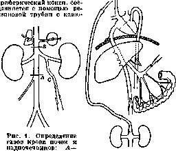
2. Kidneys. The method was developed by Barcroft and Brodie. The gastrointestinal tract is removed together with the spleen and pancreas after preliminary ligation of the corresponding vessels, first the arteries and then the veins; the abdominal aorta is ligated and a cannula is introduced into the inferior vena cava. In some cases (Barcroft, Straub), the cannula is introduced not into the vena cava, but into the ovarian vein, and then there is no need for ligation of the aorta and the vena cava (Fig. 1). It is sufficient to clamp the renal vein during the taking of blood. For a normal kidney, the absorption of O2 at rest is equal to 0.026–0.030 cm3 per 1 g in 1 min., during diuresis—up to 0.30 cm3.
3. Adrenal glands. The method is approximately the same as for the kidneys, with the addition of ligation of the renal vessels. The circulation is equal to 6–7 cm3 in 1 min., it is increased by the injection of adrenaline. The absorption of O2 is equal to 0.045 cm3 per 1 g in 1 min. After the injection of adrenaline, it increases 3 times.
4. Liver. The method was developed by Barcroft and Shore. The majority of experiments were done on cats under anesthesia and artificial respiration. The gas exchange in the region of the portal vein and the gas exchange in the region of the hepatic artery are studied separately. In the inferior vena cava, slightly above the entry of the adrenal veins, two cannulas are introduced. The cannula is introduced into the liver.
Figure 2. Determination of liver blood gases. A—place for introduction of cannula; B—place for clamp; C—cannula in the spermatic vein.
and the intestine. [A cannula is inserted] into the central end of the vena jugularis, so that blood from the inferior vena cava, with the exception of blood from the vena hepatica, flows into the superior vena cava. Blood from the vena hepatica is obtained through a cannula inserted into the central end of the inferior vena cava after clamping the vena cava slightly above the diaphragm or between the liver and the diaphragm. Blood from the vena porta is obtained from a cannula inserted into the vena lienalis or into the vena gastro-duodenalis (Fig. 2). To determine gas exchange in the region of the arteria hepatica, venous blood is taken from the vena hepatica, as indicated above, after preliminary clamping of the vena porta. Arterial blood is taken from any artery. O2 absorption (in the region of the arteria hepatica and vena porta) during fasting is equal to 0.011 cm³ per 1 g per 1 min, and during feeding—0.035 cm³. 5. Pancreas. Determination of the gas exchange of this gland was performed by Barcroft and Starling mainly on dogs. After ligation of the vena lienalis, the tail part of the gland is isolated (approximately 1/4 of the entire gland) and a cannula is inserted into the lateral branch of the main vein. When taking blood, a clamp is applied to the main vein above the exit of the indicated branch. To increase secretion, secretin is injected into the vena jugularis. O2 absorption for 1/4 of the dog's pancreas is equal to 0.06-0.6 cm³ per 1 min at rest, and during secretion—from 2 to 6 times more. The amount of flowing blood is 1.9-10 cm³ per 1 minute. 6. Spleen. Gas exchange of this organ was studied exclusively by Verzar. After ligation of the small veins heading toward the stomach, a cannula is inserted into one of the two main splenic veins. When taking blood, a clamp is applied to the main trunk of the vena lienalis. The blood obtained corresponds to a specific part of the spleen, the weight of which is easily determined at the end of the experiment. O2 absorption is equal to 0.05 cm³ per 1 g per 1 min, with a blood flow rate equal to 0.51 cm³ per 1 g per 1 min. 7. Intestine. The first experiments were performed by Brodie and his colleagues. A fairly significant part of the intestine is isolated (about 125 cm). A cannula is inserted into one of the veins. A cannula is also inserted into each of the ends of the isolated intestine for the introduction of the substances being studied. An onnometer is inserted into the intestine, and volume changes are recorded. By clamping the vein, an increase in volume is obtained, allowing the calculation of the amount of blood flowing into the vessel of the given organ per unit of time. This technique can be successfully replaced by the method developed by Barcroft and Shore for the liver. One can insert a cannula into the vena lienalis or into the vena pancreatico-duodenalis and, when taking blood, clamp the vena porta. Thus, blood from the intestine would flow entirely through the indicated vein. 8. Brain. Gas exchange of this organ was studied by Hill and Nabarro and subsequently by Alexander and Czerna. Trepanation is performed in the region of the torcula Herophili of the sinus longitudinalis and a cannula is inserted into the sinus. By this method, comparative data can be obtained. The absolute value of gas exchange cannot be determined by this method, since only a part of the venous blood of the brain is determined. 9. Muscles. The first satisfactory results were obtained by Punz (1878), who determined the blood flow rate simultaneously with the gases of arterial and venous blood. Subsequently (1887), Chauveau and Kaufmann studied the influence of normal work (chewing) on the masticatory muscles of a horse. Experiments on gas exchange were subsequently repeated on the musculus gastrocnemius of a cat by Verzar, who used Barcroft's technique. After ligation of all lateral femoral veins flowing into the vena femoralis, a cannula is inserted into the vena saphena. When taking blood, a clamp is applied to the vena femoralis above the entry of the vena saphena. Barcroft and Kato use a special pipette (Fig. 3), which serves as one

Figure 3. Pipette for blood gases.
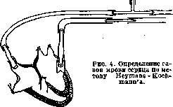
simultaneously as a cannula, and instead of a clamp, they use a loop of wool thread, which is lifted when taking blood. O2 absorption at rest is equal to 0.0032-0.0086 cm³ (according to Verzar). The disadvantage of all these studies lies mainly in the short duration of the experiment, as a result of which the results are not always satisfactory, especially in relation to the respiratory quotient. Instead of this direct determination of the gas exchange of a given organ, Tangl and his school use a method of indirect determination of the gas exchange of an individual organ. The principle of this method is that in curarized animals, gas exchange is compared before and after the extirpation of one or another organ. By the decrease in gas exchange after this extirpation, the gas exchange of this organ is determined. The methodology is briefly as follows: after preliminary tracheotomy, a 1% solution of curare is injected into the vena jugularis and then artificial respiration is established. To avoid cooling, the animal is left in a thermostat during the entire experiment. Instead of curare, some authors use urethane or chloretone or combine both substances. Using this methodology of Tangl, gas exchange was investigated after the removal of the kidneys, pancreas, liver, spleen, intestine, and individual limbs. The exclusion of the kidneys was performed by ligation of the renal vessels. The spleen was excluded in the same way. The pancreas was removed completely. To exclude the liver, the vena porta is ligated at the liver itself and then connected to the vena cava, so that blood from the portal system flows directly into the vena cava without passing through the liver. The decrease in gas exchange after the removal of both kidneys is equal to 8.7%, after the removal of the pancreas—8%, after the exclusion of the spleen—0.7%, and after the exclusion of the liver—12%. Studies using this method provoke serious criticism. It is hardly possible to assume that the removal of a specific organ remains without any influence on the activity of the remaining organs, and on the other hand, an increase in the activity of a specific organ by introducing specific stimulating substances is hardly without influence on the rest of the organism. II. Investigation of isolated organs under artificial circulation. The advantage of this method lies in the wider possibility of changing experimental conditions, especially in relation to the composition of the nutrient fluid, and simultaneously excluding the influence of the remaining organs on the activity and gas exchange of the given tissue. To determine the respiratory gas exchange of the tissue being studied, it is sufficient to determine the content of O2 and CO2 in both the inflowing and outflowing fluid, while simultaneously establishing the flow rate. A necessary condition is the freshness of the tissues, and therefore artificial circulation must be performed as soon as possible after the removal of the organ being studied, especially when it comes to tissues that quickly lose their respiratory capacity, such as, for example, the liver, brain, heart, and others. On the other hand, it is known that a longer cessation of circulation also increases the tendency to edema. Therefore, it is necessary to isolate the organ only after all preparations are completed, avoiding cooling and overly prolonged washing of the organ with physiological solution, as this often leads to a decrease in the respiratory capacity of the tissue and causes edema. The nutrient fluid can be whole blood (non-coagulating due to the addition of hirudin, heparin, or Germanin Bayer 205) (Bryukhonenko and Chechulin), defibrinated blood, a suspension of erythrocytes in physiological solution, or finally, an oxygen-saturated salt solution (Ringer-Locke solution, Tyrode solution, etc.). The use of salt solutions presents a whole series of disadvantages, such as, for example, the appearance of edema due to the insufficient viscosity of the fluid used, the transition into solution and leaching of certain substances contained in the tissue, and, moreover, the insufficient supply of O2 when it comes to the tissues of warm-blooded animals. To increase viscosity, protein, gelatin, or gum arabic is added to the nutrient perfusion fluid. Vernon recommends the addition of serum in the amount of 2%. To increase the activity of a given organ, corresponding substances are added to the nutrient fluid (urea for the kidneys, glucose for the heart, etc.). When investigating the gas exchange of organs of warm-blooded animals, the temperature of the nutrient fluid, as well as the temperature of the vessel containing the organ being studied, must be close to body temperature (37°-38°). When studying the organs of cold-blooded animals, one can be satisfied with ordinary room temperature (18-22°). Recently, the method of Heymans and Kochmann has been widely used—the organ being studied is included in the circulation circuit (Fig. 4) of another animal—and the method of Starling (the organ being studied is fed by blood flowing through an isolated heart and lungs—a "heart-lung preparation"). With these latter methods, conditions closest to physiological are achieved: constant arterialization and mixing of blood and, in addition, the prevention of the accumulation of vasoconstrictor substances in the blood—a factor that is very important, since with the usual method of artificial circulation, vasoconstriction often occurs and, as a result, the blood flow in the organ being studied is significantly slowed down.
The Jacoby apparatus (Jacoby; Fig. 5) also deserves attention. By means of artificial circulation, the gas exchange of a whole series of organs and tissues of warm-blooded and cold-blooded animals has been studied. The best results were obtained using the Heymans-Kochmann methods and the Starling heart-lung preparation. The following tables indicate the intensity of gas exchange in various tissues. The numbers given denote the quantity of absorbed O2 (cm3) per
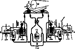
Figure 5. Jacoby apparatus: M-pulley; Z-double eccentric; L, Ea-vessels containing the lungs and the organ under study; Cv, Ca-rubber balloons serving as regulators; Dv, Da-vessels into which blood is collected; Bv, Ba-coils for arterial and venous blood; F-reservoir for blood; Av, Aa-cardiac pumps. 100 g of tissue per minute. The results obtained relate partly to isolated organs, partly to organs in situ. Some of the results were obtained by comparing the total gas exchange of an animal before and after the removal or exclusion of a specific organ, the so-called indirect method. III. Experiments on minced tissues. By the above-mentioned research methods, it was possible to determine the intensity of respiration, i.e., the gas exchange of various tissues and organs, and also to establish the influence of a whole series of physiological and pathological factors on tissue respiration. To study the mechanism of respiratory processes itself, i.e., the nature of oxidizable substances, as well as oxidation catalysts, these methods prove insufficient. To resolve these tasks, it is necessary to simplify the conditions of the experiment and, if possible, isolate those factors to which 60a Respiration of tissues at rest. Organs Heart ..... Gastrointestinal tract Kidneys ...... Liver ..... »
..... Salivary glands ....... Pancreas . . . . Muscles ..... Viscera (without kidneys) . . O2, Part of total exchange in % 3.5 1.8 8.5 7.0 2.0 2.5 4.0 0.4 4.4 7.4 7.1 12.4 0.65 1.34 24.1 24.6 Method Author Direct Indirect Direct Indirect Barcroft-Dixon, Rohde Brodie (and collaborators) Barcroft, Brodie Verzar Masing Barcroft, Mailer, Piper Barcroft, Starling Chauveau-Kaufmann, Verzar Tangl Respiration of tissues during work. Organs O2 35.0 3.6 34.0 5.0 17.5 16.0 3.2 Part of total exchange in % Method Author Heart ..... Gastrointestinal tract Kidneys ...... Liver ..... Salivary glands ....... Pancreas .... Muscle..... Viscera (without kidneys) . . 30.6 13.9 21.0 22.5 1.7 5.1 70.0 41.5 Indirect Direct » Indirect Direct & » Indirect Loewy-Schroetter, Plesch, Brodie (and collaborators) Barcroft, Brodie Verzar Barcroft Barcroft, Starling Chauveau-Kaufmann, Verzar Tangl a certain role in the respiratory process is attributed. This is the goal pursued by experiments on isolated minced tissues. The first attempts to determine gas exchange in fragments of tissue excised from the organism were made by Spallanzani and resumed by Hermann and P. Bert. In this process, pieces of tissue were introduced into a vessel of a certain capacity containing air; after a certain time, the composition of this air was studied, and in this way, the quantity of absorbed O2 and released CO2 was determined. The data obtained in this manner are far from satisfactory. The gas exchange is very insignificant, which is explained by the insufficient contact of the respiring mass with O2. Better results were obtained using the technique developed by Batelli and Stern, the principle of which consists in suspending finely minced tissue in a liquid of a certain composition with constant agitation of this mixture in the presence of O2 and at body temperature. The application of this method, in addition to significant intensity of gas exchange, makes it possible to analyze the mechanism of tissue respiration more accurately and deeply and to investigate the influence of a whole series of physical and chemical agents on individual factors of the respiratory process. Most of the methods used now are based on this principle, which in essence represent only a modification of the Batelli and Stern method. The essence of this method is as follows: the tissue being studied is taken immediately after the death of the animal, minced with scissors or in a meat grinder, and a certain quantity is introduced into a vessel (Erlenmeyer flask) containing several volumes of a liquid of a certain composition (blood or a 1% solution of Na2HPO4). With the help of a pump, the air is evacuated, the flask is filled with O2, and after the temperature has equalized, the flask is connected to a manometer or eudiometer, and agitation begins (Schüttelapparate, Fig. 6).
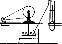
Figure 6. Batelli-Stern Schüttelapparat (shaking apparatus).
The change in gas volume in the vessel is noted by an eudiometer or manometer. The formed CO2 is absorbed by an alkali, which is contained in a special small vessel suspended or attached to the wall of the vessel containing the tissue. In cases where it is desirable to simultaneously determine the absorption of O2 and the release of CO2, at the end of the experiment, a specific amount of alkali solution is introduced into the vessel to absorb all free CO2. The amount of absorbed O2 is determined by the decrease in the total volume of gas in the vessel. Then, a specific amount of acid is added to release the bound CO2. The amount of released CO2 is determined by analysis of the gas mixture or is calculated directly from the increase in gas volume after the addition of the acid. In both cases, it is necessary to add the CO2 present in the solution to the CO2 present in the gas mixture, and the pre-existing CO2 must be subtracted from the total amount. The ratio of the CO2 obtained in this way to the absorbed O2 constitutes the respiratory quotient, the value of which in the experiments of Battelli and Stern is very close to the normal respiratory quotient of warm-blooded animals. To avoid the influence of the bacterial factor, it is necessary to reduce the duration of the experiment as much as possible, since the use of antiseptics does not remain without influence on the course of respiratory processes. In most experiments, one can therefore limit oneself to 30 minutes. This period can even be reduced to 10-15 minutes, especially since the intensity of respiration gradually decreases. When using tissues of cold-blooded animals, as well as tissues of warm-blooded animals that do not require preliminary grinding to facilitate the access of O2, such as, for example, the diaphragm of small animals (mice), respiration retains its initial intensity for a longer period, especially if serum is used instead of a saline solution. Warburg replaced the eudiometric measurement of gas with a manometric one, which allowed working with very small amounts of tissue. The test tissue in the form of thin sections ensuring sufficient oxygen diffusion or in the form of a cell suspension (erythrocytes, eggs of lower animals) is placed in small vessels of one shape or another (respirometers) connected to a manometer. By tightening the screw on the rubber reservoir of the manometer, it is achieved that the gas volume in the respirometer remains constant; the absorption of O2 is judged by the change in pressure (the CO2 released during respiration is absorbed by alkali placed in a special reservoir of the respirometer). The respirometer is immersed in a water thermostat and set into continuous oscillating motion by an electric motor, which ensures the equilibrium of gases between the medium and the free space of the respirometer. Experiments by Battelli and Stern established that most tissues quickly lose a significant part of their respiratory capacity after the death of the animal. This decrease in respiratory capacity for some tissues proceeds very rapidly at first, and then respiration remains at a certain minimum. In other tissues, a slower and more prolonged decline in respiratory capacity is noted until its complete disappearance. The gas exchange values obtained in this way by Battelli and Stern for various tissues of warm-blooded animals are given in the following table. The given numbers indicate the amount of absorbed O2 (in cm3) per 100 g of tissue during 30 minutes at 38° in an atmosphere of pure O2. Tissue: Muscle (Dog, Rabbit, Pigeon, Chicken), Kidney (Dog, Rabbit), Brain (Dog, Bull), Spleen (Dog). Attempts to establish the nature of those substances that are directly oxidized in the respiratory process have shown that the addition of certain substances to respiring tissue increases the absorption of O2 and often also the release of CO2, whereas the addition of other substances remains without influence or lowers the absorption of O2 and the release of CO2. It has also been established that an increase in gas exchange in these cases sometimes corresponds to a decrease in the amount of the added substance and the appearance of certain oxidation products. Thus, the work of Battelli and Stern established that the addition of succinic acid increases the absorption of O2 and that malic acid appears simultaneously. Einbeck subsequently showed that the first product is fumaric acid, which then converts into malic acid. The addition of malic, fumaric, and citric acids increases the absorption of O2 and the release of CO2 (respiratory quotient = 1.33), whereby the corresponding amount of added substances disappears. The work of Meyerhof established that the addition of lactic acid to muscle tissue significantly increases gas exchange and that simultaneously with the disappearance of lactic acid, an increase in glycogen in the tissue is noted. The gas exchange of some tissues (for example, kidney, liver) increases with the addition of uric acid. Simultaneously, allantoin, i.e., the product of its oxidation, appears at the expense of the disappeared uric acid. The addition of ethyl alcohol also leads to an increase in gas exchange. Simultaneously with the disappearance of alcohol, aldehyde and acetic acid appear. Since the listed substances and a whole series of others are products of intermediate metabolism in the organism, it can be assumed that they are part of the material that burns in the tissues in the aerobic phase of respiration. Speaking of respiration, one has in mind only the aerobic final phase of the respiratory process. The anaerobic phase, preceding oxidation, presents a great analogy with the process of fermentation. Factors influencing the intensity of respiration can, of course, act either on the anaerobic or on the aerobic phase of the process. (Regarding the anaerobic phase, see Anaerobiosis and Glycolysis.) As for the aerobic phase of respiration, according to Battelli and Stern, one should distinguish two types of respiration: main respiration and accessory respiration. Main respiration is observed in all tissues of higher animals, where it constitutes a substantial part of the respiratory process. It disappears more or less quickly after the death of the animal and is distinguished by instability and sensitivity to various physical and chemical factors (Fig. 7). Figure 7. Curve of the decrease in respiratory energy of the liver after the death of the animal. On the abscissa - time after the death of the animal in minutes. On the ordinate - absorption of O2 in cm3. 15 30 45' 60' 76' 90 105' 120 135' 150. Two types of substances participate in the process of main respiration: 1) the actual catalysts of main respiration, which Battelli and Stern call oxidones, and 2) a water-soluble heat-resistant substance (or rather a mixture of substances), which Battelli and Stern called pnein (pneine, Pnein) and subsequently Meyerhof - "Atmungskorper". By extracting ground muscle with water, it is possible to separate these two agents from each other; thoroughly squeezed tissue retains only an insignificant part of its activity. The addition of an aqueous muscle extract (fresh or boiled) restores the initial intensity of respiration. The active substances contained in this extract are thermostable, easily soluble in water, less so in alcohol and acetone, insoluble in ether, chloroform, and benzene, are not destroyed by acids, alkalis, or digestive enzymes, are not oxidized by H2O2, but are destroyed under the influence of H2O2+FeCl3. Pnein in all probability plays the role of a coenzyme, and, as Meyerhof subsequently established, this coenzyme presents a great analogy with the coenzyme of zymase. The catalysts of main respiration (or the "basic respiratory process" of Battelli and Stern) are not extracted from tissues without preliminary destruction of the cell structure. These catalysts, "oxidones," are closely linked to the cell structure, in all probability adsorbed by the structural elements of the cells, and only upon their destruction do they pass into the extract. The catalysts of the basic respiratory process are distinguished by particular lability, lose activity after the death of the animal in different tissues at different speeds, are sensitive to high temperature (optimum of their action at 40°), to the action of alcohol, acetone, acids, and alkalis, to the action of proteolytic enzymes and a whole series of poisons, especially lipoid-soluble narcotic substances, arsenic acid, aldehydes, etc. These substances do not act on accessory respiration. Along with pnein, it is possible to extract from most ground tissues a special substance (or mixture of substances), "antipneumin," which possesses the ability to lower the intensity of main respiration but remains without influence on other oxidative catalysts (Fig. 8). Figure 8. Curve of the action of antipneumin. On the abscissa - time in minutes. On the ordinate - amount of absorbed O2 in cm3. Thick line - tissue without antipneumin, dotted - with antipneumin. Antipneumin is quickly destroyed under the influence of high temperature, acids and alkalis, alcohol, acetone, etc. Antipneumin does not dialyze and is carried away during the precipitation of extracts (curve of the action of antipneumin).
Accessory respiration, which constitutes a comparatively insignificant part of the respiratory process in the majority of tissues of higher animals, is preserved without obvious change in tissues for quite a long time after the death of the animal. Battelli and Stern classify the catalysts of accessory respiration as a type of oxidases or oxidoreductases (in contrast to the catalysts of main respiration—oxidones); they are comparatively easily extracted from comminuted tissues and, apparently, are not connected with the structure of the cell. Battelli and Stern attribute to accessory respiration, which is most pronounced in glandular organs, mainly a protective function—the detoxification of substances poisonous to the organism by means of their oxidation. During accessory respiration, in addition to the absorption of O2, the formation of CO2 also takes place, but the respiratory quotient is significantly lower than during main respiration. The intensity of accessory respiration represents a significant value only in a few tissues, such as the liver and kidney; in the majority of others, the intensity is insignificant, and in muscle, for example, it is completely negligible. The following table shows the amounts of absorbed O2 and released CO2 in 100 g of tissue during 1 hour at 38° in an atmosphere of O2.
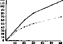
Tissue Animal O2 (in cm3) CO2 (in cm3) Liver...... Horse 45 1 » ...... Bull 42 | » ...... Ram » ...... Dog 37 1 Kidney...... Horse » ...... Bull » ...... Dog 26 ! Spleen .... Bull 21 ; » .... Horse 17 ! Lung ...... » Ram Brain....... Dog Ram Battelli and Stern provide the following characteristics of main and accessory respiration: 1. Unlike accessory respiration, main respiration exists in all tissues of higher animals. 2. Main respiration gradually decreases in tissues after the death of the animal and finally disappears completely, whereas accessory respiration remains unchanged for a comparatively long time. 3. The catalysts of main respiration cannot be extracted from tissues without prior destruction of the cellular structure, whereas the catalysts of accessory respiration are extracted very easily. 4. Under the action of alcohol and acetone, even in a concentration of 30-40%, main respiration is completely destroyed, while accessory respiration is not disturbed under these conditions. 5. Trypsin very quickly and completely destroys main respiration and remains without influence on accessory respiration, at least during short-term action (1/2-1 hour). 6. The optimum temperature for main respiration is 40°; heating to 52° completely destroys it, whereas the optimum for accessory respiration lies between 50° and 55°. 7. The pressure of O2 exerts a great influence on the intensity of main respiration. In an atmosphere of pure O2, main respiration occurs more intensely than in ordinary air. Accessory respiration is not enhanced by replacing air with pure O2. 8. Main respiration depends to a significantly greater degree on the reaction of the medium than accessory respiration. 9. Pnein enhances main respiration but remains without influence on accessory respiration. Antipneumin suppresses main respiration but does not affect accessory respiration. Subsequently, it was established by Stern that the essential difference in the mechanism of action of oxidones and oxidases consists in the fact that oxidones can use only active oxygen as a hydrogen acceptor, whereby water is formed, whereas oxidases can use as a hydrogen acceptor both molecular O2, forming H2O2 in the process, and other bodies like methylene blue, nitrates, etc. Indirect confirmation of this theory is the abundance of catalase in those organs in which the respiratory process has an oxidase character, and the absence or insignificant amount of catalase in those organs in which exclusively oxidone processes take place.
A special method for determining tissue respiration is used by Thunberg and his school. Proceeding from the principle widely developed by Wieland that biological oxidation consists in the removal of hydrogen from the substance being oxidized (the "donor") and in transferring it to a substance playing the role of an acceptor (see), Thunberg replaces O2, which according to Wieland's theory is the main H acceptor, with methylene blue, which becomes decolorized upon combining with H, turning into a leuco base. The speed with which this decolorization occurs indicates the intensity of the oxidative, or rather, dehydrogenating process. Using this methodology, the Thunberg school studied not only substances capable of being oxidized by animal tissues but also the influence exerted on tissue respiration by various physiological factors, including products of endocrine substances, vitamins, etc. (Ahlgren et al.). However, the results obtained cannot serve

Fig. 9. Thunberg vessel for studying the reducing capacity of tissue.
as a basis for general conclusions in view of the insufficiency of the methodology itself. It has been established that there is no parallelism between the decolorization of methylene blue and the absorption of O2 under these same conditions; therefore, the intensity of methylene blue decolorization cannot serve as a measure of the oxidative energy of a given tissue (Bach and Mikhlin). This lack of parallelism between these two processes is explained, according to the theory proposed by Stern, by the fact that a part of the catalysts participating in respiratory processes, namely oxidones, can transfer hydrogen only to activated O2, while another part of the catalysts, namely oxidases, or rather oxidoreductases, is capable of transferring active hydrogen both to molecular oxygen and to other H acceptors, including methylene blue. Thus, the decolorization of methylene blue reflects only an insignificant part of the respiratory processes, namely, oxidase processes, which constitute accessory respiration. The most significant part of the respiratory processes (main respiration, oxidone processes) has nothing in common with the decolorization of methylene blue. In the mechanism of respiratory processes, the structural factor plays a significant role. Through the work of Battelli and Stern, it was established that the catalysts of main respiration are closely connected with the structure of cells. Warburg subsequently showed that there is a close connection between the specific structure of cells and the intensity of respiratory processes. Thus, nucleated erythrocytes of birds absorb significantly more O2 than the non-nucleated erythrocytes of mammals; on the other hand, younger erythrocytes, the protoplasm of which represents a reticular structure, exhibit greater respiratory energy than older erythrocytes. Destruction of the structure by grinding the cells leads to the complete disappearance of respiration. Experiments performed on developing sea urchin eggs showed that simultaneously with fertilization and the development of structural cellular substance, respiratory processes are significantly enhanced. Already in the first minutes following fertilization, respiration increases 80-fold; in this case as well, destruction of the structure causes respiration to decrease strongly. The strong inhibitory effect of narcotic substances on respiration is in close connection with their adsorption on the surface; the reduction of the surface, which occurs as a result of its blocking by narcotic substances, leads to a decrease in oxidative capacity. Along with the structural factor, which does not possess any specificity, Warburg also assumes the participation in the respiration process of a specific catalyst in the form of a special organic compound of iron. This "respiratory enzyme," which is inhibited even by a negligible amount of HCN, has, according to the latest research by Warburg, a structure similar to hemochromogen; at least the absorption spectrum of the CO-compound of this enzyme presents complete identity with the absorption spectrum of the CO-compound of hemochromogen. This enzyme is, according to Warburg, identical in all aerobic organisms. Its role reduces to the activation and transfer of oxygen.
L. Stern. Respiration, pathological,
see Dyspnea, Asthma, cardiac, Metabolism, basal, Respiratory quotient—clin., study.
Related articles
Mentioned in
Cite this page
“Respiration.” Soviet Medical Encyclopedia. English translation of Bolshaya Meditsinskaya Entsiklopediya, 1st ed. (Moscow, 1928–1936), ed. N. A. Semashko. https://sovietmedicalencyclopedia.pages.dev/article/respiration/