Bronchi
Historical document, translated for reference. It reflects medical knowledge of the 1920s–30s and is not medical advice.
Summary
This article from the 1st edition of the Great Medical Encyclopedia details the embryological development, anatomical structure, histology, and physiological features of the human bronchi.
Encyclopedia article (1928–1936)
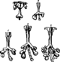
BRONCHI (from Greek bronchos - breathing tube) are the branches of the trachea that serve for the exchange of external air and the air of the pulmonary alveoli. The embryological development of the bronchi begins at a very early stage of embryonic development. Already at a length of 4-5 mm (J. Kollman, Heiss), a division into the right and left subsections of the primary primordium of the respiratory apparatus is outlined, which appears by the end of the 1st month (His) as a hollow, sac-like protrusion from the anterior wall of the digestive tube (see figure 1). The successive branching of the initial two primary, or main, bronchi ultimately leads this initially bifurcated bubble to a complex system of dichotomously diverging channels, i.e., to the appearance of a "bronchial tree." At the same time, the growth of the terminal part of the pulmonary primordium, prevailing in its speed over the other parts of the organ, creates a long "stem" bronchus throughout the entire lung, which itself gives off bronchi of the 1st, 2nd, 3rd, and subsequent orders. Anatomy of the bronchi. In the adult state, the beginning of the bronchi, i.e., their origin from the trachea, or its bifurcation, falls at the level of the IV-V thoracic vertebrae posteriorly (auscultation!) and corresponds to the 1st-2nd intercostal space anteriorly. Age-related fluctuations in the topography of the bifurcation (according to Brünings) range between the lower edge of the 1st rib (in children), the upper edge of the 2nd (in adolescents), and the upper edge of the 3rd (in adults). In addition, the position of the bifurcation is affected by movements of the head and neck and acts of respiration. During flexion and inspiration, the level of the bronchi lowers; during extension and expiration, it rises. Relative to the midline of the body, the bifurcation is shifted slightly to the right and posteriorly by the aortic arch thrown over the left bronchus. The angle of divergence of the main bronchi averages 70.4°, ranging from 56° to 90° (Aeby), but according to Brünings' research on living subjects, it rarely reaches 90° and is generally greater the shorter and thicker the neck.
it (see figure 3). In addition, the right bronchus in the frontal plane repeats the curvature of the right atrium encompassed by the right lung and has a C-shaped curvature.
Figure 1. Development of the lungs and bronchi of a human embryo (bottom middle - esophagus): A - 4 mm embryo; B - 5.9 mm; C - 6.7 mm; D - 8 mm; E - 9 mm (according to R. Heiss). The right main bronchus is wider than the left. Their calibers, according to Branne and Stahel, relate as 100 : 78.4, while according to Brünings' measurements, they have the following dimensions: Bronchi Male Female Child Newborn Right . . . Left . . . In millimeters 12-16 10-14 10-15 9-13 7-9 6-8 5-6 4-5 The right bronchus, deviating less from the midline than the left, preserves the direction of the trachea to a greater extent, and therefore, with its greater width, foreign bodies aspirated through the larynx and trachea more often enter it (see figure 2). Before plunging into the lung tissue, both main bronchi are located in the mediastinal region for a certain distance and are in contact here with large vessels and lymph glands (see below). The extent of this mediastinal segment of the bronchi is shorter on the right than on the left (6-8 cartilages on the right, 9-12 on the left). Both the main bronchus and its continuation, the intrapulmonary "stem" bronchus, in their direction, like the trachea, continue to adhere to the curvature of the spine and are located inside each lung, deviating from anterior to posterior, from superior to inferior, and, lying closer to the posterior than to the anterior surface of the lung, rush with their ending (bronchus terminalis) to the lowest part, to the posteroinferior corner
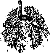
Figure 2. Bronchial tree and pulmonary artery in a newborn (from a corrosion preparation): A - a. pulmonalis; C - bronchus cardiacus; 1 - upper lobe; 2 - middle lobe; 3 - lower lobe (according to Braus).
The left bronchus is twice curved in the same S-shaped plane, in the upper bend deviating downward and to the right from the aortic arch, and in the lower bend - upward and to the left, under the pressure of the left ventricle of the heart (see figure 2). Inside the lung, the stem bronchus gives off branches of the 2nd order, longer anterior-ventral and shorter posterior-dorsal; these branches, in turn, divide into branches of the 3rd and subsequent orders. The branching of the bronchi is unswervingly followed by the adjacent and also branching vessels aa. and vv. bronchiales and pulmonales. In the left lung, the stem bronchus gives 4 pairs of branches: 4 ventral bronchi and 4 dorsal bronchi. On the right, to these same 4 pairs is added one unpaired bronchus, which, unlike others, departs from the stem bronchus at a right angle, cranially, Figure 3. Direction of the bronchus in relation to the lung. View from the side, left; in the posteroinferior corner - bronchus terminalis (according to Merkel with modifications). intended for the upper lobe of the lung and differing from all other bronchi in its relation to the adjacent pulmonary artery. It is located dorsally to the artery and generally above it, which is why it is called the "bronchus eparterialis," whereas the rest lie ventrally and below the artery and are called "bronchi hyparteriales." In addition to the above-mentioned bronchi, an additional one is distinguished on the right - br. cardiacus, which departs from the 2nd ventral bronchus and heads into the pulmonary parenchyma posteriorly, into the lower lobe. It is distinguished by its thickness and power and corresponds to the bronchus of an independent "cardiac" lobe in some animals (lobus infracardiacus). In Fig. 2, this bronchus is designated by the letter C. The successive division of the bronchi, both dorsal and ventral, leads them to terminal bronchial tubes 0.4 - 0.5 mm in diameter, bronchioles (see below - p. 74), which pass into pulmonary smooth musculature, the power of which decreases in the direction of the alveoli until it completely disappears in the latter. - Muscle fibers, embedded between the cartilaginous plates and the mucosa, have a direction relative to the axis of the tube towards
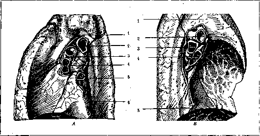
Figure 4. Roots of the right (A) and left (B) lungs. Relation of bronchi to vessels. A: 1 - sulcus v. azygos; 2 - right bronchus; 3 - right branch of pulmonary artery; 4 - right pulmonary veins; 5 - sulcus oesophageus; 6 - lig. pulmonale. B: 1 - sulcus aorticus; 2 - left branch of pulmonary artery; 3 - left bronchus; 4 - left pulmonary veins; 5 - sulcus oesophageus (according to Braus).
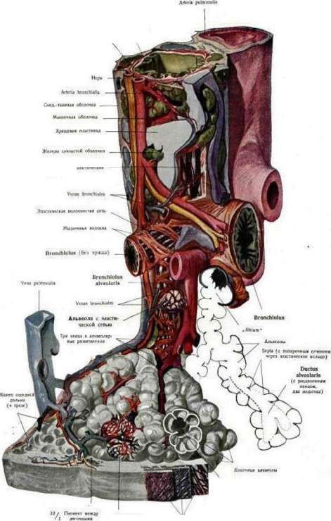
the alveolar ducts and alveoli [see sep. table (p. 107-108), Fig. 1] and correspond to the 5th-6th ordinal number of bronchial division. Their external distinguishing feature is the small alveoli visible with a magnifying glass sitting on their wall and the presence of respiratory epithelium. Histologically, the large bronchi repeat the structure of the trachea. The cartilaginous skeleton of the bronchi appears in the form of rings of hyaline cartilage almost completely surrounding the respiratory tube, with a small posterior part free from it, which is replaced by a membranous plate containing transverse bundles of smooth muscle fibers. The latter are attached to the perichondrium of the cartilaginous rings. In smaller bronchi of the 2nd-3rd order, the cartilaginous rings are replaced by closely adjoining cartilaginous plates. As the tube divides, the sizes of the plates become smaller, and the intervals between them increase (see figure 5). In small bronchi of the 5th-6th order, there is almost no cartilage, and in bronchi less than 0.85 mm in diameter, it disappears completely. - The mucous membrane of the bronchi with a strongly developed loose fibrous submucosal layer is usually gathered into folds. The ciliated epithelium sitting on its membrana propria contains a large number of goblet cells. In the submucosal layer, among loose fibrous connective tissue, mucoserous glands are embedded, and accumulations of leukocytes and lymphoid follicles are encountered. In this same layer, along the entire extent of the bronchial branches, right up to the alveolar ducts, the bronchi are supplied with juice (see color table), as a result of which their contraction causes not only a narrowing of the lumen of the respiratory tube, but also its simultaneous slight shortening. Thus, by reducing the capacity of the respiratory passages, the bronchial musculature has an expiratory significance. It is complemented by the reverse contraction
of the elastic tissue stretching with each inspiration, embedded in large numbers in the form of longitudinal cords in the fold of the muco-submucosal layer. - The arterial supply of the bronchi and venous outflow occur not only at the expense of aa. and vv. bronchiales (vasa privata), but, apparently, Figure 5. Bronchial cartilage: C - cartilaginous plates (dark part - hyaline cartilage, light outer part - elastic cartilage); M - contour of the mucous membrane. also through aa. and vv. pulmonales (vasa publica), since ligation of the bronchial arteries does not have a harmful effect on the nutrition of the organ. - The lymphatic outflow from the bronchi proceeds through the bronchial lymphatic system, i.e., through the lymph nodes located in the angles of bronchial division (lgl. pulmonales), through the glands of the root of the lung (lgl. broncho-pulmonales), and empties into the bifurcation glands. - Small bronchus
interstitial

Lymphatic vessels of the bronchi (lgl. tracheo-bronchiales), whence through lgl. paratracheales thorac. and truncus broncho-mediastinalis they are directed to the angulus venosus.-Innervation of the bronchi is connected with the pulmonary plexus and originates from the anterior and posterior plexuses (plexus pulmonalis ant. and posterior). The greatest part of the nerve trunks is concentrated on the posterior surface of the main bronchi (necessity of cocainization during lung amputation!).- Syntopy of the bronchi. Over the right main bronchus loops from behind Figure 6. Right main bronchus
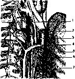
and its surrounding organs: 1-n. recurrens n. vagi d.; 2-trachea; 3-v. cava superior; 4-v. azygos opening into it; 5-nerve fibers departing from the vagus and plexus pulmonalis post. to the lung; 6-right bronchus; 7-lung pulled outwards.
the terminal part of the vena azygos, before its opening into the v. cava superior lying anteriorly. The trunk of this section of the azygos vein touches part of the posterior and upper wall of the bronchus. Medially from the vein, toward the posterior side of the bronchus, lies the right n. vagus descending along the right side of the trachea and the adjacent lymph glands (lgl. tracheo-bronchiales). Laterally-partly the lung, partly the lymph glands of its root. With enlargement of the lymph glands, both the vein trunk and the vagus nerve can be displaced and pushed away from the bronchus. The anterior side of the right bronchus touches the right pulmonary artery;
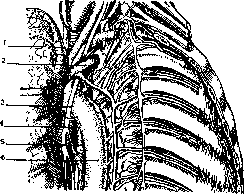
Figure 7. Left main bronchus and its surrounding organs: 1-lung pulled outwards; 2-v. anonyma sin. with v. hemiazygos opening into it; 3-n. vagus sin. and n. recurrens departing from it under the aortic arch; 4-left bronchus; 5-esophagus; 6-aorta.
medially and above it, closer to the bifurcation, the v. cava superior adjoins the bronchus (see figures 4-A and 6). The lower surface is touched by the lower lymph glands of the bifurcation. Over the left main bronchus loops from front to back the aortic arch and, laterally from it, the left pulmonary artery. Between the bronchus and the vessels are located the lgl. tracheo-bronchiales and the n. recurrens sin. departing below the aortic arch from the vagus nerve (see figure 7). Posteriorly, the descending part of the aortic arch adjoins the bronchus and, external to the latter, the trunk of the vagus nerve. From below, the bronchi are touched by the bifurcation glands and w. pulm. (see figure 4 B). Anteriorly, the left bronchus is covered by the posterior parietal leaf of the pericardium (corresponding to the posterior wall of its sinus transversi) and lymphogl. broncho-pulmonales. The relationship of the bronchi with neighboring organs in the roots of the lungs is shown in fig. 4. All the organs listed around the bronchi are separated by loose cellular tissue, allowing significant mutual displacement of them during pathological changes in the organ volume. This cellular tissue is continuously connected with the peribronchial and perivascular interstitial connective tissue of the lungs and can serve as a pathway for the spread of inflammatory and other processes (emphysema, hemorrhage, etc.) from the lung to the organs of the mediastinum and vice versa. Pathological processes in the bronchi. Aside from foreign bodies (see Bronchoscopy), inflammatory lesions originating from various layers of the bronchial wall (see Bronchitis, Pneumonia), and secondary changes caused by them (see Bronchostenosis, Bronchiectasis), traumatic injuries and tumors of the bronchi may have practical significance for the physician. Due to their elasticity, the bronchi, together with the trachea, possess high extensibility; nevertheless, under the action of gravity, compression from without, during extreme stretching of the respiratory tube (for example, with Fig. 8. Lipoma on a sharp forcible knife po... sctoetter...), cases of its rupture have been observed not only in the region of the trachea, but also of the bronchi. Clinical symptoms in injuries have a severe, but not always clear character (retrosternal pain, sometimes audible cracking during respiratory movements, progressive emphysema, pneumothorax, etc.). The prognosis, with very rare exceptions (Krinitsky), is unfavorable. Patients are ruined by complications: emphysema, pneumothorax, bronchopneumonia, gangrene of the lung, and phlegmonous infection of the mediastinum. (On gunshot wounds of the bronchi-see Lungs.)-Tumors of the bronchi can be benign and malignant. Of the former, papillomas, adenomas, lipomas, fibromas, chondromas, osteochondromas are encountered; the tumors are both solitary and multiple, developing in various sections of the bronchial divisions. Sometimes they have a wide base, sometimes a more or less short stalk. Their size can range from a lentil to the size of a hazelnut, reaching in some cases the volume of a fist and a child's head (fibromas and chondromas). During life they may not cause noticeable disorders and are sometimes an accidental finding of the pathologist at autopsy. In other cases, protruding

«3
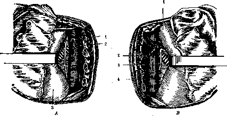
into the lumen, they cause narrowing of the bronchi and disruption of normal breathing (see figure 8). With the development of bronchoscopy, the number of cases of intravital recognition of such tumors is progressively increasing. Their treatment is operative; with particularly favorable locations-using a bronchoscope (see Bronchoscopy), with very large sizes-by a bloody approach to the bronchi (see below). In addition to the listed types of benign tumors, cystic tumors originating in connection with developmental disorders of the bronchi deserve separate mention (see figure 10). Lined on the inside with cylindrical epithelium and connected with the bronchi, they, increasing in size, can reach very large dimensions (up to a child's head in Sauerbruch's case) and turn out to be filled with a transparent mucous mass, which can become infected and produce the clinical picture of an abscess.- Malignant tumors. Depending on the section of the bronchus from which the tumor originates, malignant neoplasms of the bronchi can be localized either in the mediastinum or in the lung parenchyma. Sarcomas (round-cell or spindle-cell from the peribronchial tissue or from the bronchial wall itself) are comparatively rare. Exhibiting energetic growth, they can reach very large sizes, easily reaching the mediastinal spaces, growing through the pleura, heart muscle, and pericardium. More frequently (up to half of all bronchial tumors) cancerous tumors are encountered (see below-p. 75). Operations on the bronchi. Operative approaches to the bronchi. For the removal of foreign bodies or tumors, when the bronchoscopic method is impossible, the bronchi can be approached in two ways: transpleurally (thoracotomia transpleuralis), passing through the lung tissue (see Thorax), or extrapleurally-through the mediastinum (bronchotomia extrapleuralis mediastinalis). According to anatomical conditions, the latter path is most expedient through the posterior section of the mediastinum. Despite all the severity of the intervention due to subsequent possible complications (empyema, local gangrene, emphysema, etc.), both methods nevertheless have successful outcomes with stable results. The transpleural method is usually carried out with an apparatus :left (A) and right (B). Left: 1-n. vagus; 2-aorta; -n. vagus; 4-esophagus (after Garre-Quincke). of increased pressure according to the general rules of such opening of the chest and pneumotomy. The posterior mediastinal method, first developed and proposed in 1888 for access to the esophagus by Russian surgeons Nasilov and Dobro
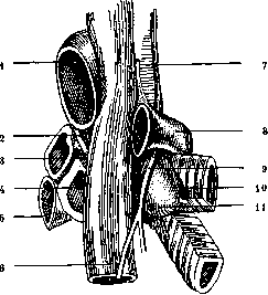
Figure 10. Bifurcation of the trachea. Congenital cyst of the right bronchus (author's observation). View from behind. 1-aorta; 2-n. vagus sin.; 3-a. pulmonalis sin.; 4-left main bronchus; 5-v. pulmonalis sin.; 6-esophagus; 7-trachea; 8-v. azygos; 9-n. vagus dextr.; 10-right bronchus; 11-bronchial cyst.
myslov, was later used also for the approach to the bronchi. It requires subperiosteal resection from a longitudinal paravertebral incision of 4-5 ribs (from III-IV to VII
BRONCHI
rib), over an extent of 4-6 cm, at the level of the transverse processes. After this, the soft tissues of the chest wall are cut (with ligation of vessels), which opens the peripleural cellular tissue (fascia endothoracica), and access to the bronchi is established by
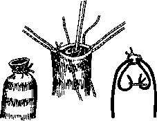
Figure 11.
careful blunt stripping of the parietal pleura and the posterior section of the lung covered by it (see figure 9). When pulling the latter with a hook, the pleura should be protected from tearing and damage. At the end of the operation, a gauze drain is placed at the sutured bronchus. - Incision (bronchotomy) and suture of the bronchi. In operations on the bronchi, the subsequent

Figure 11.
suture of the wall or closure of the bronchial stump, due to specific local conditions, are especially responsible. Rhythmic air jolts in the area of the suture or the stump of the transected bronchus during breathing require high strength of bronchial closure in order to avoid air forcing (subsequent

Figure 13.
emphysema, pneumothorax) and the penetration of infection from the lumen of the bronchus. Therefore, it is advisable to perform bronchotomy layer by layer. The wall of the bronchus is incised longitudinally down to the mucosa, and, having stretched the wound to the sides, a transverse incision of the mucosa is made. The wound is sutured layer by layer, but in the reverse order (Melnikov). For closing the stump of the bronchus during its transverse transection, 3 types of methods can be applied: 1) the gaping bronchus is ligated, and its stump is closed with a second suture with invagination of the ligated end (see Figure 11); in addition, it can be further reinforced with fascia, omentum, pleura, etc.; 2) the bronchus can be ligated and its end closed with the surrounding lung tissue, as schematically indicated in Figure 12, and, finally, 3) in the case of several transected bronchi, there is applied
Figure 14.
suturing them end-to-end (see Figure 13) or by invagination (see Figure 14). When performing operations on the bronchi, it is necessary to take into account the poor conditions of their nutrition, especially in large-caliber bronchi, due to which all kinds of excessively strong tension by sutures easily lead to necrosis and complications arising from it.
V. Karpov.
Bronchial carcinoma, bronchial cancer, is the most frequent of the malignant diseases of the lung; like most cancers, bronchial cancer occurs in presenile age, and is relatively rarely observed in young and very advanced age. Most statistics indicate a preferential incidence in men (2:1; 3:1). On the right, bronchial cancer is found somewhat more often than on the left. The etiology of bronchial cancer, like cancer in general, is unclear; in cities, it apparently occurs more frequently, which some authors associate with a high content of quartz, granite, and coal dust in the air, as well as various combustion products of coal, gasoline, and benzene (in connection with the development of automobile traffic); the significance of roentgenization and radium emanation is also suggested, for example, during ore mining in Schneeberg, where the ore is rich in radium emanation and where bronchial cancer is a major disease of the workers. Regarding pneumoconioses in general, there are no definite data; the significance of Ni, Co, As has also not been clarified; for example, in the same Schneeberg, frequent occurrence of bronchial cancer is noted among tobacco factory workers as well. Theoretically, however, it should be admitted that any pulmonary process (tuberculosis, pneumoconioses, etc.), insofar as it is accompanied by significant changes in the normal tissue relations of the bronchi, can serve as a basis (with appropriate predisposition) for the development of bronchial cancer. Many authors, especially German ones, pay great attention to the influenza epidemic (1918-19),
Figure 15. Cancer of the bronchus in the form of an obturating
polyp. Atelectasis of the lung (observation by I. V. Davydovsky). after which the frequency of cancer increased particularly; on the other hand, the increase in the number of cancers began even earlier than the said epidemic, and moreover, in the anamnesis of patients, influenza is far from a constant phenomenon. Bronchial cancer is also found in many animals, especially in dogs. Statistics indicate a significant growth of this disease. If at the end of the 19th century bronchial cancer accounted for no more than 2% of all cancers, then by 1914 it already accounted for 5%, and in 1924 over 10%. Pathologico-anatomical mortality statistics of the Moscow population note the same thing: out of 500 cancers (per 3,880 corpses), bronchial cancer accounts for 56 (data for 1925). The same applies to an even greater extent to 1926-27. Löwy-Lenz in 1919-23 had 0.7% bronchial cancer out of all autopsy material; in 1913-14, only 0.1%. Hanf's large statistics indicate the same. Pathologico-anatomically, bronchial cancer is characterized by a large number of variants both in terms of the size, location of the tumor, its relation to the lumen of the bronchus, metastasis, and in terms of its histological structure. Most often, bronchial cancer lies not far from the hilus of the lung, at the lower end of the stem bronchus, at the beginning of the branching of its last branches. The pale gray or white tumor may have the appearance of a clearly demarcated node,
Figure 16. Bronchial cancer (bronchoscopic picture): 1—tumor in the lumen of the bronchus; 2—
either protruding like a polyp into the lumen of the bronchus (see Figures 15 and 16) or surrounding it in the form of a ring; in both (especially in the first) cases, the process is accompanied by stenosis or closure of the lumen of the bronchus with corresponding consequences; such clearly outlined cancers have a small size (sometimes the size of a small cherry or pea), and are sometimes found with a certain difficulty even at autopsy, especially when located along the course of the secondary bronchial branches. Another variety of bronchial cancer is large nodes, lying also mainly near the hilus and formed in the manner of rapid peribronchial spread, as if branching, of the tumor (see Figure 17); the bronchi surrounded by cancer take on the appearance of thick-walled white tubes with a narrow, sometimes barely noticeable lumen. If the growth of the tumor proceeds predominantly along the course of the lymphatic vessels (retrogradely), one notices a copious number of small whitish strands extending from the main node deep into the lung, which, upon reaching the pleura, tend to produce lush growth on the surface here, which is usually accompanied by a fibrinous-hemorrhagic effusion into the pleural cavity. Finally, in rarer cases, bronchial cancer takes on the character of a diffusely infiltrating process, when extensive areas of the pulmonary parenchyma (see Figure 18), sometimes entire lobes of the lung, resemble the lung in gray hepatization or carnification, or simulate caseous pneumonia, which may also be facilitated by the presence of cheesy breakdown, cavity formation, etc. The growth of the tumor occurs here mainly in the parenchyma itself, along its air-bearing system, and, apparently, multicentrically, i.e., the tumor develops simultaneously in several points of the organ. Some authors believe that in such cases we are dealing with so-called lung cancer, i.e., cancer originating directly from the alveolar epithelium; at present, the possibility of such a genesis of the tumor is not denied, but it is more generally accepted in these cases as well to speak of bronchial cancer, especially since even here the presence of initial foci in small bronchi can be proven. The histological structure of bronchial cancer is diverse: glandular (adenocarcinoma),
Figure 17. Cancer of the lung root compressing the right main bronchus: 1—trachea; 2—tumor of the bronchus.
squamous cell (sometimes keratinizing), medullary, basal cell carcinoma; the latter often bears the character of small round cell carcinoma simulating round cell sarcomas. The starting point of bronchial cancer is the surface or glandular epithelium of the bronchus; since a significant part of bronchial cancers belong to squamous cell carcinoma (which is unusual for a mucous membrane lined with cylindrical epithelium), the concept of precancerous metaplasia of the bronchial epithelium arose, i.e., its transformation from cylindrical to squamous, from which bronchial cancer subsequently develops. This metaplasia indeed frequently occurs in the bronchi in various acute and chronic inflammations; here, special importance is currently attributed to influenza and gas poisonings. Bronchial cancer is also observed in the walls of tuberculous cavities, bronchiectases, in anthracotic scars; this must be borne in mind so as not to draw a direct conclusion about the absence of bronchial cancer in the presence, for example, of tubercle bacilli in the patient's sputum. The main changes on the part of the bronchus affected by the tumor

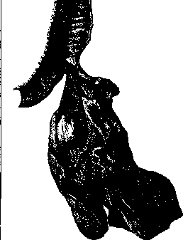

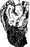
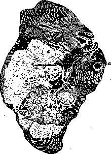
Figure 18. Large (light) tumor node (cancer) with incipient breakdown in the center; a—region of the lung root, narrowing of the bronchus by the tumor and its ulceration. In the part of the lung lying below the stenosed section of the bronchus, atelectasis develops; if the main bronchus is stenosed, the entire lung becomes atelectatic; if the upper lobe bronchus is stenosed, vicarious emphysema develops in the lower sections of the lung. In addition to atelectasis and emphysema of the corresponding lobes of the lung, bronchial cancer produces a number of pathological processes in the lungs that often completely obscure the underlying disease; most frequently appearing here are chronic pneumonias with carnification, purulence (lung abscesses), bronchiectasis, atelectasis, and gangrene; the latter sometimes encompasses the main mass of the tumor itself. All these pulmonary phenomena can also occur with very small bronchial cancers, for example, with cancers that polypously protrude into the lumen of the bronchus. Sometimes bronchial cancer invades the mediastinal spaces, the esophagus (with ulceration, which gives cause for confusion with esophageal cancer), the pericardium, the heart muscle, and the large vessels of the latter. With abundant breakdown of the tumor, erosion of vessels with fatal hemorrhage can be observed; small hemorrhages are a common occurrence. In bronchial cancer, the pleura is often involved in the inflammatory process in the form of either fibrinous deposits on it or the formation of an exudate, serous at first, subsequently acquiring a hemorrhagic character. The clinical picture in these cases initially often differs in no way from ordinary exudative pleurisies. The exudate sometimes persists throughout the entire disease or undergoes complete resorption, encapsulation, and the formation of adhesions. Cytologically, this exudate also often differs in no way from ordinary exudates. Typical giant cells with vacuoles and a nucleus shifted to the periphery, characteristic of carcinomatous exudate, are found in cases where there are metastases to the pleura, and even then not always. Bronchial cancer frequently, and usually early, gives metastases, first to the regional bronchial glands, then to the tracheobronchial glands (at the bifurcation) and, finally, to the glands of the anterior and posterior mediastina. The latter metastases are extremely important, because they sometimes cause compression of large blood vessels and physiologically important nerve trunks. Compression of the phrenic nerve causes paresis of the diaphragm, compression of the recurrent nerve causes paresis of the vocal cords, and compression of the superior vena cava or its large branches entails a circulatory disorder, the result of which is edema of the upper half of the body or one side of it and cyanosis. Some clinicians distinguish a special type of bronchial cancer—"mediastinal cancer"—but in essence, the entire clinical picture is caused by metastases to the mediastinal glands, and not by the bronchial cancer itself, which may be very small in this case. The clinical picture in these cases differs in no way from the picture of mediastinal tumors of another origin. Among the distant foci that result from the metastasis of bronchial cancer, the most important are tumors in the liver, the brain and its membranes, the bones, the adrenal glands, and more rarely in the soft tissues of the limbs, especially the thighs (simulation of thigh sarcoma). Metastases to the cranial cavity often mask the main symptomatology of the affliction, leading to incorrect diagnoses of brain tumor or apoplexy, etc. Cases of metastasis to the pituitary gland with symptoms of diabetes insipidus are encountered; metastases to the vertebral bodies, with invasion of the spinal cord and its roots, simulate primary myelitis, neuritis, etc. Clinically, therefore, it is important to remember that bronchial cancer, being sometimes inconspicuous at the site of its development, can produce extremely variegated and diverse nervous and other symptoms by way of metastasis, which, like the pulmonary complications indicated above, sometimes make the clinical diagnosis completely impossible. As can be seen from all of the above, bronchial cancer can produce a very diverse clinical picture of the disease; some of its main types have already been indicated: stenosis and ulceration of the bronchus; pleurisy obscuring the underlying disease; mediastinal tumor as a result of metastasis, also often obscuring the underlying process. In the initial stage, bronchial cancer generally produces a very scanty clinical picture. One of its early symptoms is cough in the form of paroxysms of a pertussis-like character. The cough may be dry or with sputum (mucopurulent). Sometimes, already in the early period, blood appears in the sputum and even profuse hemorrhage may occur. Sputum impregnated with blood in the form of "raspberry jelly" is considered characteristic. Microscopic examination of the sputum yields little diagnostic data. Relatively rarely, an accumulation of fat droplets in the form of spheres is found in it—apparently fatty-degenerated cancer cells (Lenhartz). An even rarer finding in the sputum is pieces of the cancer tumor. Finally, it should be kept in mind that in bronchial cancer, sputum and even cough may be absent altogether. Sometimes shortness of breath, dull constant pains deep in the chest, or stronger pains increasing with coughing and pressure on the diseased side, resembling pleuritic pains, appear very early; the phenomena of pleurisy are not always present in this case. Subfebrile temperature is observed almost always, sometimes even higher, depending on added complications (suppurative processes in the lung and sepsis). Cachexia is often absent. The duration, counting from the appearance of the first symptoms, is 1/2 to 1, rarely 2–3 years. Physical symptoms. Small tumors without metastases may manifest nothing; with larger nodes, and especially when infiltration spreads to the lung tissue, there are usually obtained: massive dullness to the right or left of the sternum, weakening of breathing and vocal fremitus. With slight stenosis and complication by pneumonia, breathing may be preserved, and then moist rales are auscultated. With stronger stenosis, the phenomena of stenotic breathing—stridor—come to the fore. As a result of tumor breakdown, resp. destruction of tissue by complicating suppurative or gangrrenous processes, characteristic cavitary phenomena are discovered upon examination of the patient. As a result of scarring and shrinking of the tissue after inflammatory processes complicating bronchial cancer in the lung or pleura, or more often as a result of atelectasis, the diseased side appears reduced in volume, and the intercostal spaces are narrowed. Diagnosis is difficult; most often one has to differentiate from pulmonary tuberculosis. When there is bronchial cancer of the upper lobe of the lung with constant temperature, hemoptysis, and chills, the clinical picture gives cause for confusing these two processes. One of the main methods in diagnosis is X-ray examination, which in bronchial cancer is based mainly on the property of the cancer infiltrate, like any dense formation, to produce a shadow on the light background of the lung field. The following features are considered most characteristic for the X-ray picture of bronchial cancer: 1) expansive growth of the shadow, 2) signs of bronchial stenosis, 3) formation of shadows characteristic of metastases in neighboring glands. According to the type of X-ray shadows produced by bronchial cancer, Assmann distinguishes the following types of it: 1) lobar, 2) lung root cancer, 3) diffuse cancer—carcinomatous lymphangitis, 4) miliary carcinosis. The first type produces the following X-ray picture: a continuous homogeneous shadow with a clear lobar boundary is visible on the screen. The primary cancer node in the bronchus is usually not visible in these cases. The darkening is a consequence of either cancer infiltration of the pulmonary parenchyma or reactive pneumonia joining the primary cancer process. And, finally, a continuous homogeneous, but less intense shadow can be the result of atelectasis of the lung tissue due to narrowing of the bronchial lumen. In the latter case, the distinguishing sign of the malignant process, namely the increase in lung volume, is absent, and, conversely, there is a narrowing of the intercostal spaces, upward pulling of the interlobar boundary, and pulling of the trachea toward the diseased side. In the presence of a reduction in the volume of the diseased lung, it is sometimes almost impossible, even on the basis of X-ray data, to distinguish bronchial cancer from a tuberculous process. The spread of the process further to neighboring lobes, the distortion of the contours of the interlobar boundary and the increase in the phenomena of stenosis, as well as the circumstance that the apex, when the upper lobe is affected, often remains free in bronchial cancer, can provide some footholds for diagnosis. Characteristic signs of bronchial stenosis are the displacement of the mediastinal shadow toward the diseased side and the paradoxical type of breathing as a consequence of the suction action on the mediastinum and diaphragm. The second type, hilus cancer, produces a dense shadow on the screen, in the form of a node, in the region of the lung root; radiant shadows depart in all directions from this shadow—a sign of the spread of the tumor along the course of the bronchi and vessels. Syphilis in the region of the lung root also produces darkening in the hilus and cords extending into the lung tissue; it is possible to distinguish it from bronchial cancer in many cases only with the help of other clinical data.
Primary tuberculosis does not show such progressive growth as bronchial cancer and has no tendency to stenosing. Actinomycosis is differentiated by sputum examination data. The third type, carcinomatous lymphangitis, is characterized by thin branching cords forming a network, occurs very rarely, and is seldom diagnosed. The fourth type, miliary carcinosis, as a consequence of hematogenic dissemination, gives small abundant spotting covering both lungs; it is almost impossible to distinguish from miliary tuberculosis, and it is diagnosed extremely rarely (Assmann). Metastases in the regional glands give nodular compact shadows in the region of the hilus and in the anterior and posterior mediastina, differ from the primary cancer node by smooth, often oval contours, and often cause compression of the phrenic nerve; in that case, fluoroscopy exclusively determines a high standing of the diaphragm dome on the affected side and a paradoxical type of respiration. Usually, metastases in the hilus are located on one side; if they are located along the midline, it is often impossible to distinguish them from a tumor of the mediastinum itself—sarcoma, lymphogranuloma. For cases with an unclear nature of the neoplasm in the chest cavity, Borak and Lenk proposed trial X-ray therapy. Sarcoma disappears quickly, lymphogranuloma more slowly, tuberculous lymphoma and chronic pneumonic infiltration even more slowly, while cancerous tumors almost completely do not respond to X-ray therapy. Recently, Lorey, Hasslinger, and Presser proposed using the introduction of 40% iodipin or lipiodol into the bronchi for the diagnosis of bronchial cancer, since it is known that the lesion of the bronchial wall with its destruction and stenosing is inherent, mainly, in bronchial cancer, whereas syphilis and tuberculosis, as a rule, do not destroy the bronchi. It is especially difficult to distinguish bronchial cancer from these two diseases if there are phenomena of decay and cavities are visible on the screen; it is precisely in these cases that bronchography, by revealing a filling defect of the bronchus with deformation of its contours, makes a correct diagnosis possible. In cases complicated by exudative pleurisy, it is recommended to release the exudate before fluoroscopy; sometimes artificial pneumothorax also helps in recognition; if there is a cancerous infiltration of a lung section, it does not collapse during pneumothorax, but protrudes against the light background of the air pocket. In general, the entire symptomatology of bronchial cancer is extremely variegated due to a mass of intervening secondary symptoms, and if the main tumor is not amenable to recognition, the clinical diagnosis often becomes only more or less probable; that is why 40%–50% of bronchial cancers are usually not recognized during life; practically, however, it is useful in all cases of prolonged indefinite pleuropulmonary or mediastinal suffering to reckon with the possibility of bronchial cancer itself. Treatment of bronchial cancer is possible only symptomatically, while surgical intervention presents exceptional difficulties and is not yet practiced. In preventive terms, it is necessary to promote the improvement of the sanitary welfare of populated areas and industrial enterprises, the occupational hazards of which contribute to the development of pneumoconioses and various chronic inflammations of the respiratory tract.
Related articles
Mentioned in
Cite this page
“Bronchi.” Soviet Medical Encyclopedia. English translation of Bolshaya Meditsinskaya Entsiklopediya, 1st ed. (Moscow, 1928–1936), ed. N. A. Semashko. https://sovietmedicalencyclopedia.pages.dev/article/bronchi/