Bronchial Fistulas
Historical document, translated for reference. It reflects medical knowledge of the 1920s–30s and is not medical advice.
Summary
Bronchial fistulas are abnormal openings or channels connecting the bronchi to other cavities, organs, or the skin surface. This article describes their classification, causes, diagnosis, and treatment approaches from the 1930s Soviet medical perspective.
Encyclopedia article (1928–1936)
BRONCHIAL FISTULAS, abnormal openings in a bronchus or abnormal channels leading to these openings, through which the cavity of the bronchus communicates with some cavity or organ, or finally with the surface of the skin. B. f. belong to complete, or true fistulas (fistulae completae). They are: 1) tubular, when they have two openings, or orifices—one external, the other internal (see Figure 1), and 2) lip-shaped, or perforating, when they do not actually have any channel, but possess only one opening (see Figure 2). Those B. f. that do not open to the surface of the skin, but have two internal openings (each in some cavity lined with mucous membrane), are called communicating, or anastomosing fistulas. Lip-shaped B. f. are also called direct or broncho-cutaneous, tubular—indirect, broncho-pleural, cavity, external-open or internal-closed. B. fistulas that take the form of a lattice or perforated tissue are called lattice fistulas (Gitterlunge Sauerbruch's, see Figure 3). In 62% of cases, B. f. are single and only in 38%—multiple (Keller). Lip-shaped fistulas and fistulas of large-caliber bronchi are round in shape, resembling the lumen of a bronchus in its cross-section and reaching a diameter of 0.5 (and more) cm. Tubular, or cavity fistulas most often occur in the form of small slit-like or oval openings. Lip-shaped fistulas are lined inside with mucous membrane, which at the opening of the fistula directly passes into the flat epithelium of the skin. Tubular fistulas are lined with granulation tissue; they usually open into cavities of different capacity and outline. Most often these B. f.
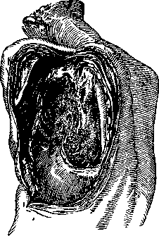
Figure 1. Tubular B. f. (according to Sauerbruch).
are associated with the cavity of empyema of the pleura. Both pleuras, under the influence of an inflammatory-purulent process, undergo fibrous degeneration, sometimes thickening to 4-6-8 cm (Timofeev) and subsequently undergoing hyaline degeneration. The discharge of B. f. in lip-shaped fistulas represents bronchial mucus; in tubular, or cavity fistulas—the contents of that cavity into which the bronchus opens. These discharges are sometimes expelled outward through the large bronchi and trachea, due to the existence of bronchial musculature, peristalsing precisely in this direction. In recent times, Reinberg, by examining the bronchi roentgenoscopically after the introduction of contrast substance into them, has visually demonstrated this. B. f. are rare; even major specialists in pulmonary surgery count them in units. No more than 150-200 operated B. f. have been described, not counting internal, closed ones, not subject in most cases to operative intervention. The causes of formation of B. f. are: 1) inflammatory processes developing both in the lung itself and in the surrounding structures; 2) open or closed trauma to the chest (lungs, bronchi) with subsequent infection and inflammatory reaction. Intrapulmonary purulent processes (abscesses, cavities, gangrene, actinomycosis of the lung, bronchiectases, disintegrating tumors, etc.) cause the formation of B. f. in 84.2% (Keller). Inflammatory processes originating from the soft or bony parts of the chest wall (hot or cold abscesses, osteomyelitis of ribs, spine,
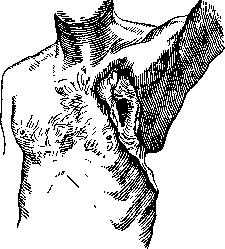
Figure 2. Lip-shaped B. f. (according to Sauerbruch).
ulcerating tumors, etc.) or from neighboring internal organs (liver, e.g., in its echinococci, esophagus, subdiaphragmatic space, stomach, etc.) cause the formation of bronchial fistulas in 2.6% (Keller). Trauma in the form of contusions, punctured wounds, and especially gunshot wounds or operative interventions (decortication, resection of the lung) constitute 13.2% (Keller) among the causes of formation of B. f. The diagnosis of B. f. reduces to detecting signs, either obvious or hidden. The first are characteristic of external, or open fistulas, the second—internal, or closed. Among the obvious signs in external B. f. are noted: 1) inhalation and exhalation through the fistula with the mouth and nose closed, 2) whistling during inhalation and exhalation, sometimes audible at a distance, 3) whistling with a gurgling or splashing sound, if the fistula opens into a cavity containing fluid, 4) fluctuation of the flame of a candle or match applied to the fistula opening, 5) the exit of smoke from a smoking patient through the fistula, 6) the emergence of air bubbles over water in a patient immersed in a bath and who has exhaled, 7) absence of negative pressure during a test with apparatuses that suck out or rarefy air, 8) staining of sputum after the introduction into the fistula of a 2-5% solution of methyl violet, indigo, etc., 9) roentgenoscopy and roentgenography. Obvious signs in internal (closed) B. f. are: the presence in sputum of bile, gastric juice, food, etc., determining the nature of the fistula, and the determination on the roentgenogram of fistula passages after the introduction of contrast substance into the bronchi with the help of a bronchoscope. Among the hidden signs in external fistulas are noted aphonia, fogging of the laryngeal mirror during examination of the fistula, and cough irritation during probing. In internal (closed) fistulas, hidden signs are aphonia and pains in the spine between the shoulder blades, characteristic of B. f. associated with the esophagus or stomach. When applying tests to determine the presence of B. f., caution is required with tests involving the introduction of various liquids into the pleural cavity, as well as when using apparatuses that suck out or rarefy air, or a high-pressure apparatus. In the literature there are indications that these tests, due to coughing attacks, caused the opening of already healed bronchial fistulas. The treatment of B. f. reduces to both conservative and operative methods. Older authors, as well as some modern ones (Graham and others), recommend leaving B. f. to their own fate for months and years until they close spontaneously. If B. f. represents a natural outflow for the discharge
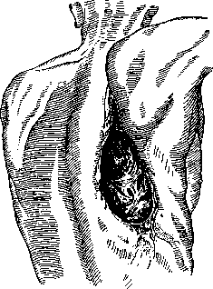
Figure 3. Lattice B. f. (according to Sauerbruch).
of large primary foci (cavity in tbc, multiple abscesses, wide bronchiectases, gangrenous foci, etc.), it is undoubtedly subject to conservative treatment. In such cases, wearing tubes similar to tracheotomy tubes and the use of a pelotte (Sauerbruch) are recommended. There are methods of treating B. f. with radium (Harris) and Beck's paste (Oschner, Lilienthal). Operative methods are extremely diverse, ranging from the simplest to such major interventions as pneumonectomy. The complexity of the operative intervention depends on the size and nature of the B. f. For fistulas of the smallest diameter, barely reaching 2-3 mm, methods in the form of cauterizing the B. f. with iodine, nitric acid, phenol, cauterization with the pacelen (Friedrich) or excising the B. f. with subsequent sutures (Garre) are quite sufficient and suitable. For B. f. of a slightly larger caliber, but not reaching a size of 0.5 cm in diameter, methods with plastic closure of the B. f. are recommended, with the plastic material being: strands (adhesions) and soft parts (Garre-Perthes), periosteum (A. Meyer), costal cartilages (Eden), lung tissue (Walther and others), muscle on a pedicle (Abragamov), and free transplantation of fascia (Kleinschmidt, Hirano, Payr and others), muscle (Smirnov), fat (Nishevich), etc. Attention is drawn to the fact that for the listed B. f. of the first and second groups with the nature of cavity fistulas, it is not so important to carefully close the fistula itself as to widely open the cavity (operations of the Schede type) with the removal of scar and degenerated tissues and with the adaptation (Esser) of the cavity with muscles on a pedicle (similar to the closure of bone cavities with muscles according to af-Schulten). Fistulas with a diameter of 0.5 cm and more require careful closure of the fistulas themselves. For this, there are methods: perichondral suture according to Tiegel (Tiegel), double ligation according to Friedrich (Friedrich), submerged stump according to Meyer (W. Meyer) and splitting of the bronchial wall according to Sauerbruch (Sauerbruch). In severe cases of multiple large fistulas, partial pneumonectomy has been proposed (Tuffier, Graham, Krause). When applying operative methods of treating B. f., it is recommended to adhere to the following rules: 1) operate on B. f. when they have become stationary, i.e., 4-6 weeks after the end of the inflammatory process that caused them; 2) exercise extreme caution in choosing the method in B. f. on the basis of tbc; 3) in multiple B. f., close the small ones first, then the large ones; 4) in severe cases of multiple B. f., operate in several sessions (Katzenstein) and 5) in major operative interventions, use general anesthesia (hedonal).
Related articles
Cite this page
“Bronchial Fistulas.” Soviet Medical Encyclopedia. English translation of Bolshaya Meditsinskaya Entsiklopediya, 1st ed. (Moscow, 1928–1936), ed. N. A. Semashko. https://sovietmedicalencyclopedia.pages.dev/article/bronchial-fistulas/