Actinomycosis
Historical document, translated for reference. It reflects medical knowledge of the 1920s–30s and is not medical advice.
Summary
Actinomycosis is a chronic infectious disease caused by Actinomyces bacteria, characterized by the formation of granulomatous lesions with sulfur granules. The disease can affect various parts of the body, with cervical and pulmonary forms being most common.
Encyclopedia article (1928–1936)
ACTINOMYCOSIS, actinomycosis (from Greek aktis - ray and mykes - fungus), a radiomycotic disease caused by the introduction of actinomycetes (Actinomycetes), ray fungi, into the human or animal organism. Actinomycetes are a group of lower plant organisms, close to bacteria, but in some features of their organization resembling lower fungi. The body of actinomycetes consists of the finest branching threads, 0.5-1.2 microns thick, which in cultures spread radially over the substrate (see Figure 1). Branching, as in fungi, is always monopodial; branches depart from the threads mostly at right angles. The length of threads and density of branching vary greatly. Often actinomycetes develop in the form of separate non-branching or weakly branching rods, indistinguishable from bacteria (e.g., tubercle bacilli). No partitions are observed in the threads, all branches represent one cell (see table for article 287-288, Figure 1). By the absence of true nuclei, properties of the shell, reserve substances, and relation to dyes, actinomycetes differ little from bacteria. Spores retain the ability to germinate for up to 15 years. Pathogenic actinomycetes form characteristic "drusen" in affected tissues from densely interwoven threads with club-shaped thickenings at the ends. Actinomycetes are extremely variable, and the establishment of sharply delimited species is impossible. Most actinomycetes lead a saprophytic existence and are widely distributed in soil (causing the characteristic "earthy" smell of it), in water, and on the surface of plants. A number of actinomycetes are parasites, pathogenic for humans and animals, as well as for plants. Actinomycetes are the cause of one form of the so-called "scab" of potatoes, causing warts and crusts on the surface of tubers containing actinomycetes. - In preparations - smears stained by Gram's method, when examined under immersion, the following features of a typical ray fungus appear: in the center, gram-positive (+ Gr.) branching threads of mycelium in the form of rays directed toward the periphery and ending in club-shaped thickenings. Part of the threads breaks down into +Gr. rod-shaped formations and short threads; in the loops of the mycelium there are many small coccal +Gr. formations (ectospores of actinomycetes). The terminal clubs mentioned, which with difficulty take the stain (+Gr., +Gr., sometimes -Gr.), represent a phenomenon of degeneration of the fungus as a result of the protective reaction of the surrounding tissue of the body. The features of the drusen also appear in the unstained state under weak magnification (100-200 times). - The described picture is characteristic of a fully developed process; in its reverse development, due to calcification or under the influence of iodine, gram-negative, poorly staining, swollen fragments of the fungus are found. In the so-called atypical A., drusen are not visible to the naked eye in the pus, as the fungus is located diffusely. It differs, in addition, by the absence of clubs and a certain degree of acid resistance, i.e., when treated with carbolfuchsin by Ziehl, the threads of the fungus take a red fuchsin stain, not decolorized under the influence of acid. The typical ray fungus and the atypical one (Streptothrix) do not represent different species, but only variants of the basic species, which are the result of special conditions of a given organism, since experimentally, in the mouse peritoneum, Streptothrix sometimes turns into a typical Actinomyces with drusen (Finkelstein). For detailed study of the fungus, aerobic and anaerobic cultures on various media are necessary. For successful culture, pus is taken, if possible, without external contamination. When seeding material containing drusen, they are first washed in a sterile sodium chloride solution and then applied to the nutrient medium. The actinomycosis fungus grows on almost all media, the acidity of which should be about 6.8 (pH) at t° 37°. For pathogenic actinomycetes, media with ascitic fluid or liver serum are preferable. Anaerobic culture (in broth with a piece of liver, according to Tarozzi, under a layer of sterile paraffin) is mandatory, since most pathogenic actinomycetes are conditionally anaerobic. The fungus develops slowly: when seeded on solid medium, round colonies (the size of a millet seed), dry, whitish-gray, are noticeable after 6-8 days; the center of which after 2 weeks appears colored yellow-red or yellowish-brown; after a month the culture appears wrinkled, dry and resembles the growth of tuberculous culture. Aerobic variants, isolated from the surface of bread plants, fruits, from soil, water, differ in variety of color (white, yellow, red, blue, violet). Actinomycetes are very resistant to drying, sunlight, quartz light, as well as X-rays. Pathogenic fungi (from the human organism) die at t° about 60°. Some saprophytic species are thermophilic (optimum about 60-70°). Of chemicals, corrosive sublute kills Actinomyces in 10-30 min. in dilution 1:1,000; 5% carbolic acid is ineffective. Some dyes [methylene blue, methyl violet (1:200-300 thousand)] have a strong effect; iodine in vitro does not kill the fungus. When Actinomyces grows on liquid artificial media, a whole series of metabolic products is found: a) of the endotoxin type, causing marasmus in animals, and b) various enzymes - proteolytic, lipolytic, amylolytic, hemolytic, and bacteriolytic (staphylolysin according to Lieske). All the above physiological properties of various variants are not constant and change depending on environmental conditions. The reactions of immunity and anaphylaxis are little studied. The pathogenicity for animals of fungi isolated from the human organism is not constant; in this respect, anaerobic cultures (of the Wolf-Israel type) have the greatest constancy, which when injected into the peritoneum of guinea pigs and rabbits sometimes give rise to the formation of typical drusen there. Routes of penetration and pathology of A. Bollinger (in 1887) first gave a detailed description of A. and its causative agent. In the field of pathology of A. in humans, the most important works belong to Israel, Bostrom, and Ponfick. From Russian works, Berestnev's dissertation should be mentioned. The following ways of penetration of the ray fungus into the organism are distinguished: through the mouth, skin, lungs, intestines, and by an unknown route. Most often in humans (and in animals) infection occurs through the mouth or skin, due to the entry of grains (especially barley) on which colonies of ray fungi live saprophytically. In many described cases of A. (Brenner and others), people who had the habit of chewing ears and grains of cereals, or in whose skin infected splinters entered (E. Muller), became ill. Such are the descriptions of Soltmann and Berth, where actinomycosis developed at the site of introduction into the soft parts of a cereal ear. A case of A. after swallowing a piece of straw is described by Lunow. Similar cases are those of Schartau, Bostrom, Illich, and others. Proof of this mode of infection is the finding in the center of the inflammatory focus of an object infected with the fungus: a pumpkin seed in Zilz's case and a beard of an ear of grain in Schwartz's case. An interesting case is that of I. Israel, where pulmonary A. apparently occurred due to a piece of a carious tooth that entered the respiratory tract. On the contrary, transmission of A. to humans by infected animals, through direct contact with them, or by eating their meat or milk seems very unlikely. Anatomically, the most pronounced and typical picture of A. is in the region of the neck and lungs, whereas with other localizations it often changes depending on various additional circumstances. In A. of the neck region, a sharply expressed, clearly delimited, often roller-like, bumpy, board-like dense on palpation swelling, dark red or bluish in color, is observed. In places on the surface of the tumor, individual (sometimes several at once) openings are noticeable, connected by fistulous passages, from which when pressed a very limited amount of pus is secreted. As a rule, no clear fluctuation is observed. On section, the tissue of the tumor is yellow-red in color, in places penetrated by nests of softening, but in general has a fibrous consistency. From the area of the section, a cloudy serous fluid is secreted. In other cases of A., on the contrary, it can proceed in the form of diffuse phlegmonous inflammation with abundant purulent decay. Path-histologically, A. proceeds according to the usual type of chronic infectious granuloma, showing this or that feature in connection with the nature of the affected organ. On microscopic examination of the peripheral zone of a typical fresh inflammatory focus, significant proliferations of young granulation tissue are found, penetrated by dense small-cell infiltration, among which a small number of polymorphonuclear leukocytes are visible. Epithelioid and plasma cells are encountered here in small numbers, as well as giant cells, not of very large size, arranged in negligible numbers along the periphery of the foci.
As one moves from the periphery to the center and approaches the yellow focus of softening, the tissue of the tumor becomes looser, the connection between its individual elements is disrupted, and the cells, accumulating, lie completely freely, as in an abscess. But even here, primarily, it is a matter of disintegrating degenerative and fatty degenerated connective tissue cells, while lymphocytes and leukocytes are relatively few. In the center of such a softened focus or 'abscess' lie the druses of the ray fungus. In older foci, already showing a tendency to scarring, one can see, going from the center to the periphery, gradual shrinkage and obliteration of the vessels of the granulation tissue, which passes through the stage of dense fibrous connective tissue into a scar. When searching for druses of the ray fungus, they are particularly easily found in fresh inflammatory foci and only with great difficulty in old foci. A. usually begins insidiously, after a fairly long incubation period, and runs a chronic course. At first, especially with a local focus, it does not essentially affect the general state of health. A distinction is made between external and internal A.- Primary external actinomycosis is rare. Up to 1926, about 70 cases of A. with more or less reliable localization of the primary focus of the disease on the skin had been collected (A. Rau-ber). As a rule, A. appears on the skin as a secondary disease, following the involvement of internal organs: jaw bones, lungs, intestines and liver, which is why it is usually localized on the neck, face, chest and abdomen. External A. for a long time runs as a purely local disease, and only occasionally, in rare cases, it can give metastases to internal organs. More often the reverse is true, and A., primarily starting in internal organs, spreads to the skin per continuitatem or hematogenously. The ability of the fungal infiltrate to grow into the bloodstream, cause thrombosis of veins in the affected area, give emboli, metastases, etc., is the reason that the process from local, limited, becomes generalized. Thus, in one of Sonnenburg's cases, the process spread simultaneously to the lungs, pleura, large glands of the abdominal cavity and to the skin of the chest, abdomen, back and thighs, while the site of infection entry could not be established. In such cases, the general condition of the body worsens immediately, often passing into true cachexia, which leads to the premature death of the patient. Sometimes generalized A. can run extremely rapidly, with high temperature, according to the type of a true septic process. In other cases, it runs under the guise of chronic pyemia accompanied by multiple abscesses. At this time, amyloidosis of internal organs can also develop. One must think that this does not occur without the influence of mixed (often pus-forming) infection, as Partsch and Moosbrugger believe, since one ray fungus is hardly likely to be the cause of an acute or purulent disease. The admixture of secondary infection can partly explain some of the atypical clinical picture of local actinomycotic lesions. This should include septic suppurations on the neck of the type of Ludwig's angina, which was sometimes mistaken for A., since the characteristic density, in other cases observed with it and distinguishing it from other abscesses of the floor of the mouth, is also very characteristic of true A. Finally, it has been proven that diseases caused by other microorganisms can run under the guise of A. One must remember that in true A., without the admixture of secondary infection, the local lymph glands, as a rule, do not swell. From what has been said it is clear: 1) that A. always begins in the form of a limited local disease, originating from and gradually spreading from the site of infection entry, 2) that the ray fungus can affect all tissues and organs; however, some of them are affected more often, and therefore the disease of these organs acquires greater practical significance. The clinical forms of A. vary, depending on the place of entry of the microbe and its virulence (see above for varieties).-A. of the face (see Figure 2). Christ described three cases of development of A. after injuries: in one case-a kick in the cheek, in two-fractures of the lower jaw. The disease begins in the form of an inflammatory infiltrate (often fragments of awns of grain, grains, etc. were found in the center), spreading into the soft parts of the face-superficial (cheek) and deep (temporal area under the temporal muscle, orbit, etc.); sometimes a cord can be palpated going from the site of infection, for example, from the alveolus of a tooth. ACTINOMYCOSIS The inflammatory process, running chronically, gives rise to the formation of abscesses and long-lasting fistulas, little inclined to healing. The density of the infiltrate is characteristic, in which abscesses with scanty liquid content (in which druses are sometimes found) form only in places. The process sometimes extends to the bones, giving rise to erosions under the periosteum or deeper, if, for example, the infection comes from a dental cell; then on X-ray examination, rarefied foci are visible. Spasm of the jaws (inability to open the mouth) appears early due to inflammatory edema of the masticatory muscles. From the lower jaw, the process can penetrate into the floor of the mouth, the submandibular area and further down the neck, even to the mediastinum; from the upper jaw-into the nasal cavity, orbit, pterygopalatine fossa, further through the base of the skull into its cavity, giving rise to meningitis, encephalitis, etc. (see below). Limited processes in the salivary glands (infection through the ducts) have been described. It is also necessary to mention infections through the tear
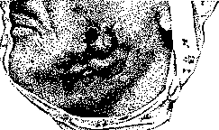
Figure 2. Actinomycosis.
the ear canal and Eustachian tube (middle ear). Actinomycosis of the face may be mixed with various inflammatory processes, especially osteomyelitis; any prolonged inflammation prone to spread is suspicious for actinomycosis; but even limited processes, for example, of the gums or cheek, may be of actinomycotic origin. The lymph glands are usually not affected in this process or only swell in mixed infection, which is common in opened inflammatory foci.--Actinomycosis of the neck. The process spreads to the neck either from the face, or the portals of entry are the pharynx, throat, less frequently the external coverings, larynx, and esophagus. Characteristic is a diffuse, very dense (as if wooden) infiltration, often with transversely bluish-red ridges of the skin. Here also, in different places, foci of softening appear, opening both on the skin and in the oral cavity, pharynx, etc., which leads to the formation of prolonged and complex (sometimes branched) fistulas. The diffuse infiltration not only impedes movement of the neck but sometimes constricts swallowing as well as breathing, which can even lead to suffocation (necessity for tracheotomy). The process can destroy vertebrae, descend into the mediastinum, etc. However, limited processes are also encountered on the neck, which are often confused with ordinary inflammatory processes, adenitis, etc. In limited processes the prognosis is good (with proper treatment); but they can be prolonged in time and recur (hidden infection).- Actinomycosis of the tongue deserves special mention. Although the process is rare (Erb in 1925 collected from the literature 8 cases of primary actinomycosis of the tongue and added his own 2 cases), it causes diagnostic difficulties. In this form, a limited infiltration develops, more often in the front part of the tongue; the process is little inclined to diffuse spread; it is easily confused with chronic abscess, gumma, etc. Actinomycosis of the tongue responds well to treatment (excision, etc.). Limited processes (infiltrates) on the tongue, as well as on the lip and floor of the mouth, may also be mistaken for cancer; here microscopic examination is important.-Actinomycosis of the chest. In the lungs the process can be primary and secondary. Primary gives a picture of prolonged pulmonary disease with the formation of bronchopneumonic foci. These foci, breaking down inside, lead on the one hand to destructive processes (opening into bronchi, cavities, etc.), and on the other to scarring. The peculiarity of the process, which is easily confused with tuberculosis, is that it does not remain limited to the lungs but quickly spreads to neighboring parts (pleura, chest wall, mediastinum, pericardium and even the heart), then descends into the abdominal cavity, sometimes as far as the pelvis, etc. Pleurisy can be serous (sometimes with hemorrhagic exudate), but more often there is adhesion of the visceral and parietal layers of the pleura, with the formation of callus-like masses, which are penetrated by purulent tracts connecting the pulmonary foci with the surface; and here prolonged, persistent fistulas form. It should be noted, as clinical features of pulmonary actinomycosis, the mainly lower lobe involvement and severe chest pains (radiating to the scapula, sometimes to the neck and arm), which is probably explained by compression of the nerves by the callus-like tissue. The process can be accompanied by hemoptysis, expectoration, in which the fungus is sometimes microscopically detectable (and other microbes in mixed infection). The febrile reaction is of varying degrees; sometimes exhaustion, night sweats, etc. The prognosis is very serious, but recovery has sometimes been observed. Primary pulmonary actinomycosis must be distinguished from secondary, which occurs either metastatically (metastases not through lymph vessels, but through blood), or due to the spread of the process from neighboring areas (neck, abdomen). In the latter case, the lung disease is masked and recedes into the background.-Actinomycosis of the mammary gland is rare: in 1926 one Italian author collected in the literature only 16 cases. The process appears in the parenchyma of the gland in the form of dense infiltrates, which then soften (fistulas); the process can involve a considerable part of the gland. Sometimes in the mammary gland the process is secondary, having spread from the chest wall, lung, etc. (such cases were observed by Veliaminov and Efron). The process can be confused with ordinary chronic abscess and with tuberculosis. Primary cases respond well to treatment.- Actinomycosis of the abdomen. Here first of all it is necessary to point to the frequently encountered actinomycosis of the abdominal wall (in the USSR such cases were described by Efron, Gorelik, Spizharny, and others). It can be primary and secondary; the latter more often spreads from the ileocecal area. A dense (sometimes like wood) infiltration is the main characteristic feature (sometimes only on the basis of this sign is it possible to make the correct diagnosis). Focal softening is also common. The prognosis for primary diseases is good with appropriate treatment (see below). Secondary diffuse process, when the entire thickness of the abdominal wall, abdominal organs, retroperitoneal cellular tissue are affected, is a very severe form of the disease.-Actinomycosis of the abdominal cavity. The favorite site for actinomycosis of the abdominal cavity is the ileocecal area. The process begins with the intestinal mucosa (nodules under the mucosa, then ulcers) and for a long time can remain hidden. Gradually the entire thickness of the intestinal wall, peritoneum, subserous cellular tissue, etc., become involved in the process,-dense infiltrates form, sticking the intestines together and with the abdominal wall, and in them-foci of softening; the latter open outward and into the cavities of the intestines, bladder, etc.; strictures, symptoms of ileus, etc., can develop; the process can descend into the pelvis, to the perineum, giving a picture of paraproctitis, or ascend retroperitoneally to the perirenal cellular tissue, spine, etc. Sometimes the process initially gives a picture of appendicitis. The diagnosis is not always easy; confusion with tuberculous process, osteomyelitis, etc., is possible; repeated examination of granulations and tissue pieces is necessary (drusae in the discharge are rare). The prognosis is very serious: death can occur from exhaustion, pyemia, intestinal obstruction, etc. (mortality up to 80%). Sometimes metastases occur in the liver (penetration of the roots of the portal vein) and in distant organs. In the literature cases of actinomycosis of the stomach (infection through ulcer), gall bladder and bladder (among other things, after intertrigo), female genital organs (primary on the vaginal portion), kidney (according to Kleinschmidt-primary, there was an unsuccessful attempt at extirpation) and others are described. Royle described a case of recovery in actinomycosis of the stomach (opening of the focus in the omental bursa).-Actinomycosis of the brain and its membranes. Anders in a comprehensive work collected from the literature 59 cases, adding to them 2 of his own. Usually the process is secondary (per continuitatem or by metastasis); all described as primary, Anders is inclined to explain as secondary, in which the primary focus was either overlooked or there were no sharp anatomical changes at the site of infection (pharynx, teeth, tonsils, ear, etc.). Processes in the brain are more often expressed as abscesses; localization is very varied; tumors (actinomycomas), meningitis, more often secondary, accompanying changes in the brain, base of the skull, etc., are also described; and here mixed infection is often noted. In the literature cases of involvement of the Gasserian ganglion are also described. The prognosis in all these processes is, of course, extremely serious.-Primary actinomycosis of the skin is observed after injuries (infection through a wound), in the form of nodules (resembling lupus), infiltrates, ulcers, fistulas. The process can spread inward, reaching the bones (Bergmann's case on the thigh, after a kick from a horse). Cases of actinomycosis on the forearm, foot (Tansini) and finger phalanx (Willems) are described.-Primary actinomycosis of bones is doubtful: the described cases (for example, actinomycosis of the lumbar spine), probably belong to those where the primary focus was overlooked. Course of actinomycosis. The disease proceeds either as local (limited forms) or with general, various (depending on localization) symptoms. In general the course is chronic, with moderate fluctuations in temperature. There can be a temporary stoppage of the process and recurrences after apparent recovery. In long and diffuse processes-exhaustion, anemia, mainly in mixed infection: then there can be purulent fever, pycemic phenomena, especially when foci open into blood vessels or the heart cavity (such cases are described). Sometimes the disease in mixed infection takes an acute pycemic course; for example, 17 abscesses (metastases) were described within four weeks. Cases of miliary actinomycosis (Petrov) are described. With prolonged course death from exhaustion, amyloidosis is possible; more rarely from complications,-peritonitis, suffocation, etc. Diagnosis of actinomycosis. Among the symptoms, the density of the infiltration is characteristic, which is only found also in Reclus's phlegmonous induration. Also characteristic is the chronic course, formation of abscesses with a small amount of liquid pus (sometimes with drusae). Without a microscope, however, the diagnosis can still be difficult: the histological picture by itself is not characteristic, it is necessary to find drusae or filaments, which is not always easy (in one case an experienced bacteriologist-microscopist found them only on the third examination).
According to Clermont, the complement fixation reaction gave positive results in 90% of cases. - The prognosis is especially serious in pulmonary and abdominal forms. But here too, many cases of recovery have been described. The prognosis is not hopeless even in pyemia (mixed infection). In cerebral forms of actinomycosis, the prognosis is poor. Treatment. As early as the last century, the works of Berard, Frei, Prutz, and others established the specific effect of potassium iodide in A. In some cases, Efron observed good results - admittedly, mostly in combination with surgical treatment. Iodine must be administered for a long time (sometimes for many months, with breaks). Dose: 2.0-3.0 per day; some authors gave up to 12.0. It is necessary to keep in mind the possibility of intoxication (irritation of the respiratory tract, diarrhea, palpitations, etc.). In some cases, treatment with injections of potassium or sodium iodide into infiltrates gives good results: Figure 1. Median section of an actinomycosis of the jaw of an ox through a fully developed druse (pre-staining with Bismarck brown, Gram staining with aniline-methyl violet, subsequent staining with eosin): a - dense network of mycelium; b - a finer network, from which strongly branched roots (c) grow into the tissue; d - peripheral outgoing bundles of threads; f - clusters of spore-like grains; the fungi are surrounded by polynuclear leukocytes, epithelioid cells, and large, often multinucleated cells (pre-stages of giant cells g).
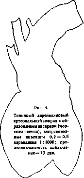
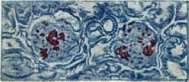
Figure 2. Amyloid infiltration of the kidney glomeruli and afferent vessel. Reaction with iodine.
Figure 5 and 6. Blood picture in pernicious anemia; aniso- and poikilocytosis (top right - normoblast; bottom left - megaloblast; top left - lymphocyte). In Fig. 6 - normal red blood cells for comparison of volume.
Figure 3. Amyloid infiltration of the kidney glomeruli. Staining of amyloid red by Congo red.
Figure 7. Blood picture in severe chlorosis: aniso- and poikilocytosis, oligochromemia.
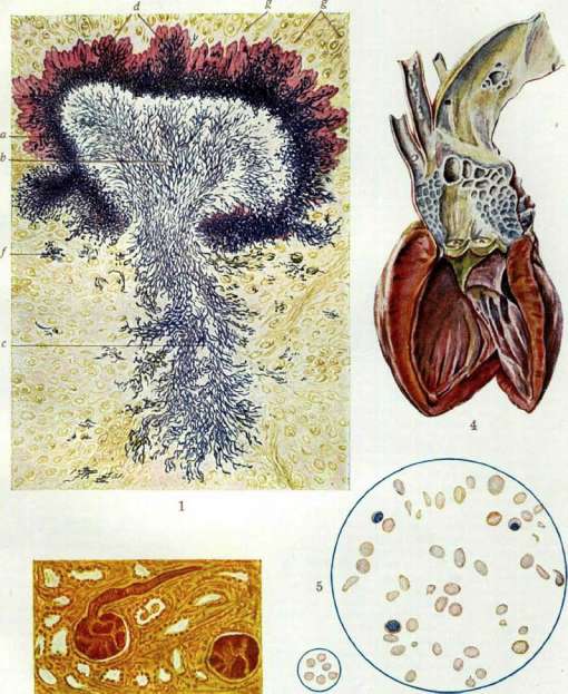
o < A' st. Actinomycosis. Amyloid degeneration. Atherosclerosis of the aorta, Angina. This method is quite painful, but novocaine can be injected beforehand. Surgical treatment, the importance of which was proven in Bergmann's clinic as early as the last century (works by Schlange and others), should consist of incisions and curettage: sometimes parts of organs were removed (resection of intestines, piece of lung, appendix, kidney, etc.)- In recent times (1916) the clinic of Kiittner (Melchior) enriched the therapeutic arsenal with a new powerful means - radiotherapy, which, according to Melchior, is especially effective in combination with iodine. In the USSR, Grigoriev has extensive material on radiotherapy of external A.; radiotherapy should always be applied in A. Radium was also successfully used (Jungling). Eckert reported in 1922 a case of recovery from a severe form of cervical A. with yatren, in which treatment with iodine and radiotherapy was ineffective; he injected a 5% solution of yatren into the veins. Neosalvarsan and protein therapy (Beck) were also used. In chronic cases, strengthening treatment is also important; Efron saw benefit from kumys. For various surgical complications - operative therapy (tracheotomy, anus praeternaturalis, etc.). A. in animals. It is observed in cattle, less frequently in pigs, horses, dogs, and cats. Infection occurs through the mouth, respiratory tract, and skin. A in cattle is most often localized in the jaw. Here, slowly increasing, hemispherical, dense tumors form, growing into the skin in the form of greasy, mushroom-like formations of grayish-yellow color. From them, pus with characteristic yellow grains is squeezed out. Subsequently, the nodes disintegrate and cause tissue necrosis. Self-healing is occasionally observed. Sometimes, due to the infiltration of blood vessels, the process generalizes with the formation of metastases. Transmission of infection through the lymphatic system usually does not occur. In horses, a peculiar localization of A is observed in the spermatic cord, after castration. In pigs, A runs peculiarly, in the form of scattered degenerated foci in the muscular tissue with subsequent calcification. In this case, the fungal grains of A in pigs are smaller than in human A. In addition, pigs may also have true actinomycosis.
L. Kursanov, V. Gaponopel'ny, Yu. Finkel'shtein, N. Efron.
Related articles
Mentioned in
Cite this page
“Actinomycosis.” Soviet Medical Encyclopedia. English translation of Bolshaya Meditsinskaya Entsiklopediya, 1st ed. (Moscow, 1928–1936), ed. N. A. Semashko. https://sovietmedicalencyclopedia.pages.dev/article/actinomycosis/