Neutrophilosis
Historical document, translated for reference. It reflects medical knowledge of the 1920s–30s and is not medical advice.
Summary
Neutrophilosis is the increase in the percentage of neutrophils in the blood, often accompanied by an increase in total leukocytes. It occurs in physiological states and in various pathological conditions, including infections and inflammation. The condition is a significant diagnostic and prognostic marker in many diseases.
Encyclopedia article (1928–1936)
NEUTROPHILOZ, an increase in the percentage of neutrophils in the blood; in this case the total number of leukocytes is also more often increased, but it can be normal and even decreased. N. is characteristic both for physiological (for example, digestive leukocytosis, especially after a large amount of protein food, leukocytosis of newborns, etc.) and for all pathological leukocytoses, infectious and inflammatory, toxic, etc. (see Leukocytoses, pathological leukocytoses). In clinical practice N. has great significance in infectious diseases, since in most infectious diseases N. occurs regularly, and only some of them proceed as a rule without N. (for example, typhoid fever, chronic malaria); finally, in the same disease, depending on the severity of the latter, N. may be differently expressed or entirely absent. N. is observed in lobar pneumonia, catarrhal pneumonia, epidemic cerebrospinal meningitis, actinomycosis (especially in chronic purulent processes), rheumatic polyarthritis, septic processes, tuberculosis, most often cavitary, complicated by secondary infection, Asiatic cholera, smallpox (in the first and again in the pustular stage), chickenpox (usually without an increase or even with a decrease in the total number of leukocytes), epidemic parotitis (mostly not strongly expressed), whooping cough (in the catarrhal period), diphtheria, typhus (in the initial stage), scarlet fever, angina, measles (only in the first stage, later only with complications), tetanus, relapsing fever, malaria (during attacks), often in lymphogranulomatosis. No less significance is N. in purulent processes: empyema, peritonitis, perityphlitis, abscesses of the liver, lungs, purulent meningitis, etc. In these processes N. often has great diagnostic and prognostic significance. Very often, by the degree of neutrophilic leukocytosis in appendicitis, one can judge the catarrhal or purulent character of the process. N. with a significant general increase in leukocytes sometimes even without a characteristic general clinical picture makes one think of a purulent process. N. in diseases that usually proceed with lymphocytosis (for example, typhoid fever, measles, in later stages, etc.) often indicates complications by some inflammatory process. As for the prognostic significance of N., then with a very large general leukocytosis (< 40-50,000) both in general infectious and in purulent processes it usually serves as a poor prognostic sign; the same can be said about N. with a normal or even decreased total number of leukocytes, if such is observed in purulent and those infectious diseases that usually proceed with an increase in the total number of leukocytes (for example, lobar pneumonia, peritonitis). The diagnostic and prognostic significance of N. becomes clearer when taking into account not only the total amount of leukocytes, but also the character of the nuclear shift of neutrophils. (For details see Blood Leukocyte Formula - neutrophilic leukocytosis.)
E. Gedeynstein. NEKATOR, a disease caused by the nematode Necator americanus (Stiles; 1902), belonging to the family Ankylostomidae. Its place of localization is the small intestine. From Ankylostoma (Ankylostoma duodenale), causing a clinically similar disease, N. americanus is distinguished by the following details of its anatomical structure: 1. Its oral capsule is less powerful and, in contrast to Ankylostoma, is armed not with teeth, but with a pair of cutting plates. 2. The copulatory bursa of the male Necator is significantly narrower than that of Ankylostoma and has the shape of a tulip (in Ankylostoma the shape is a bell). 3. The spicules of Necator have at the end appendages in the form of knitting needles (in Ankylostoma they end sharply). 4. The vulvar opening of the female Necator lies in the anterior half of the body (in Ankylostoma at the border of the posterior and middle thirds). 5. The caudal end of the female Necator does not have a needle-like pin, which is present in the females of Ankylostoma. For the first time both these species were distinguished in 1902 by Stiles (Stiles), who pointed out that the diseases called tropische Chlorose, Gottardtunnelanemie, anemie des mineurs, cachexia africana and others (see Ankylostoma) are caused not only by Ankylostoma, but also by a worm close to it in anatomical structure, widely distributed in the USA and therefore received the name Necator americanus (see figure). The primary source of N. apparently is Central Africa, where it was found among dwarf tribes living completely isolated. From Africa Necatoriasis was introduced into the USA during the active slave trade and thanks to suitable climatic conditions it took root there firmly. According to old data, N. is spread in the zone lying between 30° south and north latitude. This zone includes the states of Missouri, Oklahoma, Texas, California. Mexico is quite strongly affected and Puerto Rico very strongly. In South America N. is spread in all tropical and subtropical areas, and Paraguay is especially strongly affected. Besides this, N. is registered on a whole series of islands: Sumatra, Java, Samoa, etc. Among immigrants from Eastern Asia, Glover discovered 63% Necator carriers. The degree of infection of the listed areas by ankylostomids is different: from 60% to 100% of the population, and pure infection by Necator americanus falls on 80-90% of the total number, and the remaining 10-20% relate to pure infection by Ankyl. duodenale and mixed Ankyl. duod. + Necator am.
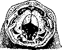
Necator americanus. Starting from 1923, Necatoriasis is also registered on the territory of the USSR, first in western Georgia (Parcvanidze), then in Abkhazia (Blazhin; 1925), Ajaria (Mchedlidze; 1925) and Azerbaijan (1927). In Abkhazia, Ajaria and Azerbaijan, with a general infection by ankylostomids from 40% to 90%, Necator americanus (pure infection) gives from 75% to 95% of the total number of ankylostomids. In western Georgia, especially in Zugdidi and Senaki districts, a fairly significant admixture of Ankylostoma duodenale is observed. The biology of Necator americanus and Ankylostoma duodenale and the clinic of N. and ankylostomiasis are currently considered almost identical (see Ankylostoma). It is noted only that ankylostomiasis proceeds more heavily, and the greater power of the fixation and injuring apparatus and greater resistance to antiparasitic drugs are significant. The most favorite therapeutic agent is a mixture of CCl4 and 01. Chenopodii; with a single dose, 99.7% Nee. am. and 80.0% Ank. duod. are expelled (Soper; 1925). Prescription: CCl4-2 volumes + 01. Chenop.- 1 volume, for adults 2.5-3.0+40.0 English salts in a concentrated solution. For children 1/18 of the adult dose per year of age; treatment is repeated after 10-12 days. The Abkhazian Tropical Institute applies Thymol 8-10 g pro die for an adult, 3 days in a row by 2.0 g every hour. After the last dose, an hour later a laxative salt of 30-40 g. For children 5-7 years - 2.0 g; 7-9 years - 3.0 g; 9-12 years - 4-5 g; 12-15 years - 6.0 g.
Related articles
Cite this page
“Neutrophilosis.” Soviet Medical Encyclopedia. English translation of Bolshaya Meditsinskaya Entsiklopediya, 1st ed. (Moscow, 1928–1936), ed. N. A. Semashko. https://sovietmedicalencyclopedia.pages.dev/article/neutrophilosis/