Mammary Gland
Historical document, translated for reference. It reflects medical knowledge of the 1920s–30s and is not medical advice.
Summary
This article from the 1928–1936 Soviet Great Medical Encyclopedia provides a detailed anatomical and histological description of the mammary gland. It covers the structure of the gland, its development, the role of the nipple and areola, and the vascular and lymphatic systems associated with the organ.
Encyclopedia article (1928–1936)
MAMMARY GLAND, or breast (glandula mammaria s. mamma), is a paired organ located in humans, just as in all mammals, on the anterior (abdominal) surface of the body and serves in women and female animals for nursing offspring. Functionally, the mammary glands are closely connected with the reproductive organs; based on their developmental history (see below), they belong to the group of cutaneous glands, specifically large sweat (so-called apocrine) glands.

Anatomy and histology. In an adult woman, the mammary glands are located as two hemispherical formations on both sides of the chest, at the level between the III and VII ribs. Laterally, each breast extends to the axillary line, and medially it does not quite reach the midline of the body, so that a space remains between both mammary glands (sinus mammarum). At the height of the IV intercostal space or V rib (approximately in the center of the hemispherical protrusion of the mammary gland) is the nipple, papilla mammae, a small pigmented protrusion on which a row of small openings (pori lactiferi) is visible. The nipple is surrounded by a pigmented circle, called the areola (areola mammae). The size and shape of the mammary glands are extremely variable and depend on age, degree of nourishment, the number of previous births and nursing periods, etc. In appropriate cases, one speaks of macromastia or micromastia. In men, the mammary glands do not normally develop and remain in a rudimentary state (their size does not exceed 1.5x0.5 cm), and the nipple only slightly protrudes above the surrounding skin (diameter about 2-5 mm). Only in rare cases does a large mammary gland develop in men, capable even of secreting milk (such a phenomenon is called gynaecomastia).
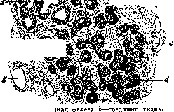
The mammary gland consists of a small number (15-20) of lobes (lobi), each of which has its own excretory duct (ductus lactiferus), 2-3 mm in diameter. All excretory ducts direct their endings toward the nipple, and, just before reaching the skin surface, each duct forms an ampullary expansion (sinus lactiferus), 7-9 mm in diameter. Further on, approaching the skin, the duct narrows again (its external opening, porus lactiferus, is no more than 0.4-0.7 mm in diameter). The individual lobes of the mammary gland are held together by a connective tissue stroma, which is more or less rich in adipose tissue. The actual suspensory apparatus of the mammary gland is the superficial fascia (lig. suspensorium-Giraldès), which attaches from above along the entire length of the clavicle. Descending downward, at the upper edge of the mammary gland, the superficial fascia divides into two leaves, forming a kind of capsule that encloses the mammary gland. In addition, connective tissue processes (retinacula mammae) serve to strengthen the entire gland, connecting it with the subcutaneous adipose tissue and with the fascia of the pectoralis major muscle.
The lobes of the mammary gland are usually arranged radially around the nipple, forming a fairly regular circle. However, it is not uncommon for some of them to protrude beyond the main mass of the mammary gland (or even detach completely, forming so-called mammae aberratae). Such "stray" lobes were most often found in the axillary fossa (see Figure 1). However, there is another, more correct explanation for the development of stray lobes of the mammary gland, namely in the order of metamorphosis of the so-called apocrine glands (see). From the concept of accessory or stray lobes of the mammary gland, one should distinguish the concept of "accessory mammary glands": the latter always have a nipple and an excretory duct (see below).
Each lobe of the mammary gland consists of smaller formations—lobules (acini s. lobuli), and the latter—of alveoli (alveolae). Thus, the mammary gland is a complex alveolar gland. The alveoli are lined with a single-row cylindrical glandular epithelium. The height of the cells of this layer varies; it depends on the presence and quantity of secretion and fluctuates between 2 and 15 microns. The nuclei of the cells are round or oval, sometimes slightly flattened, contain 1-2 nucleoli and a delicate chromatin network.
Figure 1. Accessory lobule (axillary) of the mammary gland of a nursing woman. (According to Seitz.)
Figure 2. Alveoli of the mammary gland in a resting state; a'-alveoli preparing for secretion; m-large duct. (From Koelliker.) The alveoli, during secretion, are filled with fat droplets of various sizes. The lumens of the alveoli at this same time expand and fill with a homogeneous mass containing leukocytes and fat droplets. In a resting state, the alveoli remain solid in a significant part, i.e., devoid of a lumen (see Figure 2). Around the alveoli is located a membrane, which according to some authors consists of smooth muscle fibers, and in the opinion of others—of connective tissue. This is the proper membrane of the alveolus, its membrana propria. In the layer between the epithelium and this membrane, so-called myoepithelial cells have been described. Anastomosing with each other, they form a network that apparently plays the role of a supporting device.
The excretory ducts in the depth of the gland are lined with a single-layer cylindrical epithelium and are surrounded, just like the alveoli, by a proper membrane (membrana propria). Approaching the skin, after its expansion into the sinus lactiferus (see Figures 3 and 4), the duct replaces the single-layer cylindrical epithelium with a stratified squamous one, consisting of 8-10 rows of cells, forming at the very surface so-called funnels of entry (see Figure 4).
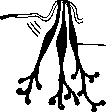
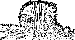
Figure 3. Scheme of the excretory ducts of the mammary gland: 1-muscles of the nipple; 2-sinus lactiferus, or milk cisterns. (According to Bonnet.)
Figure 4. Normal nipple of the mammary gland; semi-schematic designation of the bundles of smooth musculature; the excretory ducts with their expansion in the depth are visible. (According to Basch.)
The skin of the nipple and the areola is pigmented; in its papillary layer, there are many high papillae and smooth muscle fibers, especially around the endings of the excretory ducts and along the nipple itself (see Figures 3 and 4). In the circumference of the nipple, there is a row of large sweat (apocrine) and about 12 modified sebaceous glands. The latter secrete a substance that is an intermediate between milk and the secretion of ordinary sebaceous glands. These glands are called glandulae areolares s. Montgomery.
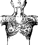
Figure 5. Anastomoses between the lymphatic vessels of both mammary glands. The mammary gland receives arterial blood supply from three sources: 1) from the internal mammary artery (a branch of the subclavian artery); 2) from the lateral thoracic artery (a branch of the axillary artery) and 3) from the intercostal arteries (from the 3rd to the 7th).
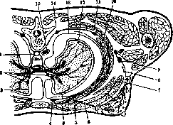
Figure 6. Scheme of the lymphatic vessels and glands of the lung and chest, including the mammary gland (horizontal section at the level of the mammary glands): 1-thoracic duct; 2-bronchopulmonary glands; 3-superficial lymphatic vessels of the lung; 4-anterior intercostal lymphatic gland; 5-lymphatic vessels from the mammary gland and pectoral muscle to the anterior intercostal lymphatic gland; 6-pectoralis major muscle; 7-pectoral fascia; 8-lymphatic vessels from the mammary gland to the axillary glands; 9-axillary lymphatic glands; 10-internal intercostal muscle; 11-external intercostal muscle; 12-visceral layer of the pleura; 13-parietal layer of the pleura; 14-posterior intercostal lymphatic gland; 15-aorta. (According to Corning.)
The network is located parallel to the arterial one, corresponding to its branches. From the medial sections of the mammary gland, blood flows out through the vv. perforantes into the intercostal veins, and from the skin and superficial tissues through the vv. subcutaneae into the vv. axillares. The lymphatic system of the mammary gland (see Figure 5) drains lymph along several pathways. The main one is directed from the lateral part of the mammary gland into the axillary fossa; the vessels of this pathway usually unite into 2-3 large trunks, which are directed toward the axillary glands, lying parallel to the lower edge of the pectoralis major muscle. Besides this main pathway, there are several additional ones: 1) the subaxillary pathway, the vessels of which are directed through the pectoralis major muscle to the deep axillary glands; 2) the subclavian pathway—between the pectoralis major and pectoralis minor muscles to the subclavian glands; 3) the supraclavicular pathway—bypassing the axillary glands, it is directed directly to the supraclavicular ones; 4) the intrathoracic pathway—2-3 small trunks piercing the intercostal spaces and draining lymph into the anterior mediastinal glands (see Figure 6). Besides these pathways, another one has recently been described, directed from the lower-medial quadrant of the mammary gland into the epigastrium and then anastomosing with the lymphatic pathways of the pleura, the subdiaphragmatic space, and the liver (Handley). The superficial lymphatic vessels form a dense network around the nipple, while widely anastomosing with the vessels of the opposite mammary gland. The sensory nerves of the mammary gland originate from the supraclavicular and intercostal nerves (from II to VI). Branching in the skin and in the gland itself, they terminate in the nipple with Vater-Pacini corpuscles, and around the larger lactiferous ducts in the form of Krause end bulbs. Furthermore, sympathetic nerves also approach the mammary gland, forming plexuses around the blood vessels (mainly arterial). Development of the mammary gland. In the early stages of fetal life in mammalian animals (in humans at a fetal size of 9.5 mm), a skin fold appears, extending from the axillary fossa to the inguinal fold (symmetrical on both sides). This is the so-called milk line (or
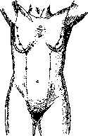

Figure 7. Diagram of the arrangement of accessory mammary glands. (According to Merkel.)
Figure 8. Bilateral accessory glands that secreted milk during nursing. milk ridge), which in humans develops only in the upper part of the trunk. By the end of the 2nd month of fetal life, in the human embryo, a series of bead-like thickenings forms on this line (in animals, they correspond to the future mammary glands). Of these thickenings, one pair (according to some authors, the fourth, counting from the top) begins to develop gradually, while the others normally disappear. Only in rare cases do accessory mammary glands (hypermastia or polymastia, see Figures 7 and 8) or only nipples (hyperthelia or polythelia) form from them. The actual development occurs in such a way that in the 5th-6th embryonic month, solid cellular cords begin to extend from the epithelial primordium (corresponding to the site of the future mammary gland) (see Figures 9, 10, and 11). In the 7th-8th month, lumens begin to appear in these cords, initially bordered by several layers of epithelium. From the same epithelial primordium from which the initial cords of the future mammary gland develop, the primordia of both the hair follicle and the sebaceous gland also originate; however, the development of hair in the area of the nipple and the areola usually does not occur. By the end of fetal life, the gland consists of 12-15 not yet

No. "Ch ^ch Figure 9. Primordium of the mammary gland in a 21 cm fetus. Solid epithelial cord (primordium) plunging deep into the skin. (According to Basch.)
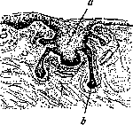
fully developed lobules and has a tubular structure. The nipple in this period appears undeveloped; its area is usually retracted, contains keratinized masses, forming a shallow nipple pocket (see Figure 11). Subsequently, the formation of the nipple occurs by the eversion of the nipple pocket outward due to the development of connective tissue and smooth muscle (true nipple). Another method of nipple formation is observed in various mammals (ungulates, carnivores). In this case, the skin around the glandular area rises in the form of an annular ridge, at the bottom of which the excretory ducts open. Macroscopically, the mammary gland of a newborn is a round formation, sometimes even larger, Figure 10. Primordium of the mammary gland in a 25 cm fetus. From the epithelial cord (a), individual outgrowths (b)—the future lactiferous ducts—extend deep into the skin. (According to Basch.)
ducts. (According to Basch.)
ranging in color from tender pink to dark red. The glandular lumens are lined with 1-2 rows of cuboidal epithelium; the lobules are enveloped in loose, highly hyperemic connective tissue. An interesting detail of the structure of such glands is the development in them of significant cellular infiltrates (hematopoietic foci) with the presence of even megakaryocytes (the so-called mastitis of newborns). This phenomenon, just like the secretion of the mammary gland of infants (Hexenmilch), represents a physiological process and is observed in children of both sexes in the overwhelming majority of cases. The morphological and chemical nature of the secretion of such glands is extremely similar to human colostrum. Figure 11. Diagram of the development of the mammary gland: new "pocket". Sometimes an infection, especially with mechanical intervention, can penetrate inside and cause true inflammation, e.g., purulent, as a result of which the death of the gland is apparently possible. Since the secretion of infants is not excreted outward, subsequently, due to retention, the lumens of the gland expand, and it degenerates, as it were, "cystically." Following this, the lumens begin to collapse again, dense connective tissue develops, and by the 5th-7th month, the gland reaches complete involution (the "resting gland of the child" type). Further development of the mammary gland until the period of puberty proceeds extremely slowly. During the period of puberty in most boys, a transient swelling of the mammary gland is observed, when the glands sometimes reach a significant size—the so-called "mastitis" of youths. Histologically, in such a gland, hyperplasia of the glandular lumens and a histiocytic reaction are observed. In girls, the mammary gland during the period of puberty does not microscopically differ from the pubertal mammary gland of boys. Sexual differentiation in the mammary gland occurs no earlier than the onset of menstruation, when in girls the mammary gland begins to acquire an alveolar structure. Subsequently, with age and pregnancies, the number of lobules continues to increase. The mammary gland increases in size. Increased fat deposition occurs around it. Individual lobules grow, and their number gradually increases.
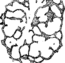
Figure 12. Mammary gland during the lactation period: a—dilated alveoli; b—excretory duct.
However, the mammary gland reaches its full development only during nursing. Already starting from the 2nd month of pregnancy, the areola enlarges and darkens. The vessels of the mammary gland proliferate, and the gland itself increases sharply in size. Already from the first months after conception, clear hyperplasia of the parenchyma, the formation of secondary lobules, and a histiocytic reaction are microscopically detected. Subsequently, the alveoli are strongly distended and contain fluid with a large number of fat droplets (ready alveolar milk). The interstitial connective tissue gradually atrophies, giving way to the multiplied and enlarged glandular lobules. The structure of the lobules of the mammary gland at this time very much resembles lung tissue (see figures 12 and 13). In men, however, the mammary gland remains in a state of regressive development throughout—it retains a tubular structure; however, the presence of even weak secretion in it can be noted up to old age.
Secretion. Significant secretion of the mammary gland begins in the 2nd half of pregnancy. The fluid secreted from the nipples by this time (colostrum) is not yet true milk. It is rich in proteins and differs from milk by a higher specific gravity and the presence of special colostrum corpuscles (leukocytes loaded with fat droplets). Only a few days after childbirth does true milk begin to be secreted. This is an alkaline fluid containing 1-2% protein, 3-4% fat, 5% sugar, and 0.6% lecithin, with a specific gravity of 1.028-1.034. Milk is an emulsion of minute fat droplets (1-5 μ in diameter), which are surrounded by a casein membrane, which prevents them from coalescing. In addition, a small number of lymphocytes are found in milk. The secretion of the mammary gland is predominantly apocrine in nature and occurs in such a way that the inner part of the cell, in which the formation of fat droplets occurs, is pinched off (the phenomenon of 'decapitation') and ends up lying freely in the lumen of the alveolus. With the onset of the climacteric period, the mammary gland begins to gradually atrophy: the glandular lobules die and are replaced by connective tissue; the ducts undergo cystic degeneration, and the fatty lining disappears. This process progresses all the time, and senile mammary glands consist of highly atrophic excretory ducts surrounded by connective tissue; such glands are rich in elastic tissue. Regarding the latter, it should be said in general that its amount in the mammary gland increases with age in both sexes, reaching a maximum in senile mammary glands, especially with a large number of births in
the anamnesis. In the mammary gland of women who have given birth, elastic tissue is determined in negligible amounts by various reactions for elastin. The female mammary gland, among a whole series of organs
occupies an exceptional place. If one does not count the mucous membrane of the uterus, then no other organ is subjected to such constant physiological irritations as the mammary gland. Already at the moment of the child's birth, as mentioned above, the mammary glands begin, as was indicated, to swell rapidly, increase in volume, and secrete.
Further, the life of the mammary gland subsides, only to receive irritation again by the time menstruation appears (a push towards the beginning of the development of the mammary gland). A constant change of irritations begins, which accompany menstruation monthly, and this physiological cycle of the mammary gland runs parallel to the physiological cycle in the genital sphere. Rosenburg and Moschkowicz distinguish 4 periods in the vital activity of the mammary gland: premenstrual, menstrual, postmenstrual, and the interval state.

They also note certain histological changes in the gland tissue corresponding to each period and differ only in details. At the same time, one can ascertain the swelling of the breasts and then their reduction; this often causes a whole series of painful sensations; sometimes a discharge appears from the nipples, occasionally taking on a bloody character (vicarious menstruation). Sufficient attention should be paid to such discharges, as they can also occur on a pathological basis of a developing neoplasm.
Figure 13. Alveolus during the lactation period. Pregnancy is a particularly strong irritant for the mammary gland; the hormonal influence of the genital organs manifests itself here by the intensified proliferation of milk lobules at the expense of the connective tissue base—the mammary glands are prepared for a new postpartum function—the secretion of milk. Puerperal mastitis (which can also be encountered during pregnancy) is encountered here quite often (see Mastitis); the scars remaining after them, like the gland itself, can subsequently also be subjected to irritations and give cause for degeneration. Abundant lactation of the mammary gland can be observed in certain diseases of the glands of internal secretion, for example: with tumors of the anterior lobe of the pituitary gland (in acromegaly), with tumors of the ovaries, with castration. Upon cessation of nursing, the involutionary period of the gland's life begins in the gland after the phenomena of rest, and during the next pregnancy, the described changes are performed again. Thus, with each pregnancy (full-term or abortion), the mammary gland is subjected to a new irritation each time, until the climacteric period arrives. But even then, changes characteristic of senile involution begin to develop in the mammary gland, which already present a fairly significant irritant for the gland. Thus, throughout a woman's entire life, the mammary gland is subjected to constant physiological traumas (not to mention mechanical ones), and in this circumstance, many authors are ready to see the cause of a whole series of pathological processes of both an inflammatory (see Mastitis) and neoplastic nature.
I. Silberberg. Fissures of the nipples. Feeding the infant frequently leads to nipple fissures. Their usual cause is traumatism brought about by this act. However, there are other predisposed factors depending on both the mother and the infant. The skin of the nipples and the areola is generally tender and easily injured. The less pigmented it is, the more frequent are the fissures. Insufficient body hygiene often entails the formation of scups and crusts from the epidermis on the skin of the nipples. Upon their shedding, excoriations remain. But overly frequent washing of the nipples also contributes to the formation of fissures (Jaschke), as well as a flat, retracted, as well as tight nipple that is poorly grasped. Eczema of the skin of the nipples caused by galactorrhea, as well as scabies, lowering the skin's resistance, predispose to the development of fissures. A mixture of saliva and milk that is not washed off but retained in the skin folds of the nipple can irritate the skin. Inflammatory processes in the oral cavity and pharynx of the infant, infectious stomatitis, thrush, and diphtheria can cause nipple fissures in the mother. The technique of feeding plays a prominent role in the genesis of fissures. Grasping the nipple itself instead of a part of the areola often leads to the formation of fissures (Czerny, Keller). Feeding in a lying position predisposes to the formation of fissures more frequently than in a sitting position (Moll). According to Marfan, erosions and fissures of the nipples are encountered in almost 50% during the first days of feeding, and according to Platzer in 51.5%. Dietrich and Frangenheim consider that almost 40% of all nursing mothers suffer from nipple fissures. In primiparae, fissures are encountered more frequently than in multiparae; in the latter, more frequently than in grand multiparae. But this is not an absolute law. In blondes, especially redheads, fissures are encountered more frequently than in brunettes; they have less pigment in the areolae. In urban women, fissures are encountered more frequently than in rural ones; in affluent individuals more frequently than in the poor: according to Cazeaux's statistics as 20:1; the skin in affluent women is apparently more vulnerable. Nipple fissures begin with the desquamation of the epidermis, the exposure of the corium surface, and the production of an erosion. If the skin is torn somewhat deeper in the folds, a fissure is produced, i.e., a small linear wound with parted and thickened edges. Its bottom is red, moist, and easily bleeding during nursing. Sometimes these fissures penetrate more deeply into the skin layer, forming wedge-shaped open fissures with inflamed, thickened sebaceous edges. All forms of fissures bleed during nursing. According to their location, one can distinguish fissures at the apex of the nipple and at its base. Fissures can cause such severe pain during feeding that the mother sometimes refuses breastfeeding. Fissures at the base of the nipples are more sensitive than those at the apex. Usually, the pain is more sharply expressed at the beginning of the nursing act. In the intervals, it may even be absent. Fissures can also exert a bad effect on the infant: as a result of pain, the quantity of milk decreases, and possibly its quality as well. Therefore, a slight dyspepsia and weight stagnation are often observed in such infants (Marfan). Fissures can serve as an entrance gate for the formation of lymphangitis, galactophoritis, and mastitis. Therefore, every fissure must be treated as early as possible. Treatment aims to achieve three moments: 1) to cicatrize the fissure, 2) to prevent the formation of milk stasis and thereby reduce the predisposition of the breast to infection, 3) to prolong breastfeeding as much as possible. Aside from various devices (bandages, nipple shields, milk-sucking apparatuses), many remedies were locally proposed. Wet antiseptic bandages, various ointments, irritating agents, as well as toxic substances should be avoided. In mild cases, one can resort to no remedies, but only wash the nipple after feeding with hydrogen peroxide (1:2 of boiled water), dry it thoroughly with sterilized cotton, and apply a dry sterilized gauze. In more severe cases, Marfan recommends, after the aforementioned washing and drying of the nipple, to lubricate the fissure with the following composition: Aquae rosarum 45.0, Glycerini 15.0, Natri biborici 5.0, T-rae Benzoes 10.0. A 3% aqueous solution of methylene blue acts well. In small fissures located at the apex of the nipple, one can apply the infant directly to the breast. But if feeding causes severe pain and bleeding, the nipple must be protected from trauma. Then protective shields are resorted to. The best of them is "Infantibus". When fissures are located at the base of the nipple and feeding even through a shield is extremely painful, the infant has to be taken away from the breast for 24-36 hours, but during this time it is necessary to express the milk so that its stasis does not form. With such deeply running wedge-shaped tears, careful lubrication with tincture of iodine or 5-10% silver nitrate is applied. In almost all cases where there are no sharp deformations of the nipple of the mammary gland, the best results are obtained from the treatment of fissures with a quartz lamp (Chatin, Tamarina, Jaschke). Prophylactically, it is necessary to raise the resistance of the nipple skin before childbirth. If the nipples protrude sufficiently, it is necessary from the end of pregnancy every 2-3 days to slightly rub the nipples with a thin cloth moistened with cold water, and then dry them thoroughly with a sterilized cotton ball. In this way, epidermis scales and small particles of colostrum are removed, which can contaminate and irritate the skin. If the areolae are poorly pigmented, an intensification of pigmentation should be induced with a quartz lamp. In the postpartum period, one must warn against very frequent washing of the nipples. Caution is especially needed if the nipples were not prepared before childbirth. Despite all the aforementioned measures, one cannot avoid fissures with certainty. Under no circumstances are fissures a contraindication to breastfeeding. The only moment preventing feeding in the presence of nipple fissures is pain, but it can also be eliminated by treatment with pads and a quartz lamp.
E. Sklovskiy. Anomalies of the Mammary Gland. A significant, sometimes insurmountable obstacle to the nursing act is presented by the retracted nipple, called, due to its resemblance to the navel, umbilicated (umbilication of the nipple - umbilicatio). Due to the impossibility of sufficiently thoroughly disinfecting such a nipple, inflammatory processes easily develop in it, leading ultimately to mastitis (see). In mild degrees of nipple umbilication, the application of appropriate suctions generally succeeds in gradually pulling out the nipple, while its severe degrees are subject to surgical treatment. The most widespread method until recently was Kehrer's method. By a circular incision around the nipple, two half-moon flaps are formed above and below the nipple, and the corresponding parts of the skin are excised, as a result of which defects are obtained, the suturing of which leads to the narrowing of the integuments around the nipple, which protrudes forward from this. In 1917, Sellheim proposed a method yielding a well-protruding nipple. A rare anomaly is the absence of nipples (unilateral or bilateral), as well as the absence of mammary glands (see Amastia). More often, on the contrary, there is an excessive formation of nipples (polythelia, hyperthelia) and mammary glands (polymastia, hypermastia). They are usually located within the so-called milk (see above) line (see Figures 7 and 8), but rare cases of their location on the middle of the anterior abdominal wall, in the region of the shoulder, back, and outer surface of the thigh have been described. The maximum number of excessive mammary glands of a single person described by authors reaches 8 (Neugebauer). Hereditary transmission of this deformity is observed, which is encountered (according to the statistics of various authors) on average in 3.8% of all males and 7.6% of all females. Frequently, in subjects burdened with a deformity of this kind, one also has to observe various other anomalies, which gave Leichtenstern grounds to consider it a manifestation of atavism. Excessive mammary glands are often equipped with excretory ducts secreting milk in women (during pregnancy and lactation), sometimes in very large quantities. Seitz observed a woman nursing an infant with four mammary glands (2 normal and 2 accessory; see Figure 8). Accessory lobules of the mammary gland (mammae aberratae, see above) should be distinguished from excessive mammary glands (hypermastia). They are located near the mammary gland, for the most part along the outer edge of the pectoralis major muscle and in the axillary fossa. They have no nipples. The clinical significance of excessive mammary glands and accessory lobules thereof is great, since they can become the site of development of an inflammatory process (mastitis) or a neoplasm (adenoma, carcinoma), as a result of which some authors propose removing them upon detection (for prophylactic purposes). This radical view, having weighty grounds in relation to "accessory mammary glands, which more frequently undergo malignant degeneration, should hardly be extended to excessive mammary glands and nipples, which have to be removed mainly for cosmetic reasons.
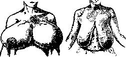
Figure 14. Hypertrophy
Figure 15. Sharp de-
gree of pendulous breast
in a 22-year-old girl.
in a 19-year-old girl. The size of the mammary gland varies and depends both on fat deposition and on racial characteristics and hereditary transmission. Excessive enlargement of the mammary gland (macromastia) is caused more often by large fat deposition (lipomatosis of the mammary gland), less often by diffuse true hypertrophy of the gland; the latter cases are characterized by a uniform increase in the volume of the mammary gland, sometimes reaching colossal sizes and hanging down to the navel, crista ilii, and even to the thighs (see Figure 14). The weight of such hypertrophied mammary glands reaches 30 kg (Bouyer, Iordansky, et al.). In the majority of cases, the disease is bilateral (according to Symens, in 31 cases, hypertrophy was bilateral in 26 cases; according to Kirchheim, in 55 cases, 13 cases were bilateral). A distinction is made between hypertrophy of the mammary gland during the onset of puberty (according to German authors, Pubertatshypertrophie) and another variety of hypertrophy in young women during pregnancy, mainly the first one (Graviditatshypertrophie). The former is observed more often, at the age of 14 to 25 years. It is characterized by the persistence of phenomena (in contrast to the second form, which undergoes regression with the end of pregnancy) and the uncertainty of etiological factors. Regarding the function of hypertrophied mammary glands, it should be said that in cases of hypertrophy developing during pregnancy, the secretion of colostrum and milk may be observed during the puerperal period; in cases of hypertrophy of a different nature (Pubertatshypertrophie), there is no secretion. Significant degrees of hypertrophy of the mammary gland cause pain (mastodynia) and a feeling of tension, which increase during menstruation and the onset of pregnancy. Cases of unilateral hypertrophy of the mammary gland can be confused with a malignant neoplasm, since the consistency of the hypertrophied mammary gland can be quite firm, and the surface nodular. Due to the significant weight of the hypertrophied mammary gland, its skin stretches and thins, the apparatus suspending the mammary gland (see above) relaxes, and so-called hanging breast (see Figure 15) (mastoptosis, mamma pendula) develops, which is not only a cosmetic defect but also causes pain, edema of the mammary gland, and in obese women, intertrigo of the transitional skin fold, and sometimes severe eczema. It should be noted that a hanging breast develops not only on the basis of hypertrophy of the mammary glands, i.e., due to mechanical causes (weight of the mammary gland), but also due to the loss of tissue elasticity, which depends on multiple births and breastfeeding. Often, a hanging breast is one of the symptoms of general ptosis. For the treatment of hypertrophy of the mammary gland, it is necessary to remove a larger or smaller part of its tissue. In mild degrees of hanging breast, binding of the mammary gland and the wearing of appropriate brassieres or corsets are recommended; more pronounced forms are subject to surgical treatment, to which many works have been devoted and for which many methods have been proposed. The most commonly used are the methods of Lexer, Mornard, Hollander, etc. The prototype for many other methods can serve as the method of Lexer (see Figure 16): a circular incision around the nipple, removal of a tongue-shaped section of skin and mammary gland tissue, the nipple is transferred to a new, higher-lying place where it is sutured to the surrounding skin, and the defect of the mammary gland and skin is closed with a row of sutures. The mammary gland is held in the new position due to the displacement of the nipple and with it the entire mammary gland, as well as by the scar of the mammary gland. Mornard and others make an incision along the transitional skin fold, thanks to which a hidden scar is obtained. The nipple, when moved, is passed through
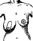
Figure 16. Plastic
On the right is the incision line, on the left is the final result. A window in the skin flap covering the upper half of the mammary gland. With such nipple transpositions, the milk ducts are not injured and the nursing function is preserved. The results of these operations are in most cases quite good; recurrences are observed mainly in cases of congenital weakness of the elastic tissue. In the rarely observed congenital atrophy of the mammary gland, adipose tissue can be transplanted under the skin of the mammary gland for cosmetic reasons. Injuries to the mammary gland during the menstrual period or during pregnancy (especially during the nursing period) can cause very severe bleeding; contusions lead to the formation of extensive, very painful, poorly resorbing hematomas prone to suppuration. Rupture of the milk ducts leads to the formation of milk cysts (see Galactocele). (On the significance of mammary gland trauma in the etiology of tumors, see below.) Among the severe, sometimes very persistent afflictions of the mammary gland is mastodynia (mastodynia, neuralgia mammalis), which is sometimes the first symptom of chronic mastitis developing deep in the mammary gland; it is observed in hypertrophy of the mammary gland, in pendulous breasts, and finally appears in hysterical girls and women (from 20 to 40 years old) without any organic causes. Neuropathologists consider the latter cases a special type of neuralgia of the mammary gland. By its nature and localization, mastodynia is close to intercostal neuralgia. The pains are spasmodic, radiate to the shoulder and shoulder blade, and are sometimes caused by the slightest touch of clothing. Treatment is causal: removal of scars, tumors; in the purely neuralgic form—rest (supporting the mammary gland), heat and light procedures, and ionization. Often, the pains cease with the onset of the climacteric. Mastodynia is often one of the causes of so-called cancerophobia. Tuberculosis of the mammary gland. Among chronic inflammatory diseases of the mammary gland, tuberculosis of it occupies the first place. According to Scott's statistics, tuberculosis of the mammary gland occurs approximately 40-50 times less frequently than cancer. The process usually affects only one side; it is most often observed at the age of 20 to 40; it is very rare in men. Infection penetrates the mammary gland by the metastatic route (hematogenous), sometimes via lymphatic pathways or directly by extension from a neighboring organ (e.g., breakthrough of a carious process in a rib, a process in the pleura). Rare cases of direct infection through the nipple (sucking, kissing) have been described. Regional lymph nodes are affected secondarily. The process usually begins with the formation of one or several nodules, which subsequently undergo cheesy decay or purulent melting; foci of fibrous-indurative tissue are observed less frequently. Subsequently, non-healing fistulas and ulcers characteristic of tuberculosis of the mammary gland form. The mouths of the fistulas are surrounded by pale granulations, liquid pus is discharged, and secondary infection easily sets in, from which patients often perish (sepsis). The entire mammary gland appears dense and shriveled. The course of tuberculosis of the mammary gland is very protracted. The diagnosis is easily made in the presence of fistulas, but in the initial stage, tuberculosis of the mammary gland is often confused with a malignant neoplasm, and the mammary gland is amputated without performing a preliminary biopsy. In severe forms that do not respond to conservative treatment (general treatment, curettage of individual foci and fistulas, heliotherapy, quartz lamp), the only reliable method is still amputation of the mammary gland with removal of regional lymph nodes, following the type of operation for mammary gland cancer; Braendle reports 93.75% good long-term results in 16 patients he operated on. Some authors (Kaufmann, Zabludovsky) consider it necessary for prophylactic purposes to perform amputation of the mammary gland in all cases of tuberculosis of the mammary gland, as experience shows that cancer, distinguished by a particularly malignant course, easily develops in a mammary gland affected by tuberculosis. The secretion of tubercle bacilli with milk is observed in cases where cheesy decay penetrates into the lumens of the excretory ducts. Syphilis, actinomycosis, and echinococcus of the mammary gland. Syphilis can affect the mammary gland in all three of its stages. The primary stage is often observed in the form of a hard chancre of the mammary gland on the basis of extragenital infection. Among 536 cases of extragenital infection, Rille found the primary affect (hard chancre) on the mammary gland in 14 cases, most often as a consequence of infection from a sick child while nursing. In this case, there is usually significant enlargement of the regional lymph nodes, which easily leads to diagnostic and therapeutic errors. In the secondary stage, papules are observed, which have no surgical significance. The mammary gland is least often affected by syphilis in the tertiary stage, which is observed here in two forms: 1) in the form of diffuse infiltration of the gland, proceeding under the guise of ordinary chronic mastitis (mastitis simplex), and, more often, 2) in the form of a gummatous form, mainly in the late period of acquired syphilis, as well as in hereditary syphilitics (more often in women than in men). A gumma of the mammary gland usually appears near the nipple in the form of a rounded, insensitive nodule sitting deep in the glandular tissue, the size of a chicken egg, often with a bumpy surface. Subsequently, purulent melting of the nodule and its breakthrough to the outside may occur, with the formation of an ulcer; sometimes the regional nodes enlarge. The entire process proceeds completely painlessly, in contrast to tuberculosis of the mammary gland, for which severe pain in the mammary gland is characteristic. The tertiary stage of syphilis of the mammary gland is easy to confuse with cancer of the mammary gland with all the consequences of such an error. In cases of known syphilis, a correct diagnosis does not encounter difficulties; conversely, in cases of latent syphilis, a diagnosis is possible only in the presence of other signs of tertiary syphilis (as described in the case of Blumenthal). Treatment is specific. Primary actinomycosis of the mammary gland is a very rare affliction; it most often joins actinomycosis of the pleura as a secondary disease due to the breakthrough of fistulas to the outside. The clinical picture of the disease is a diffuse infiltrate of the mammary gland, on the surface of which there are fistulas surrounded by characteristically altered skin (see Actinomycosis). Treatment in initial forms is conservative (curettage of fistulas, large doses of iodine); in severe cases—amputation of the mammary gland. Bergmann described 15 cases of echinococcus of the mammary gland, which is observed in the form of solitary cysts, usually interpreted as non-parasitic cysts; their true nature becomes clear only after surgical removal. The course of echinococcus of the mammary gland does not present anything characteristic.
N.
Blumenthal. Neoplasms of the mammary gland. Neoplasms of the mammary gland should be sharply distinguished into benign and malignant, but one should not forget that some benign neoplasms can undergo malignant transformation. In view of the fact that the resolution of such a question is extremely important, it is necessary to combine clinical examination of the tumor with laboratory examination. If in some cases recognizing both benign and malignant neoplasms (especially in far-advanced cases) presents no difficulty, then in other cases it becomes very difficult (sometimes, based only on clinical presentation, even impossible); histology, the examination of the tumor, must come to the rescue. This examination can be performed either after excision of the entire tumor or after excision of a small piece of it for biopsy. This method, however, is not safe: in the event that the tumor is of a malignant type, this procedure opens the way for the introduction of malignant tumor elements into both the lymphatic and the circulatory system (such cases have been observed); therefore, either the entire tumor must be removed with the surrounding healthy tissue (but even this does not guarantee against seeding) or pieces of the tumor are excised with a thermocautery. The histological report must be obtained as soon as possible so that, in the event of an unfavorable report, radical intervention is not delayed for long. However, one cannot be limited only to microscopy without paying attention to the clinical presentation; in many cases, a correct decision can be formed only on the basis of a discussion of data obtained by both methods. But the clinic must also remember that "long-established views on the benign nature of some mammary gland tumors are now shaken" (v. Saar). In many tumors, transitional stages are observed, which greatly complicates diagnosis. Even in the same gland, in different parts of the existing tumor, areas of both benign and malignant character can be encountered; hence it is clear that parts accidentally obtained for biopsy, even if they turn out to be of a benign character, cannot serve as proof of the absence of a malignant area somewhere. The conclusion suggests itself that in doubtful cases, both from the point of view of the clinic and the laboratory, it will be a lesser error to treat the tumor as malignant than the opposite. One thing remains certain: every mammary gland tumor, especially in a woman aged 30 to 40, must be unconditionally removed and subjected to microscopic examination, depending on the results of which one decision or another will be made. Both benign and malignant tumors of the mammary gland can be of epithelial and connective tissue origin. The predominant group is that of mixed fibro-epithelial formations (v. Saar). - "Pure" tumors of the epithelial type include adenomas, which are dense grayish-red nodules of homogeneous or finely granular structure, resembling the pancreas, sharply demarcated from the healthy tissue of the gland. From simple hypertrophy of the gland, they differ microscopically in that they do not have excretory ducts, and their passages appear blind. They are surrounded by a membrana propria, which often undergoes hyaline degeneration and becomes greatly thickened. Fibroadenomas are encountered predominantly at a young age, from 20 to 30 years, do not cause any pain, and are usually discovered quite accidentally; they are located in different parts of the gland; if they lie near the skin itself, the latter is usually thinned, but is never fused with the tumor. Sometimes several tumors of different sizes are encountered in the same gland; they are also encountered simultaneously in both glands. By their dense consistency, they are sharply separated from normal tissue, have their own capsule (this constitutes their characteristic feature), are nodular, are quite mobile in the tissue of the gland, and can grow very slowly, while usually not reaching large sizes. Many authors attach a certain significance to this mobility in the tissue of the gland, mobility under the skin, and mobility together with the gland in the sense of the possibility of establishing the benign nature of the tumor on this basis. But some forms of cancer in the early stage of their development can also possess such mobility (Salomon). For the removal of these tumors, local anesthesia is quite sufficient. If the tumor is located deeply or if there are several of them, then for cosmetic purposes the Bardenheuer-Morestin incision is used: along the lower semicircle of the mammary gland, at its border, in the skin fold, an incision is made up to the exposure of the fibers of the pectoralis major muscle. The entire gland is flipped upward, and then on the posterior surface of the gland, incisions are made in the glandular tissue in the direction of the tumors, which are enucleated from here, after which the tissue of the gland is sutured with catgut; the gland is returned to its place, and skin sutures are applied, but here it is necessary to insert a small drain for 24-48 hours. These tumors do not give recurrences, and their malignant transformation is encountered very rarely. On cross-section, the removed tumor appears matte-whitish in color, its consistency is significantly denser than normal, the tumor tissue protrudes somewhat from the plane of the section; sometimes branched slits are noticed in the form of excretory ducts, sometimes long, narrow, sometimes wider, branched. Not only the external appearance and consistency of these tumors, but also the microscopic picture appears somewhat different depending on the ratio of epithelial and connective tissues. With a predominance of glandular elements, one speaks of fibro-adenoma, with a predominance of connective tissue, of adeno-fibroma; many transitional forms are also encountered. Histologically, two forms of fibroadenomas are distinguished: f. pericanaliculare and f. intracanaliculare. In fibroadenomas of the first type, the glandular passages are tightly surrounded by a connective tissue sheath rich in cells and fibers. The glandular passages are lined for the most part by a double-layered epithelium. In the second form, the picture of glandular ducts predominates, the connective tissue sheath of which penetrates into the lumen in the form of nodular, roller-like or leaf-like polyps. At the same time, they are pushed forward by cylindrical and cuboidal epithelial cells, so that the glandular lumens form strongly branched slit-like spaces. Often both types of fibroadenomas are encountered simultaneously in the same tumor. In rare cases, total fibroadenomatosis of the mammary gland is observed, which gives a strong enlargement (hypertrophy, see above) of the entire gland; sometimes such a phenomenon is designated as "mammoma" or "mastoma". Besides adenomas, cystadenomas are also encountered. They represent cystic glandular passages, which expand under the influence of increased pressure accumulating within the secretion. They are of various sizes, from a pea to a goose egg. Due to the growth of cysts and the disintegration of the septa between them, many cysts connect with each other. With the formation of many papillary growths, a form is obtained bearing the name papillary cystadenoma (cystadenoma papillare s. papilliferum). If the entire lumen is filled with such leaf-like or cauliflower-like formations, then one speaks of cystadenoma phyllodes. Abundant development of connective tissue with insignificant development of interstitial substance gave rise to the name of such a formation cystosarcoma phyllodes, but these formations do not give any metastases, even when they reach fairly large sizes during rapid growth. True sarcomas develop from them in very rare cases. On cross-section, these tumors are soft, moist, with slits in which cauliflower-like growths are clearly visible; the boundaries of these tumors are unclear, the axillary glands are free. The prognosis for such tumors after surgery is comparatively favorable. Clinically, all these formations, with the exception of the latter, do not give special deviations, but in view of the fact that they can still more often undergo cancerous degeneration, operative assistance is unconditionally indicated here. Among cystadenomas, there stand completely apart formations which were long known to Cooper, Billroth (Astley Cooper, Billroth) and others, but which were studied and first described in detail by Reclus in 1860 under the name "maladie kystique des mamelles". This disease attracts the attention of many surgeons, hence the multitude of names that have been given to it. Some authors take this disease for a neoplasm of the cystadenoma type (v. Saar, Brissaud, Schimmelbusch, and others), others (led by Virchow and König) believe that a chronic inflammatory process lies at the basis (mastitis chronica cystica); they see this mainly in the fact that inflammatory infiltration is always present here. But some authors (Tietze and others) do not recognize this disease as having either an inflammatory or a neoplastic character, but believe that all changes relate to senile involution of the mammary gland (involutional cysts). In the opinion of Tietze, senile involution, cysts, and carcinomas are related processes.
Krompecher, noting the similarity of the cyst epithelium to the epithelium of the apocrine sweat glands (see below), which are genetically close to the mammary gland, argues for the origin of the cysts in this disease from the aforementioned large sweat glands. Most authors at the present time believe that what is taking place here is only diffuse fibromatosis of the mammary gland, which has a certain relation to senile involution. It is characteristic of this disease that both mammary glands are involved in the process, either simultaneously or sequentially. It is usually encountered in women between 30-40 years of age, in the period close to menopause. The process develops very slowly. The gland swells during this, often taking on a nodular appearance. The skin over the gland is slightly mobile; upon palpation, one gets the sensation of a bag filled with rounded objects. These spherical cysts are of various sizes; the large ones sometimes give a sensation of fluctuation, are painless, and develop either in the entire gland or in one part of it. Often, upon pressing (in rare cases also spontaneously), fluid appears from the nipple in 2-3 places, sometimes transparent, sometimes turbid, sometimes liquid, sometimes thick; its color is yellowish, sometimes grayish-green; reddish indicates an admixture of blood; sometimes the discharge is completely bloody (bleeding gland, blutende Mamma). Pathological-anatomical cysts consist of enlarged glandular lobules, separated by fibrous connective tissue, with cystic dilation of the excretory ducts; the number of lobules increases significantly (up to several hundred). In the lobules, there is proliferation of the epithelium, and then, due to the disintegration of the epithelium, primary cysts appear, which, by merging, form secondary, large ones, where remnants of the walls that separated them are still visible. A peculiarity of the cysts in this disease is that the epithelium lining them is often very tall, with light eosinophilic protoplasm, resembling the epithelium of the so-called apocrine glands (see). Often, this epithelium forms papillary outgrowths. The main clinical interest in this disease lies in the fact that a significant number of authors attribute a precancerous character to it, and this undoubtedly leaves a certain mark on the therapeutic measures for this disease; the frequency of cancer development here is determined by authors at figures from 7% to 50%, therefore some authors set indications for amputation of the gland quite broadly, based on the considerations that it is better to sacrifice several mammary glands than to leave one that will give rise to cancerous degeneration in the near future (de Quervain). Upon examination of the patient, both mammary glands must be investigated; it is necessary to carefully palpate not only one surface of the gland, but necessarily bimanually the entire glandular mass of it, as well as the gl. aberrantes, if such are present; then sequentially both axillary fossae and the sub- and supraclavicular regions. Such detailed examinations must be performed no less frequently than every two to three months; therefore, these patients must be under constant supervision, and at the slightest signs of impending cancerous degeneration (induration, retraction of the nipple, appearance of even the smallest glands in the axillary fossa, especially if they are of a denser consistency), a radical amputation must be proposed. All cases of 'blutende Mamma' must be operated on. Partial excision, as some suggest, is completely inexpedient (Zilberberg), because the remaining part of the gland will also begin to undergo malignant degeneration in the near future. The question of the bleeding mammary gland is given especially much attention. While the majority of authors, both domestic and foreign, see a precancerous period in this disease and on the basis of this advocate for the same radical intervention as in malignant neoplasms, on the other hand, some (representatives of the Viennese school - Erdheim, Risak) say that the bleeding mammary gland is only a symptom, which is also observed in benign tumors, especially in papillomas of the lactiferous ducts; cases of bleeding mammary gland should be individualized, and depending on this, either perform a partial removal of the mammary gland or a radical operation. This question was also discussed in the USSR at surgical congresses, and the majority hold the point of view that a bleeding mammary gland indicates a precancerous period and therefore requires a radical operation (Tovbin et al.). In the absence of indications for a radical operation, some authors (Warren) have proposed performing an operation for fibromatosis of the mammary gland, consisting of the following: by a semilunar incision according to Bardenheuer-Morestin (see above), the mammary gland is dissected from bottom to top, reflected, and on its surface facing the pectoralis major muscle, a whole series of incisions is made in a radial direction; in this way, a large number of cystic formations are emptied. Some authors (Zilberberg) still believe that it is unlikely that any effect can be achieved by such an intervention. It is undoubtedly the case that the remaining small cysts will in turn begin to develop and completely annul the result of the Warren operation. If such formations reach large sizes, if pain in the gland (mastodynia) appears, if, in particular, early signs of incipient cancerous degeneration appear, then amputation is of course indicated—either simple amputation by two converging semilunar incisions, without excision of the lymphatic apparatus of the axillary fossa, or, if there is suspicion of carcinomatous involvement of the axillary glands, a radical intervention. Such radical interventions are mandatory once the diagnosis of cancer of the mammary gland has been established. The mammary gland, in terms of the frequency of malignant tumor diseases in women among similar diseases of other organs, occupies second place (the uterus is in first place). And among diseases of the mammary gland, carcinoma occupies the first place—a little more than 80%; the rudiments of the mammary gland in men are also not spared (2-3% of all cancers of this gland). From the combined statistics of a number of authors, both foreign and domestic, one can derive an approximate figure for the annual mortality of women from cancer of the mammary gland. In the USSR, this figure is approximately about 13,000 people per year, and since the average life expectancy of each patient from the moment of the appearance of the tumor is 3 years, it follows from this that during the year on the territory of the USSR there are about 40,000 women suffering from cancer of the mammary gland. Such is the morbidity; as for mortality, the successes of surgical intervention have significantly reduced it: for the years 1923-27, out of 21,383 autopsies of adults in Moscow prosectoriums, only 63 cases of death from cancer of the mammary gland were noted. Cancers develop predominantly at the age of 40-50 years, which coincides with the time of the cessation of menstruation, and many authors see in this one of the causes of its development. Statistics at the same time indicate that cancerous neoplasms appear more often in women who have given birth and breastfed* than in virgins. Previous mastitis also apparently has some influence. Mortality statistics show that among other cancers, cancer of the mammary gland in women in Moscow is present in 5-6% (in relation to the entire population - 2-2.5%); these data of Arkhangelsky for 1892-1911 are also confirmed by sectional data of Moscow prosectoriums * In married women, three times more often (76.4% and 23.6%; Dietrich, Frangenheim). In men, cancers of the mammary gland account for about 2% of all cancers of the mammary gland for 1923-27 (2.2%). In other European cities, mortality from cancer of the mammary gland is significantly higher: in Munich 11.6%, in Berlin 12.6%, in large centers of England even 16.6% (Arkhangelsky). In recent decades, statistics note two points: 1) cancer diseases have become significantly more frequent, and the morbidity figure continues to grow, and 2) the age of maximum disease has decreased and falls on the period from 30 to 40 years. Other authors dispute this assertion. The question of hereditary predisposition is currently being given a great deal of attention, but statistics have so far been very unsatisfactory, and therefore unconvincing. However, some observations, and especially experimental data (Maud Slye), force one to consider that hereditary predisposition under favorable circumstances plays a certain role in the development of cancer in general, and cancer of the mammary gland in particular. In the opinion of a number of authors, trauma plays not the last role in the etiology of cancer of the mammary gland. The literature on this question is rich; a whole series of cases is cited which should convincingly attest to this connection—both of a single trauma (Konig) and chronic; the percentage of the latter is calculated by authors from 2.6% (Theilhaber) to 44.6% (Williams). However, when examining some of the cited cases, supposedly proving an undoubted connection, it turns out that the possibility is not excluded that the trauma was inflicted on a gland in which pathological changes were already present. Such a mammary gland is, of course, much more sensitive to trauma than a healthy one. It is especially important to establish a causal link between the development of cancer and trauma in legal cases: in claims for damages, etc. According to Thiem, traumatic cancer is a great rarity; one can with difficulty recognize this connection in barely 2% of cases.
It has not yet been possible to experimentally induce the formation of cancer by trauma. Cancer of the mammary gland develops either from its glandular lobules or from the lactiferous ducts. The nipple can also be the starting point of a neoplasm (Paget's disease). Billroth established the following 4 types: carcinoma tubulare, acinosum, scirrhosum, and gelatinosum; but the name tubulare was soon replaced by solidum, and acinosum by the word medullare. In addition, other forms were introduced and studied, such as carcinoma adenomatosum, cysticum, etc. The anatomical structure of cancerous tumors is of great practical importance for their clinical manifestation and course. Hard cancer, scirrhus (carcinoma scirrhosum), appears to be the most frequent form of cancer of the mammary gland. Although scirrhus usually grows very slowly, spreading into the adjacent tissue in the form of radiating strands, it can early on give metastases to the lymphatic glands; it is distinguished by its density and is usually of small size. As a consequence of cicatricial shrinkage of the stroma (which is considered a healing act), the nipple retracts (see figure 17). Kaufmann classifies scirrhus of the mammary gland as one of the most malignant forms of cancer (see below). The second form is carcinoma medullare—medullary cancer. The tumors are distinguished by fairly rapid growth; they often reach quite significant sizes, often grow through the skin, ulcerate, and early on give metastases. Regressive changes are observed in the tumor—fatty degeneration and necrosis. On a cross-section, it is easy to obtain so-called cancer juice from these foci by scraping. Carcinoma tubulare, acinosum, and adenomatosum are also classified as soft cancers. As for adenocarcinoma, it is rarely encountered in pure form, being combined more often with other forms, e.g., with cystocarcinoma, sometimes with the development of papillary growths in the cavities of cysts (carcinoma or cystocarcinoma papillare). In late periods, and specifically in the central parts of medullary cancer, scarring areas of the scirrhus type often arise. Mucous degeneration of epithelial cells (in women it is encountered in 1.66%, in men in 5% of all cancer cases) is the dominant manifestation of the fourth group—mucous cancers (carcinoma gelatinosum s. colloides). Delbet and Mendaro called these forms epitheliomes mammaires secretants and recognize, on the basis of the examination of these patients, that the prognosis for these forms of cancer is significantly more favorable. The same is noted by Klose and Sebening: "These cancers grow very slowly, the tendency to metastases is weak, but after 5 or more years, late recurrences are nevertheless observed." Squamous cell cancers of the mammary gland are also observed quite rarely—typical cancroids with keratinization, and finally carcinoma psammosum—cancer with calcified spherical formations. Localized and diffuse tumors are distinguished. Localized nodular forms grow slowly, while diffuse ones can infiltrate large sections of the gland; the latter are not sharply defined, sometimes present great difficulties for diagnosis, and clinically are distinguished by a more malignant course. Even with a very radical operation, it is not always possible to remove all the diseased tissue; they more often give both recurrences and metastases. According to Salomon's statistics, carcinoma solidum is encountered in 48% of all cancers of the mammary gland, carcinoma medullare accounts for 16%, infiltrating—20%, adenocarcinoma—9%, carcinoma cysticum—3%, carcinoma gelatinosum—1.5%, and finally Paget's disease—2.5%. Not all sections of the mammary gland become diseased with equal frequency; the site of development of the cancerous node also has some prognostic significance. Most often, tumors develop in the upper-lateral section of the gland; a neoplasm is also frequently encountered in the gl. aberrans, at the site of attachment of the lower edge of the pectoralis major muscle to the humerus. The closer to the axillary fossa the node develops, the earlier the lymphatic glands (gl. thoracales anteriores) are involved in the process, from where more and more new lymphatic glands are gradually seized along the course of the v. axillaris up to the clavicle. Sometimes the glands reach much larger sizes than the initial node; in such cases, the prognosis is particularly unfavorable. On the other hand, the lymph flow also rushes through the pectoralis major muscle, which is also involved in the sympathetic process. According to some authors, nodes developing in the medial section of the mammary gland give metastases to the lymphatic glands of the mediastinum, as well as the pleura. Regarding the supraclavicular glands, it should be pointed out that due to the too weak anastomoses of the lymphatic pathways between the sub- and supraclavicular glands, the latter are infected relatively slowly. Since the lymphatic connection between both mammary glands is relatively weakly expressed, transfer to the opposite gland is observed relatively rarely. The growth of the tumor can proceed in the direction of the skin integuments; here, initially, a multitude of separate small nodules appears, which grow in both volume and number; gradually they merge into one common mass of wooden hardness. This mass encompasses the entire half of the chest, passes to the abdomen, and strongly hinders, and sometimes completely stops, breathing on the corresponding side; such a picture bears the name cancer en cuirasse (Velpeau). If this picture develops unusually quickly, it is called erysipelas carcinomatosum (Kuttner). Similar processes are observed both in non-operated cases and after operations—more often in the latter. Symptomatology, recognition, prognosis. At the beginning of their development, cancerous tumors, just like benign ones, are completely painless; patients do not consider it necessary to consult a doctor about this, and the tumor itself is usually discovered quite accidentally, while washing, while palpating for some other reason, or upon injury. Correct information about these diseases is relatively little disseminated; even if a patient has found a tumor in herself, she sometimes delays consulting a doctor for advice, fearing to hear his verdict about the necessity of an operation. This explains why, in the vast majority of cases, patients only seek advice when the process has already gone more or less far. A doctor must accustom himself, with any tumors of the mammary gland, to resolve the question: is this not cancer? When there are already enlarged lymphatic glands, a retracted nipple*, the neoplasm is fused with the skin (of the orange peel type), with the underlying pectoralis major muscle, the tumor is not very mobile, sometimes even ulcerated (see figure 18), the mammary gland has reached large sizes or, conversely, has strongly shrunken—then diagnosis is not difficult, but therapy is difficult. Careful but gentle palpation not only of the node, but of the entire gland (also on the opposite side) and the lymphatic glands of all regions related to this, can provide a great deal of valuable data for recognition. The general condition of such patients can be excellent; this, however, does not contradict the fact that a cancerous tumor can develop in such patients. From the point of view of prognosis and obtaining good long-term results, early recognition in the early period of the process is important, but errors can easily occur here: one can mistake a benign tumor for a malignant one, one can confuse a tumor with tubercular infiltrates in the mammary gland, etc. There are proposals in some doubtful cases to perform a trial excision for biopsy (see above). * A retracted nipple is not pathognomonic for cancer of the mammary gland; it is also encountered in other diseases of it.
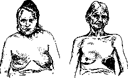
Figure 17. Carcinoma mammae dext.: significant retraction of the nipple.
Figure 18. Ulcerated cancerous tumor of the left mammary gland.
Regarding the prognosis, in cases where an operation has not been performed, the disease, with rare cases of self-healing, ends fatally. The average life expectancy fluctuates between 32.9 and 38 months (Neugebauer). On the question of prognosis depending on the histological form, opinions are directly opposite: some (Iselin) believe that cases of carc. medullare have the most favorable course, then carc. solidum, and the worst prognosis is with scirrhus; others (v. Eiselsberg) posit the worst prognosis for carc. medullare and the best for scirrhus (the epitheliomes secretants of Delbet and Mendaro have already been mentioned above). For prognosis, the clinical course is significant: the location of the tumor, the relationship to neighboring tissues, or lymph nodes, the speed of growth, etc. Statistics can acquire great significance, but they must be compiled in a uniform form, and patients must be followed up over a very long period. Treatment. Moving on to the question of the treatment of malignant tumors of the Mammary Gland or cases where such a disease is merely suspected, a firm position must be established: any tumor accessible to operation (and the decision on accessibility is made by the surgeon depending on a detailed examination not only of the diseased area but also of the patient's general condition, and on his personal experience and technique) must certainly be operated on. To make concessions, to wait, to offer the patient any other methods of treatment whatsoever—this absolutely worsens the prognosis; the indication for treatment lies in the diagnosis itself: ubi carcinoma, ibi excisio. Since the first proposal to remove not the tumor but the entire gland in cancer (Moore in 1867), 60 years have already passed; during this time, the pathways of cancer spread have been studied in detail, and thanks to accumulated experience, a fairly detailed and accurate, anatomically grounded technique has been developed. Nevertheless, the long-term results are still very disappointing, and the main evil lies in the fact that patients arriving at the operating table consult a surgeon (or are sent to a surgeon by doctors) relatively late. Doctors must conduct broad propaganda about the enormous importance of early operation for patients. Since it is impossible here to provide descriptions of all methods proposed for the radical operation of amputation of the Mammary Gland, the main principles underlying a correct radical operation will be indicated below. Regarding the skin incision, as a rule, it is necessary to remove the covering skin along with the entire gland, and the boundaries of the incisions, according to most authors, should be no closer than 3 finger-breadths on both sides of the tumor. Some authors (Halsted, Tansini) consider it necessary, in view of the fact that in their opinion cancer spreads along the lymph pathways of the skin, to remove significantly more of it, even at the cost of leaving part of the wound surface uncovered. Some, believing that cancer spreads along the fasciae (Handley), consider it necessary to remove the subcutaneous tissue over a large area around the Mammary Gland from under the dissected skin. Furthermore, along with the gland (everyone agrees on this), the pectoralis major muscle must certainly be removed together with the fascial layers; as for the pectoralis minor muscle, Silberberg removes it in all cases without fail; some (S. P. Fedorov, A. M. Zabludovsky, Dietrich, Frangenheim) consider it necessary to remove it only when enlarged nodes are palpable beneath it. When, thus, the entire neurovascular bundle is opened, they proceed to remove the adipose tissue together with the enlarged lymph nodes contained within it, starting from the subclavian fossa downwards, up to the tissue covering the anterior scalene muscle (m. scalenus major), while the tissue covering the anterior surface of the subscapularis muscle is also removed. All these parts, together with the gland and the skin covering it, must be removed entirely in one piece (en bloc), and not separately, to avoid unnecessary exposure of the lymph pathways. On the question of supraclavicular nodes, there is still disagreement. Some authors, when these nodes are involved in the process, consider such a case already inaccessible to operation and decline it; the prognosis is indeed very unfavorable here, but cases of good long-term results have also been described in the literature. Sawing through the clavicle is considered unnecessary; it is better to make a special separate incision. Technically, this is usually quite difficult, especially on the left (injury to the duct. thoracici). To close the wound, if the available skin coverings are insufficient, a large number of plastic operations have been proposed, starting from Thiersch grafts up to the transplantation of a healthy Mammary Gland (Raug). Leaving wound surfaces uncovered is not recommended, as recurrences easily develop in these places. The wound is sutured, and drains are placed in the dependent parts. Operative mortality at the present time is close to zero. As for the postoperative course of wounds, it is usually normal; but the most important thing is the long-term results, which depend on the clinical preoperative picture; in general, it can be stated that as operative technique develops, depending on the study of cancer spread pathways, the percentage of good long-term results increases; even for the same surgeon over time, as his personal technique improves and his handling of the tumor becomes more and more "gentle," the results also improve. The first local recurrences are usually observed within the first year after the operation; with constant observation of patients, with examination of all adjacent areas, nodules or nodes that have appeared can be found very early, which should be immediately radically removed whenever they are noticed, wherever they are confirmed; sometimes they have to be removed from the skin, on the scar or near it, sometimes in the lymph nodes; finally, they may sit on the sternum or on the ribs—then it is necessary to perform resections of the corresponding sections. One should not lose sight of distant bone systems, especially when patients complain of pain in them; here, the vertebrae are in the first place, then the femur, and finally the humerus. How do metastases get into these areas? According to some—through blood vessels; Handley refutes this and believes that the only pathway is the lymph system; as a weighty argument, he cites the fact that no one has ever observed the development of metastases on the lower leg or forearm: "patients do not live long enough for this." Of course, in the presence of metastases in the vertebrae or in the long tubular bones, operative assistance is no longer indicated. In the long tubular bones, these transfers often give rise to fractures, which can occur with the most insignificant trauma. Transfers to the brain are not uncommon, but the pathways through which transfers get there have not yet been studied. If, after the 3-year period, patients live without these severe complications, if only small local recurrences develop, which can be removed without particular difficulty, then they begin to speak of favorable long-term results. To collect more accurate statistical material, it has been recognized as necessary to classify the disease in some way, both according to the clinical and the pathological-anatomical picture of the development of the cancerous process that presents itself during the operation. There are several schemes—Steinthal, Jungling, Zabludovsky, and others. The distribution of patients according to these schemes already somewhat improves the correct collection of statistical material. According to the latest statistics, the percentage of postoperative mortality has decreased from 30-23 in the 70s of the XIX century to 0.9-1-2, and the percentage of three-year survival has risen from 4.7 to 40-44. As for early operations, the percentage here is significantly higher: up to 79.68 (Halsted) and even up to 100 (Anschütz, von der Hütten, König—no, Klose-Sebening). However, even after three years, the appearance of recurrences is observed, so that at the present time the 3-year observation period is considered insufficient, and the term has been extended to 5 years; however, casuistry shows that recurrences appear even after 10-20 years. Some authors in such cases speak no longer of a recurrence, but of a new disease. All the indicated statistical calculations concern one general picture of "cancer." In view of the diversity of the histological picture, attempts are being made to collect materials on long-term results according to anatomical forms (Salomon), but so far no definite data are available. To improve operative results, as well as for the treatment of cases inaccessible to the knife, recourse is had to radiant energy in the form of X-rays and radium bromide. Some use irradiation prophylactically before the operation with the aim of destroying those cellular elements that may remain in the wound, as well as those that may get into the wound from the tumor and lymph pathways (seeding); others conduct prophylactic treatment shortly after the operation; finally, others use irradiation only when incurable recurrences appear, as well as metastases in the spine and in the long tubular bones. As for inoperable cases, irradiation is also used here.
Regarding each of the listed methods, there are observations by some authors who are very satisfied with the results, but there are also observations by others who not only did not see any benefit from irradiation but directly point to significant deterioration. This must be explained by the fact that the methods of applying radiant energy differ among different authors, the dosage also differs, and finally, it seems that the circumstance that neither the histological picture of the neoplasm nor the biological properties of each individual form of cancer are taken into account is of enormous importance; the latter is still, at the present time, in a period of study. A severe complication after a radical operation is sharp edema of the entire upper extremity on the corresponding side; this is explained by the compression of the axillary vein and lymph pathways either by scars or by a recurrence of the neoplasm; collateral circulation in this case is completely insufficient. Handley proposed performing lymphangioplasty to relieve patients (thick silk threads are passed subcutaneously in several rows in the subcutaneous tissue, starting at the forearm and ending above the shoulder joint), but the results are quite sad: edema and severe pain serve as an indication for amputation of the arm. Regarding the treatment of cancer with castration, sera, and colloidal lead salts, one can only say that the results are still very dismal, and the number of observations is still very insignificant. Neoplasms of the connective tissue type (mesenchymal tumors). 1. Benign. Fibroma of the mammary gland is encountered extremely rarely; some authors even believe that those tumors which were described under this name were fibroadenomas in which the lumens of the glandular tissue are so compressed that only narrow, with difficulty recognizable cellular elements remain; one should only not confuse them with those fibromas that originate from the skin or from the nipple. Lipomas of the mammary gland are encountered somewhat more frequently; they develop either in the gland itself, or behind it in the adipose tissue, or in the axillary fossa, where they are easily confused with gl. aberrans mammae. Sometimes they reach quite significant sizes [51/2 kg - Kravchenko, cited by Dietrich and Frangenheim] and even up to 14 kg (Honigsberger - ibid.)]; upon microscopic examination, one sometimes finds in them remnants of the glandular tissue of the mamma. Angiomas develop either in the skin, from where they grow into the mammary gland, or in the gland itself, and it is sometimes very difficult to distinguish them from one another. Their histological structure and clinical picture do not differ in any way from angiomas located in other places. Besides these tumors, mixed connective tissue tumors are also observed, such as fibro-myxoma, myo-fibroma, enchondroma, osteoma, etc. 2. Malignant. Sarcoma of the mammary gland. Compared with cancerous tumors, they are encountered quite rarely and therefore have no particular practical significance. They appear as large, nodular, rapidly growing diffuse tumors; the most frequent histological picture is round-cell sarcoma; compared with the previously mentioned sarcoma phyllodes they proceed with the usual character peculiar to sarcomas. The tumor sometimes fuses with the skin, disintegrates, easily grows into the muscles, gives distant metastases, and early on causes cachexia. Besides these, spindle-cell sarcomas and polymorphic ones with giant cells, chondrosarcomas, and osteosarcomas are also observed; the lymph glands in these cases are often spared. Combinations of sarcoma and cancer (sarco-carcinomas) are encountered. Diseases of the nipple and areola. Tumors are encountered very rarely. Benign ones include cystic tumors, which, just as in the mammary gland, sometimes undergo cancerous degeneration and then require a radical operation. Warty tumors, angiomas, and fibromas for the most part already belong to diseases of the skin, and not of the nipple or the areola. Standing apart is the disease described in 1874 by Paget, which has since retained the author's name, although views on this disease have changed. This disease is encountered almost exclusively in women, specifically at the age of 40-60 years, and initially gives the impression of chronic eczema; the skin of the nipple and areola appears reddened, weeping, as if velvety, granulating; it easily ulcerates, in places as if covered by newly formed epidermis. The boundaries of this formation are sharply defined [see separate table (pp. 323-324), fig. 1]. Some authors take it for hyperkeratosis, but Paget believes that a cancerous tumor develops from this eczema, which, having started on the surface, grows into the depth. But the latest works (Jacobaeus, Ribbert, and others) have shown that in this disease there is already a cancerous node in the depth from the beginning, and the changes in the integuments of the nipple are secondary. So-called Paget cells are cancer cells that grow into the epithelium and spread at first within the epithelial covering along its plane; subsequently, the cancer begins to grow into the depth of the gland. Treatment is exclusively radical surgical: amputation and removal of the lymph glands. Ya. Zilberberg.
Related articles
Mentioned in
Cite this page
“Mammary Gland.” Soviet Medical Encyclopedia. English translation of Bolshaya Meditsinskaya Entsiklopediya, 1st ed. (Moscow, 1928–1936), ed. N. A. Semashko. https://sovietmedicalencyclopedia.pages.dev/article/mammary-gland/