Apocrine Glands
Historical document, translated for reference. It reflects medical knowledge of the 1920s–30s and is not medical advice.
Summary
Detailed historical overview of apocrine glands from the 1930s Soviet medical encyclopedia, covering their anatomy, histology, secretion mechanisms, phylogenetic distribution, and clinical significance including related diseases such as hidradenitis.
Encyclopedia article (1928–1936)
APOCRINE GLANDS, a designation that can be applied to all glands that secrete "apocrine," i.e., by the shedding of a part of the cellular protoplasm into the lumen of the gland at the moment of secretion. Most frequently this name is applied to the "large sweat" glands of the skin; the mammary gland, the glands of Moll, and ceruminous glands also belong here (as derivatives thereof); reference is also made to apocrine secretion of the kidneys, the epithelium of the bile ducts, intestinal glands, and some others. Only the apocrine glands of the skin will be considered here; they represent an organ whose secretion is rich in substances that cause the odor inherent not only in a separate genus or species of animal, race or nationality, but also in each individual. In the animal kingdom, the odor secreted by apocrine glands serves, firstly, as a weapon of defense, since it repels or, by virtue of its toxicity, kills approaching parasites; secondly, it allows animals to find each other by scent and even in the air (bats); furthermore, it serves to attract animals to each other and for mating, as it possesses the ability to act in an exciting manner on the opposite sex. In humans, the odor secreted by apocrine glands reflects the entire complex metabolism of the organism, and the sexual component is so sharply expressed in it—i.e., the ability to act excitingly on the opposite sex—that some authors call the odor of apocrine glands "sexual," and the apocrine glands themselves "accessory sexual glands" or "glands of sexual odor," especially since the final development of apocrine glands and the onset of their secretory activity coincide with the sexual maturation of the individual. In addition, a close relationship can be established between their function and certain aspects of sexual life (menstruation, pregnancy, menopause). In mammals, apocrine glands are scattered throughout the hairy coat. Their phylogenetic regression begins only in the order of primates (in monkeys) and reaches its limit in humans, equally in individuals of both sexes. In humans, apocrine glands have survived only in certain areas: in the skin of the axilla, the nipple, the lower abdomen, the pubis, the labia majora, and around the anus. In Chinese, Negroes, and Australians, their distribution extends onto the chest and even the face. In all the mentioned places, apocrine glands are located extremely sparsely, with the exception of the axilla, where they lie in a continuous layer consisting of a series of dense granular formations easily distinguishable by the naked eye, located between the skin and subcutaneous tissue. The size of this layer of apocrine glands along the plane of the skin corresponds approximately to an oval the size of a chicken egg, and its thickness varies individually (from 0.5 to 6.5 mm). Apocrine glands are derivatives of the epithelial complex from which sebaceous glands and hair also originate; originating from the hair follicle, they maintain a connection with it all the time and, having reached their full development, flow, as a rule, into the hair follicle (see figures 1 and 3). From the same rudiment, through further differentiation, the mammary gland is also formed; the hair and sebaceous gland in this case
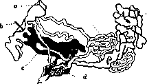
Figure 1. Topographic diagram of skin glands (sweat and apocrine): a—epidermis; b—hair; c—hair sheath; d—arrector pili muscle; e—apocrine glands; f—sweat gland (after Pinkus).
incidentally perish. The apocrine gland has an alveolar-tubular structure. Its loosely coiled glomerulus is surrounded by a connective tissue capsule containing an extensive nerve plexus and a rich capillary network. The wall of the secreting tubule consists of a dense membrane and a two-layered lining: the layer directly adjacent to the membrane is represented by a number of smooth muscle elements which, by virtue of their epithelial origin, are called "myoepithelium" or "epithelial musculature"; internal to it lies the actual epithelial lining. Non-functioning glandular epithelium has a cubic or low-cylindrical appearance, cell boundaries are well delineated, and the border is smooth and slightly rounded. The nucleus is large and rounded in configuration. The protoplasm is sharply eosinophilic and stains pale. During secretion, the cell changes its appearance: in the prefunctional period, it begins to elongate, becomes high-cylindrical, and in its distal part, eosinophilic (protein) and golden-yellow granules accumulate, the majority of which give a positive reaction for iron, and, in addition, fat droplets having the character of phosphatides and fatty acids. Figure 2. "Droplet-like" secretion of an apocrine gland (from author's microphotogram). Subsequently, the distal part of the cell elongates more and more, a constriction appears in it, pinching off a drop-like rounded end; the constriction thins out, disappears, and the pinched-off drop of secretion finds itself in the lumen of the gland (see figure 2). Therefore, the protoplasm turns out to be reduced, the cell becomes lower, and the nucleus is preserved. The cell can return to a resting state in order to become cubic again after a certain time and go through the established secretory cycle. 151 The odor inherent to an individual depends on the composition of the secretion, i.e., the presence in it of volatile fatty acids and ethereal compounds. In children before puberty, apocrine glands do not function. In newborns, they have the appearance of solid cellular clusters of tubular configuration. Only over the years do they begin to acquire a glandular configuration, first rudimentary, then typical for apocrine glands. The secretory activity that begins at the moment of puberty continues thereafter, both in women and men, throughout life and physiologically fades out only with the onset of senile decrepitude of the organism, with the climacteric also having an effect in women. Pathological regression of apocrine glands can also occur at a young age during exhaustion and lowering of general metabolism resulting from starvation, debilitating disease, and other causes. Hyperplasia and hyperfunction of apocrine glands are observed in women during menstruation and pregnancy. In the first case, they are explained by the increased sexuality of women in
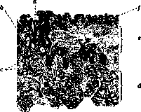
Figure 3. Topography of apocrine glands in the skin of the axilla: a—hair; b—sebaceous glands; c—sweat glands; d—layer of apocrine glands; e—cutis; f—epidermis (from author's microphotogram).
this period, which corresponds to the maximum of the secreted "sexual" odor; in the second case, by the general enhanced metabolism of the organism at this time, expressed in hypersecretion of other skin glands (mammary, sweat, sebaceous, and paraurethral). This hyperplasia sometimes reaches severe degrees and manifests as swellings in the axillary fossa. If, in the process of proliferation, the epithelium of the apocrine glands metaplastically transforms into the related epithelium of the mammary gland and if, due to duct blockage, there is a retention of secretion, tumors known as "axillary mammary glands" can form in the axilla of pregnant women. Apocrine glands can also take part in the creation of another morbid form: cystic breast (Cystenmamma, mastitis chr. cystica). In this condition, along with cysts lined with ordinary breast epithelium, an abundant number of cysts bordered by the "clear" eosinophilic epithelium characteristic of apocrine glands is discovered in the latter. Whether the designated cysts are formed by a proliferative-metaplastic pathway, or whether their presence in an inappropriate place is a developmental defect of the mammary gland, has not yet been sufficiently clarified. Hidradenitis, i.e., purulent inflammation of the apocrine glands, can occur wherever these glands are located, but most often they arise in the axilla. They differ from ordinary furuncles by their initially deep location, spherical shape, absence of hair in the center of suppuration, and absence of a necrotic core in the liquefied focus. The course is prolonged. Hidradenitis apparently arises as a result of hematogenous elimination of microorganisms or local penetration of bacteria through the ducts. Like any gland, apocrine glands can become the starting point of cancer, adenoma, cystadenoma, and polycystoma.
Related articles
Mentioned in
Cite this page
“Apocrine Glands.” Soviet Medical Encyclopedia. English translation of Bolshaya Meditsinskaya Entsiklopediya, 1st ed. (Moscow, 1928–1936), ed. N. A. Semashko. https://sovietmedicalencyclopedia.pages.dev/article/apocrine-glands/