Mastitis
Historical document, translated for reference. It reflects medical knowledge of the 1920s–30s and is not medical advice.
Summary
This article from the 1928–1936 Great Medical Encyclopedia covers the inflammation of the mammary gland, with a primary focus on postpartum mastitis, adolescent mastitis, and mastitis in newborns. It details the etiology, pathological anatomy, clinical symptoms, and historical treatment approaches of the period.
Encyclopedia article (1928–1936)
MASTITIS, grudnitsa, mastitis, mammitis, mastadenitis (from Greek mastos - female breast), inflammation of the mammary gland. Acute and chronic inflammatory processes are distinguished. Acute inflammations of the mammary gland can occur at all periods of life, but are most common in nursing women, followed by newborns of both sexes (see Newborn) and occasionally during puberty. These forms of mastitis differ significantly from one another. Mastitis of adolescence (mastitis adolescentium). During the period of puberty in girls and boys, a painful swelling of the mammary glands may be observed. The mammary gland becomes hard to the touch, of a tense-elastic consistency, and painful to the touch. The nipple protrudes somewhat, the areola is more sharply pigmented; sometimes redness and occasionally insignificant milk secretion are observed. Patients feel a dull pain in the swollen gland, and movements of the arm on the corresponding side are often painful. Swelling and tenderness of the axillary lymph nodes may be observed. Friction from clothing, a blow, or a fall on the chest can cause or aggravate the inflammatory state of the mammary glands. Usually within 2-3 weeks the phenomena undergo regression, the swelling decreases, and the pains disappear. Warm compresses and slightly pressure bandages lead to a quicker softening of the inflammatory infiltrate and a reduction of pain. In rarer cases, as with mastitis in newborns, it comes to suppuration and the formation of abscesses, which must be opened by incisions. Postpartum mastitis (mastitis puerperalis) is observed most frequently; speaking generally of acute mastitis, it is precisely this disease that is meant. Postpartum mastitis is observed in the vast majority of cases in nursing women, much less frequently in puerperas who are not breastfeeding. According to statistics by Bryant, Nunn, and Billroth, out of 218 acute mastitis cases, 171 occurred in the postpartum period in nursing women, only 13 in pregnant women, and 34 in non-nursing puerperas and non-pregnant women (more often in primiparae). The frequency of mastitis has decreased over recent decades in connection with the improvement of general hygienic conditions and the improvement of care for the nipple and the woman's mammary gland during lactation. In the 1880s, puerperal mastitis was observed in 6-13% of all puerperas; newer statistics indicate only 0.5-1.9% (Frangenheim). Etiology. The occurrence of acute purulent mastitis is associated with the introduction of infection. Most often, the causative agents are pyogenic bacteria—staphylococci (albus and aureus) and streptococci (less frequently), and in isolated cases pneumococci and gonococci. These pathogens usually penetrate via lymphatic pathways through small abrasions and cracks around the nipple. One cannot deny the penetration of infection through the milk ducts, which is facilitated by catarrhal processes within them (galactophoritis); cases of purulent mastitis with a completely healthy nipple speak in favor of this. While the pathways of infection penetration through cracks and wounds around the nipple or through the nipple itself are understood and studied, little is known about the further advancement of pathogens (staphylococci and streptococci) from the excretory ducts deep into the small ducts and lobules of the gland, where inflammatory phenomena usually begin. Finally, a third method of infection is possible—metastatic transfer of pyogenic pathogens from foci in other parts of the body. The abundance of blood vessels in the mammary gland during lactation favors the exit of bacteria from capillaries into the tissue of the mammary gland. Mastitis in typhoid fever, for example, is undoubtedly of metastatic origin. Bacterial pathogens are often found on the skin of the mammary glands, especially in the wrinkled skin of the areola. The aforementioned portals of entry for infection in the form of cracks and abrasions are also frequently observed here: 40-50% of all nursing women have such cracks and abrasions. Maceration of the skin and trauma to the nipple by the child facilitate the penetration of infection into the depths. In some cases, the infection spreads to the mammary gland directly from purulent foci on the mammary gland itself. Such sources can be boils on the chest and eczema. Cases of abscess formation on the mammary gland in scabies have also been observed: scratching in scabies wounds the nipple, and soiled hands bring infection into the formed wounds and abrasions. These scabies-related mastitides are mostly superficial and do not have a tendency to spread deeply like ordinary postpartum mastitis. Pathoanatomically, the following groups are distinguished. The first group: the inflammatory focus is nested within the milk ducts themselves (intracanalicular mastitis). The tissues surrounding these ducts and the connective tissue of the glandular lobules are also involved in the process. Some mastitides caused by milk stasis (Stauungsmastitis of German authors) also belong here, although this latter cause is disputed by some authors. In any case, it is undoubtedly true that when contents are stagnant in the milk ducts and lobules, pyogenic pathogens are more easily retained and develop. When this form transitions into suppuration, pus is secreted along with milk. More common are infiltrating mastitis and mastitis in the form of an abscess. In infiltrating and phlegmonous mastitis, hyperemia and the accumulation of serous fluid in tissue clefts (inflammatory edema) are observed first, followed by an accumulation of leukocytes, mainly around blood vessels. Such a stage of inflammatory edema and cellular infiltration can transition into recovery upon resorption of the exudate and infiltrate. In other cases, infiltrating mastitis passes into a more severe form—phlegmonous, i.e., it comes to purulent tissue dissolution. With further development of the phlegmonous stage, the swollen and enlarged mammary gland may turn out to be permeated by numerous small abscesses, which is already a transition to the next pathoanatomical group—mastitis in the form of an abscess. Multiple small abscesses merge in this case, the surrounding tissue dissolves, and thus fairly large purulent cavities are formed. These abscesses can be located in various parts of the mammary gland (Fig. 2). When they are located in the tissue of the gland itself, an intramammary abscess is obtained; upon blockage of a milk duct, a retention abscess is obtained. With further development of the purulent process, the abscesses, increasing in size, can reach the outer integuments, break through the skin, and, upon discharge of the pus, scar over, much like how an abscess heals after a surgical incision. However, such a favorable outcome is far from the rule; an abscess, especially a deeper one, can spread even deeper, locating itself behind the gland (retromammary abscess). Piercing the fascia further, the abscess can spread along the chest wall (paramastitis), and in rare cases even pass to the pleura. Further consequences of the spread of the infectious process can be lymphangitis with involvement of regional lymph nodes. A rupture of the infectious focus into the bloodstream with subsequent sepsis is also possible. In some cases, instead of the progression of the suppurative process, small abscesses of the mammary gland may undergo regression with the breakdown of leukocytes into fatty detritus, resorption of the inflammatory focus, and its transition into scar tissue. Finally, as the last form (albeit rarely encountered), gangrenous mastitis should be mentioned. In this form, the inflammatory process (apparently due to thrombosis of larger blood vessels) is accompanied by necrosis of larger areas of the mammary gland with their subsequent demarcation and rejection. The granulating surface remaining after this cleanses and scars. Course and symptomatology. Phenomena of so-called stasis mastitis can occur in the first days after childbirth with insufficient milk excretion. The mammary glands swell, the skin is tense, skin veins are dilated, and touching the breast is painful. With proper breastfeeding, milk extraction in non-nursing women, and breast-suspending and slightly pressing bandages, the painful phenomena of stasis mastitis usually disappear quickly. In other cases, as indicated above, such stasis phenomena can serve as a favorable soil for the development of penetrated infection, which leads to acute purulent mastitis. The phenomena usually begin in the first 4 weeks after childbirth. Temperature always rises and often reaches high figures—39-40°—already in the first days. Not infrequently, high temperature is accompanied by chills. Patients experience sharp pain in the mammary gland; the latter increases in volume, swells, and hardening is observed in it, which is extremely painful upon palpation. The skin over the affected area becomes edematous, shiny, and reddens. When the infiltrate transitions into softening and suppuration, fluctuation is determined. The latter is especially distinct in large and more superficially located abscesses of the mammary gland. Often, red stripes of inflamed lymphatic vessels (lymphangitis) stretch from the inflamed focus to the axillary region, where swollen nodes are palpated. All arm movements are painful. If one does not intervene in time and open the purulent focus, the pus may gradually break outward through the skin, forming one or several fistulas. The temperature drops, and the patient feels relief.
Often, however, such evacuation is insufficient: pus is retained, the fistula closes, the temperature rises again, new pains appear, the inflammatory process intensifies once more, spreads further, and can lead to a rupture and fistula in another area of the mammary gland. If surgical intervention is not undertaken even now, the appearance of further abscesses with constant temperature elevations greatly weakens and exhausts the patients, and in severe cases, the matter can end in general blood poisoning with a fatal outcome. In some cases, both mammary glands become diseased, which can drag the illness out for several months. The diagnosis of acute purulent mastitis in most cases presents no difficulties. An acute onset with elevated temperature, pain, and swelling in the mammary gland (Fig. 1) in a nursing woman suggests puerperal mastitis. At the very beginning of the disease, confusion with congestive enlargement of the mammary gland is possible, as is observed in women who have just given up, but the phenomena of congestion pass in the first days after the start of feeding, whereas puerperal mastitis is rarely observed immediately after childbirth; as indicated, it usually develops not earlier
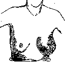
Figure 1. Postpartum mastitis.
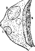
Figure 2. Schematic arrangement of abscesses in the mammary gland: 1—superficial abscess; 2—intramammary; 3—retromammary. 3rd-4th week of the postpartum period. Swelling, redness of the skin, and phenomena of fluctuation make it easy to recognize purulent mastitis in the form of an abscess. Deep retromammary abscesses (Fig. 2) can in some cases be confused with deep suppurations that originate from the affected rib tuberculosis (caries costae). In these cases, pus from the rib perforates the fascia of the pectoralis major muscle and also accumulates behind the mammary gland. The absence of inflammatory phenomena on the part of the mammary gland itself makes it possible to understand the diagnosis. With a prolonged process and the presence of fistulas with purulent discharge, recognition is also not difficult. In the case of long-standing fistulas with a thin purulent discharge, sometimes with an admixture of cheesy-like grains, tuberculosis of the mammary gland should be suspected, especially in the presence of a predisposition to tuberculosis or corresponding heredity (See Mammary gland, tuberculosis of the mammary gland). Prognosis is generally favorable; much depends on expedient and timely treatment. Evacuation of pus accelerates recovery, but the course is often still prolonged, especially with sequential involvement of several areas of the mammary gland. Only in rare cases, in the presence of severe general phenomena of blood poisoning, does fear for the lives of the patients arise. A fatal outcome is observed extremely rarely. Treatment. In incipient mastitis, the mammary gland must first of all be provided with complete rest, which is achieved by weaning the child and applying fixing bandages with the use of cold, especially in congestive phenomena. To reduce milk secretion, a laxative is given, and fluid intake is restricted. In mild cases, these simple home remedies manage to achieve the regression of the inflammatory process. If the temperature does not drop, and pain and swelling increase, which indicates a progressive phlegmonous process in the mammary gland, then it is necessary with a corresponding early incision to give an outlet to the infectious agent and relieve tension in the infiltrated tissues. There is no need to wait for clear fluctuation with the formation of an already demarcated abscess. With earlier incisions, it is possible to reduce the melting of the affected tissues of the mammary gland and thereby shorten the time of necessary treatment, but such patients do not always come to the surgeon early; often they also refuse earlier intervention; then by suspending the breast, warming compresses, it is necessary to accelerate the formation of an abscess. The incision must be of sufficient length and depth to give a free outflow of pus and relieve pressure in the infiltrated tissues, sometimes pierced by small abscesses. All incisions on the mammary gland must be arranged radially to the nipple; the latter and the areola should not be cut, because this needlessly damages the excretory ducts of the gland. With large purulent cavities or purulent streaks, one has to make several incisions. Abscesses located behind the mammary gland (abscessus retromammarius) require incisions along the outer edge of the mammary gland. After the incision, to ensure a free outflow of pus, a drain or tampon is inserted into the wound. At the same time, precisely in purulent mastitis, one should not forget the principles of tampon-free treatment of purulent diseases and leave tampons and drains in wounds for a long time, which delays healing. In any case, any traumatization of the wound after incisions should be avoided; compression of tissues, rough manipulations with a finger or hemostatic forceps must be abandoned; a sharp spoon should also not be used here. Such rough techniques only open the way for the further spread of infection. Only necrotic tissue areas can be carefully removed with tweezers and scissors. To provide a better outflow of pus, for a more complete exposure of the infectious focus, and to avoid cosmetically unsatisfactory radial incisions, Bardenheuer and Morestin proposed a special lower incision with the reflection of the entire mammary gland. A semilunar incision is made along the lower edge of the mammary gland to the fascia, the mammary gland is reflected upwards and backwards, abscesses are opened in the tissues of the gland itself, the contents of which are emptied; drains are led out into the lower incision. As a result, a scar is obtained which is hidden in the lower fold of the mammary gland and is completely invisible. Such incisions are indicated for large abscesses located behind the mammary gland, while in other cases (and they are the majority), with a more superficial location of the purulent foci, such an incision with the reflection of the entire mammary gland appears to be too traumatizing and an unnecessarily large intervention. For all more or less significant and deep incisions in purulent mastitis, short-term anesthesia is required, best of all in the form of ether intoxication. Local anesthesia in the form of infiltration anesthesia in inflamed tissues should not be used, but local anesthesia with novocaine or by means of ethyl chloride freezing may prove sufficient for superficial abscesses. A good remedy in the surgical treatment of purulent mastitis, according to many surgeons, is treatment with congestive hyperemia according to Bier (see Bier's method) with the use of special suction glass cups according to Klapp. Large cups are used in which the entire mammary gland is placed. When the air in the cup is rarefied, the latter is sucked onto the skin, congestive hyperemia is obtained in the gland, and with the help of a suction syringe, easy discharge of pus is achieved. The suction cup (see Abscess, Fig. 6) is applied daily for 15-45 minutes. With such a long session, a break of 2-3 minutes is made every 5 minutes. The advantages of treatment according to Bier are the rapid disappearance of pain, good and faster discharge of pus and stagnant milk. The method requires careful technique and strict control by a physician and therefore is not always applicable in outpatient practice. In very severe forms of mastitis with phlegmonous inflammation of the entire gland, with incessant suppuration, with a non-dropping temperature, and with a deteriorating general condition, one has to weigh the necessity of amputating the entire mammary gland. In the gangrenous form of mastitis, which fortunately is extremely rare, timely amputation can save the patient. The question of feeding the child by the mother in purulent mastitis is resolved differently by surgeons. Some allow feeding only with the healthy breast, others do not object to applying the child to the sick breast, believing that milk suction frees the mammary gland and reduces congestion. Harm to the child from the possible admixture of pus to milk has not been observed. It should be remembered, however, that in the inflamed state of the mammary gland, the latter must be given rest, and in any case, in the presence of large incisions and a significant wound surface, applying the child to the sick breast is inexpedient. Prophylaxis of mastitis consists of a number of measures that must be undertaken during pregnancy. These measures consist in preparing the nipples for feeding, in protecting them from traumatization and from the formation of abrasions and cracks (see Mammary gland, nipple cracks). Stroganov correctly recommends starting the prophylaxis of nipple disease from the first days of the girl's life in the form of aseptic maintenance and protection from any trauma. By frequent washings of the nipples with cold water and wiping with diluted alcohol, a certain coarsening of the tender and sensitive skin of the nipple and areola can be achieved. Wiping the nipples with pure alcohol is not recommended; this dries the skin too much, which can facilitate the appearance of cracks. Stroganov proposes the following mixture for wiping the nipples: Glycerini 8.0, Spiritus vini rectificati 100.0, Aq. destil. 80.0. With a retracted flat nipple, it is necessary to achieve a certain change in shape to one more suitable for sucking and feeding the child, which is achieved by carefully pulling the nipple while washing it. Even greater attention must be paid to the care of the nipple at the moments of the start of feeding, since the nipple, abrasions and cracks on it or around it are the main gates through which the infection leading to puerperal mastitis penetrates. The infant must be applied to the breast at specific times; after feeding, the nipple must be thoroughly washed with a solution of boric acid or hydrogen peroxide. In the presence of abrasions or cracks, the latter must be treated (see Mammary gland, nipple cracks). In some cases, the use of special nipple shields is recommended as a prophylactic measure. In the obstetric clinic in Würzburg, such use of shields during feeding sharply reduced the number of cases of mastitis (Frangenheim).
In addition to the aforementioned individual prophylactic measures among pregnant women and nursing mothers, health education work must be conducted with special attention given to the necessity of observing general rules of cleanliness and hygiene in the care of the mammary gland before and during feeding. As for chronic mastitis, they are of lesser clinical significance, are encountered much less frequently, and by some authors are attributed partly to neoplastic and partly to involutional processes in the mammary gland (see Mammary gland, tuberculosis, syphilis, neoplasms). Mastitis in newborns - see Newborn.
Related articles
Mentioned in
Cite this page
“Mastitis.” Soviet Medical Encyclopedia. English translation of Bolshaya Meditsinskaya Entsiklopediya, 1st ed. (Moscow, 1928–1936), ed. N. A. Semashko. https://sovietmedicalencyclopedia.pages.dev/article/mastitis/