Gynecomastia
Historical document, translated for reference. It reflects medical knowledge of the 1920s–30s and is not medical advice.
Summary
This article from the 1928-1936 Soviet Great Medical Encyclopedia defines gynecomastia as the enlargement of male mammary glands resembling female breasts. It details the histological findings, clinical presentation, and its frequent association with testicular pathology and endocrine dysfunction.
Encyclopedia article (1928–1936)
GYNECOMASTIA, gynecomastia (from Greek gyne - woman and mastos - breast), a term introduced by Galen denoting the enlargement of the mammary glands in men resembling female breasts. This enlargement occurs due to the proliferation of all tissues that make up the normal male mammary gland, i.e., the excretory ducts, connective tissue, and sometimes also adipose tissue. Such true gynecomastia must be distinguished from the enlargement of the mammary glands based on an excessive accumulation of fat in this area (in general obesity or in castrates prone to obesity). In such cases, the similarity to the female breast is merely external. Statistical data on the frequency of gynecomastia are extremely scarce. According to available data by Leveque, however, there is one gynecomast for every 200 soldiers. The condition can be bilateral and unilateral. According to some indications, the frequency of both forms of gynecomastia fluctuates within equal limits; according to others, bilateral gynecomastia occurs more often (in 80%). In unilateral gynecomastia, predominantly the right mammary gland is affected. The described enlargement of the mammary glands in the highest percentage of cases (up to 75%) is observed in young and middle ages (15-40 years); however, it is also encountered in 5-10-year-old boys and 70-80-year-old old men. The size and weight of the gland vary extremely. Its transverse diameter can be 2-15 cm, and the gland can protrude above the chest surface by 1-6 cm. The nipple is almost always enlarged; the areola is sharply pigmented and dilated (can reach 3.5x3.5 cm). The weight of the gland ranges from 1 g to 160 g. Single but reliable observations indicate the possible secretion of the mammary gland, sometimes so strong that such gynecomasts can feed children. Regarding the composition of the secreted fluid, there are only old indications: according to them, the fat content in "milk" is 1.2%. The surgically removed or autopsy-discovered mammary gland of a gynecomast is well-delineated, dense, coarsely lobular, white in color, and sometimes interspersed with adipose tissue. Its microscopic picture varies in different cases. Depending on whether gynecomastia has existed for a long time
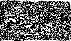
Figure 1. Section through a lobule of the mammary gland in gynecomastia. (several months or even years) or appeared only recently (several days or weeks), the proliferated connective tissue of the gland is either homogeneous and poor in nuclei, or rich in them and has an embryonic appearance. Around the glandular ducts, it is arranged in concentric layers, loosened, and sometimes permeated with a polymorphonuclear cellular infiltrate. Glandular ducts are sometimes more, sometimes less, but always multiplied. Sometimes their number in one field of view (at low magnification; objective 3) reaches 30-35. They rarely retain their tubular configuration; more often they branch and bud. Sometimes, though rarely, true lobules are formed (see Figure 1). In such a case, the mammary gland of a gynecomast is indistinguishable from a well-formed female mammary gland. In almost all cases, the epithelium of the ducts is in a state of enhanced proliferation, is often multilayered, and forms papillae protruding into the lumen. Sometimes pictures of apocrine secretion are observed in it, and round droplets of secretion are found in the lumen of the tubules. As a rule, an abundant amount of smooth muscle fibers is discovered in the nipple, imparting a pronounced erectility to the nipple of the gynecomast. The clinical picture of gynecomastia is very characteristic: painless—with few exceptions—enlargement of the mammary glands; absence of inflammatory changes; sometimes pain due to the inconvenience of male clothing or uniform. The growth of the gland has no tendency to rapid and unlimited enlargement and often stops without reaching large sizes. In many cases, gynecomasts have a female type of pubic hair and sometimes other female characteristics: voice, pelvis, skeleton, absence of beard and mustache. Often gynecomastia is combined with a violation of sexual functions or with deeper or shallower disorders of the sexual sphere: underdevelopment of genital organs, hypo-, epispadias, hermaphroditism, testicular tumors (chorioepithelioma, see Figure 2), castration and testicular atrophy (with a general female habitus, as a result of trauma, tuberculosis, mumps, typhus, etc.). At the same time, it is indicative that all the indicated changes of the urogenital sphere pre-exist, i.e., they are earlier manifestations than gynecomastia. It is well known that the closest interrelationships exist between secondary sex characteristics, to which the mammary gland belongs, and the reproductive apparatus. While puberty in girls gives an impetus to the development of the mammary gland, the growth of the latter in boys during this period is delayed. Throughout the rest of life, the male mammary gland, although it does not develop, does not atrophy either, but remains in a latent-potent state, ready to begin developing as soon as the equilibrium of the organ system that manages the preservation of sex characteristics is disturbed, especially in individuals endowed from the very beginning with a "hermaphroditic predisposition in relation to the mammary gland" (Biedl). Taking all this into consideration, a number of researchers believe that any gynecomastia is secondary, i.e., that it always develops in response to macroscopic or microscopic, congenital or acquired changes in the gonads. Recent studies show that if atrophy of the testes lies at the base of the process, the development of gynecomastia depends not on changes in Leydig cells, but on damage to the seminal epithelium. It should be pointed out, however, that some, especially older authors, recognize the dependence of gynecomastia on lesions of the sexual apparatus in only 40-50%. They note a number of cases where gynecomastia arose as a result of trauma or mechanical irritation, was even transmitted by inheritance, and was combined with apparently healthy genitals. However, to determine the state of the gonads in these cases, only the microscopic criterion is valuable, which cannot take place with "apparently healthy" testes. It is known that macroscopically unchanged testes upon microscopic examination may turn out to be deeply affected, being in a stage of advanced sclerosis with complete loss of spermatogenesis. It is well known that libido sexualis and potentia coeundi can be preserved in this process. Thus, these cases can simulate gynecomastia with "apparently healthy genitals." Gynecomastia that sometimes arises in connection with chorio

Figure 2. Gynecomastia with testicular lesions.
epithelioma of the testis deserves special interest. Here, not only abundant secretion from hypertrophied mammary glands can be observed, but also decidual transformation of their stroma. This phenomenon is regarded by authors as a specific reaction to chorioepithelioma, analogous to the reaction to "pregnancy." Gynecomastia must be differentiated from benign tumors of the mammary gland (adenofibroma, adenoma) and cystic mammary gland. Mastitis of youths, which should also be taken into account in this sense, is currently regarded as a variety or early stage of the same gynecomastia. Since gynecomastia does not pose a danger to health, the removal of the enlarged mammary gland is not an absolute necessity. It should be performed only if the enlarged glands interfere with wearing clothing or activities, as well as for moral or aesthetic reasons. In view of the fact that gynecomastia is an expression of a disorder of the functions of the sexual sphere with the loss of the corresponding hormone, good results should be expected from testicular transplantation or organotherapy.
E. Herpsiverg.
Related articles
Mentioned in
Cite this page
“Gynecomastia.” Soviet Medical Encyclopedia. English translation of Bolshaya Meditsinskaya Entsiklopediya, 1st ed. (Moscow, 1928–1936), ed. N. A. Semashko. https://sovietmedicalencyclopedia.pages.dev/article/gynecomastia/