OBESITY
Historical document, translated for reference. It reflects medical knowledge of the 1920s–30s and is not medical advice.
Summary
Obesity is defined as an increase in body weight compared to normal due to excessive fat deposition in subcutaneous tissue, omentum, mediastinum, etc. The article discusses the classification, causes, and physiological mechanisms of obesity, including the role of nutrition, heredity, and endocrine factors.
Encyclopedia article (1928–1936)
OBESITY (tuchnost', obesitas, adipo-sitas, polysarkia, lipomatosis universalis, liposis), an increase in body weight compared to the normal due to excessive deposition of fat in subcutaneous tissue, omentum, mediastinum, etc. If as a result of this there are disturbances in the functions of the entire organism or of individual organs and systems, then O. is called pathological. The degree of O. can be determined by comparing the weight of the patient with the normal weight, calculated from tables or indices (see Body Weight and Indices of Physical Development). Exceeding the normal weight by up to 25% is considered mild O., up to 35% - distinctly expressed, up to 50% - moderate degree, and above 50% - severe O. (Bichtwitz). Cases of O. weighing 490 kg and 331 kg have been described (quoted from Grafe). Usually, however, cases of O. with weight above 170-180 kg are extremely rare. In a normal person of middle age (according to Pettenkofer) the amount of fat equals 18% of body weight (44% of the weight of the dry residue), while the amount of proteins equals 20-22% of the dry residue. About 75% of fat is deposited in subcutaneous fat tissue. Fat deposits serve to accumulate energy reserves, since due to their high caloric value (9.4 large calories per 1 g) 9 kg (approximately) of fat in the body of an average person contain over 80,000 cal., which is sufficient for 1 month (approximately) of life. Proteins and carbohydrates (in the form of glycogen) can accumulate only to a very limited degree. Due to their inertness, fat in the body does not change until there is a need for its combustion, and thus is an ideal reserve substance. Fat deposits occur mainly from fats in food. In excess of foreign fats, the latter can be deposited in the body in an unchanged form. The second source of fat formation is an excess of carbohydrates introduced. Although the possibility of fat originating from proteins in food has been proven, practically this occurs in completely negligible amounts. Food fats are emulsified and broken down by splitting enzymes of digestive juices (lipases). After absorption from the intestine, where in the intestinal wall neutral fats are resynthesized from glycerin and fatty acids, the main part of fat enters the right ventricle via the lymphatic vessels of the mesentery and thoracic duct, and from there to the lungs. These latter occupy with respect to fats the same anatomical position as the liver does with nutrients absorbed into the blood. Research by Roger and Binet has shown that cells of the pulmonary endothelium are capable of capturing fats (lipopexia) and breaking them down (lipodieresis). In addition to the lungs, breakdown of fats occurs mainly in the liver, as well as in the blood and tissues through lipases, and lipodieretic ability is enhanced with an excess of oxygen. Undecomposed fat is captured and accumulated mainly by fat tissue, and according to some data, fat cells themselves have the ability to synthesize fats. With increased energy needs, fats pass from subcutaneous fat tissue into the blood, and from there to the liver, where they undergo combustion. Correct fat metabolism and maintenance of constant body weight represent the result of a very complex process of interaction between the central and peripheral nervous system, endocrine glands, and executive organs, mainly the liver and subcutaneous fat tissue. Disturbance in the activity of any part of this system affects the work of the entire regulatory apparatus, which is expressed in the occurrence of O. (or conversely, in weight loss of the body). The main role in the regulation of fat metabolism belongs to the central nervous system. The question of the etiology of O. has not yet been resolved. At first it was believed that the cause of O. lay exclusively in exogenous factors - nutrition and regimen, but when data accumulated that did not fit this scheme, they began to attach predominant importance to the innate properties of the organism and heredity, while underestimating the influence of external conditions of work and life associated with belonging to a particular family. With the development of endocrinology, the occurrence of O. began to be explained exclusively by damage to the endocrine glands, especially the thyroid and pituitary. But this was true only for some cases. Works of the last two decades have indicated the importance of the central and peripheral nervous system, and especially the independent role of subcutaneous fat tissue. At present, most researchers tend to attach decisive importance to endogenous factors, while external conditions are assigned the role of a factor that allows internal "causes" to manifest. For example, it is pointed out that in Germany during the blockade and the associated famine, the number of obese people sharply decreased, but after the blockade was lifted and when nutritional conditions improved, it again increased sharply. However, it is undeniable that the influence of external conditions, although very different in different cases, plays a decisive role in most cases, i.e., without appropriate conditions of nutrition and work, the existing tendency to O. cannot manifest. In the overwhelming majority of cases, many factors participate in the origin of O., closely related to each other. Nevertheless, for convenience, due to the predominance of factors located outside or inside the body, O. is divided into exogenous and endogenous, although more or less pure cases of either are very rare. External factors can include overeating, especially with immoderate consumption of beer and wine, as well as a transition to more complete and fat-rich food when accustomed to a large volume of it, then changes in work regimen and living conditions (for example, transition from a profession requiring heavy physical labor and mobility to office work). However, the influence of profession should not be overestimated. Thus, J. Bauer pointed out that out of 270 cases of O. there were only 3.37% of so-called professional obesity (confectioners, bakers, etc.). O. is predisposed to by chronic diseases associated with long-term immobility, for example, tuberculous coxitis and others. Finally, there is a role for decreased physical and emotional mobility, observed in some people in middle age. However, the overwhelming majority of people maintain constant weight, especially between 25-40 years. Regulators are appetite and the feeling of satiety. After severe starvation or exhausting disease, this regulation is disrupted, and such individuals eat significantly more than needed. An increase in appetite occurs with more tasty and varied nutrition, as observed in sanatoriums and rest homes, and in connection with decreased energy expenditure this leads to an increase in body weight. In such cases, after returning to ordinary conditions of work and nutrition, weight quickly returns to its original value. In healthy people, with changed conditions of nutrition and life, equilibrium is restored by changing appetite or by increased excess oxidation in the body (Luxus-consumption, see below), and weight remains stable. In cases of excessive appetite (dizoreksia, likoreksia), excessive fat deposition occurs. In the presence of mm predisposition or other internal causes disrupting the regulation of fat metabolism, fat accumulation constantly increases and can reach very large values. In such cases, excess nutrition can be even insignificant; for example, sometimes it is sufficient to excessively introduce 10 g of oil daily for weight to increase by approximately 3-4 kg in a year. One of the main causes of O., inherent in the body itself, is hereditary predisposition, under which can be understood the innate weakness or anomaly of the entire or part of the system regulating fat metabolism and maintaining constant body weight, i.e., the central and peripheral nervous system, endocrine glands, and peripheral organs. According to some authors, about 50% of obese people come from families with hereditary predisposition. According to
------г data of Liebendorf
11^ a (Liebendorf), constitutional O. is transmitted dominantly (figure 1). However, in addition to hereditary transmission, belonging to a certain family already
Fig i due to purely external conditions (nutrition, occupation, lifestyle) can lead to O. A very significant role in the origin of O. is played by the increased tendency of subcutaneous fat tissue to accumulate and retain fat (lipophilia), developed to varying degrees in different areas of the skin, for example on the forehead and abdomen. The distribution of fat is strongly dependent on the influence of the nervous system and endocrine glands, which explains the occurrence of various types of O. when one or another gland or the nervous system is affected (for example, after encephalitis), as well as the development of local fat deposits (see below). However, despite this dependence, different areas of subcutaneous fat tissue persistently retain their characteristics. For example, when abdominal skin is transplanted to the back of the hand, the transplanted piece became fatty with general weight gain of the patient, while the surrounding skin did not change. Such facts speak for the significant independence of subcutaneous fat tissue and the possibility of explaining its anomalies by various local disorders of fat metabolism, as well as such cases of O. where it was impossible to reduce body weight by any means, since with increased energy needs the body burned up carbohydrate and protein reserves, while fat deposits remained untouched. One of the main causes of O. are disorders of the central nervous system, which in close connection with the endocrine glands regulates fat metabolism. In recent times, it has been established that the main vegetative center regulating fat metabolism is located in the diencephalon in the area of the gray tubercle. Some authors (Dresel and others) consider it the center of fat metabolism, while others (Bogolyubets, Mohilytsky) believe that here there is a center of organs (liver, pancreas, muscles, etc.), reflexively connected with them and regulating their functions related to protein, fat, salt-water, carbohydrate metabolism and thermoregulation. According to these authors, there are no special "metabolic" centers. The gray tubercle is connected to the pituitary gland through the funnel, the secretion of which (according to Biedl and Raab of its middle part) directly penetrates the third ventricle through this path and acts on the vegetative center of the diencephalon. Damage to one part of this system (pituitary gland-diencephalon) or disruption of the connection between them can give the syndrome of dystrophia adiposo-genitalis or general pituitary O. due to the cessation of normal fat metabolism in the liver and subcutaneous fat tissue. Such relationships are indicated by cases of rapidly developing O. after damage to the diencephalon or the pituitary stalk in epidemic encephalitis, syphilis of the brain, meningitis, while the pituitary gland remained unaffected. On the other hand, damage to the pituitary gland gives the same syndrome without involvement of the diencephalon in the process. Recent research has more or less clarified the normal and pathological conditions of regulation of fat metabolism and distribution of fat in subcutaneous fat tissue through the nervous system, namely-impulses from the vegetative center pass through the vegetative fibers of the cervical cord, from there through the splanchnic nerve (n. splanchnicus) to the liver, as well as to other organs. The secretion of the pituitary gland, acting on the vegetative center, causes the movement of fat from the blood and tissues into the liver and its intensified oxidation there (Cooper, Chamberlain, Raab). The action of insulin is opposite to the action of pituitrin, i.e., it reduces fat burning and promotes its accumulation. When the vegetative center is destroyed, the cervical part of the spinal cord is severed, or the splanchnic nerve is severed, the action of the pituitary secretion ceases (Raab), which indicates the possibility of O. developing due to damage to the vegetative center and its peripheral pathways. The influence of the center on subcutaneous fat tissue is transmitted through the sympathetic and parasympathetic centers located in the spinal cord, and then through peripheral vegetative fibers to the subcutaneous fat tissue. This can explain the occurrence of local O. of the affected limb in sciatica and in the aftermath of infantile paralysis or in the experiment after severing the sciatic nerve. The same is indicated by cases of the occurrence of symmetrical fat deposits limited to the area of certain roots, so-called multiple symmetrical lipomatosis (for example, Madelung's fat tumors on the occiput). Unilateral damage to the vegetative center can give O. of one half of the body similar to hemiplegic. Some authors explain the occurrence of progressive lipodystrophy of Simons (Simons) by damage to the vegetative center, in which the subcutaneous fat layer of the face and upper half of the body disappears, while the lower part of the body and lower limbs become fatty. This same factor may be responsible for the occurrence of asymmetrical fat deposits in the subcutaneous fat layer, resembling tumors, while the rest of the subcutaneous fat tissue may remain unchanged (so-called lipomatosis simplex) or atrophy (lipomatosis atrophicans). In Dercum's disease such deposits are painful (lipomatosis dolorosa). However, some authors believe that these deposits owe their origin to local anomalies of subcutaneous fat tissue. According to recent research, it has been established that the hormone of the pancreas-insulin plays a major role in fat metabolism and in the occurrence of O. In many obese people, especially during the period of weight gain, hypoglycemia, increased tolerance to carbohydrates were observed, and after glucose load, the blood sugar level fell significantly below the initial value, which indicated increased insulin secretion into the blood. Experimental observations showed that the action of insulin on fat metabolism is opposite to the action of pituitrin; with insulin injection, fat in the blood increased, in the liver it decreased; the respiratory coefficient with simultaneous injection of insulin and even glucose orally rose above 1.0, which indicated the conversion of sugar into fat. Insulin also has the ability to retain water in tissues. With hyperinsulinization and the associated hypoglycemia, appetite sharply increases and absorption from the intestinal tract accelerates. All this leads to an increase in body weight due to the accumulation of fat and water in the body tissues. This action is used in the treatment of emaciation, where with insulin injections it is possible to achieve a stable increase in weight. Falta (Falta) and others believe that obesity with overfeeding depends mainly on hyperinsulinization, and call such obesity insular. However, such an explanation is suitable only for some cases. There are known cases of O. in diabetes, i.e., in a state of hypoinsulinemia. In addition to the pituitary gland and pancreas, the thyroid gland also participates in the regulation of fat metabolism. Its hormone, thyroxine, mainly regulates the basal metabolism. When the function of the thyroid gland is absent or reduced, oxygen consumption sharply decreases, heat production and general vital activity decrease. However, O. in such cases occurs relatively rarely, since fat burning is almost not disturbed (Grale). In hypothyroidism, a decrease in the specific-dynamic action of food has been found, which together with the lethargy and low activity of such patients may predispose them to O., especially with a simultaneous decrease in pituitary function.-The sex glands are closely connected with the brain appendage, and their effect on fat metabolism is of a secondary order through the medium of the middle lobe of the pituitary gland. Moreover, a decrease in their function sharply reduces the mobility and activity of the patient and thus also contributes to fat accumulation. The influence of the pineal gland, adrenal glands, and thymus on fat metabolism has not yet been clarified. Any O., both exogenous and endogenous, is possible only as a result of an excess of calories introduced with food over their consumption. In cases of overeating or lack of movement, such a result is quite understandable; however, along with such patients, there is a large number of obese people with very limited appetite and a mobile lifestyle. Bouchard found in his material that only 40% of obese people ate a lot, 50% had normal appetite. In terms of mobility, only 37% were lethargic and inactive, while 28% moved a lot. These facts show that the occurrence of O. cannot be explained by a simple violation of the energy balance. In general, the questions of energy exchange in O. are not yet fully clarified. Undoubtedly, in addition to fat deposition, which depends on overfeeding or on reduced consumption, as well as on physiological causes (for example, during a woman's sexual development), there are forms of O. where fat metabolism itself is qualitatively impaired, i.e., where obesity represents a disease of metabolism. Such are cases of O. where fat deposits almost completely do not undergo burning and in case of need, protein and carbohydrate reserves are used, as well as local O. of individual areas of subcutaneous fat tissue. However, in practice, such a precise distinction is almost impossible, since our knowledge of the pathology of fat metabolism is completely insufficient. After the discovery of the influence of the thyroid gland on basal metabolism, it was believed that fat accumulation is explained by a decrease in oxidation associated with weakening of the thyroid gland function.
It has been found, however, that in the majority of obese individuals, the basal metabolism is within normal limits, and in approximately 25% of all cases it is even elevated, and only in 15-20% below normal. On the other hand, myxedema, in which the basal metabolism is sharply lowered, is accompanied by obesity only in individual cases. It has been established that the specific dynamic action, i.e., the increase in oxidative metabolism after food intake, is significantly lowered in the majority of obese individuals compared to healthy individuals, and sometimes is completely absent. This mainly concerns proteins, since carbohydrates, and especially fats, also give a low specific dynamic action in healthy individuals, which may explain the development of obesity when these substances predominate in the diet. According to some authors (Grafe), overfeeding can cause such a strong and prolonged specific dynamic action, i.e., an increase in oxidation in the body, that fat deposition becomes impossible and body weight does not increase (so-called Luxus-consumption—increased combustion*). Other authors do not confirm this. The known weakening of combustion in obese individuals is also caused by a decrease in heat dissipation, since the subcutaneous fat layer is a poor conductor of heat. The decrease in specific dynamic action with overfeeding and especially with decreased mobility leads to enhanced fat deposition. During moderate physical work, the energy expenditure and increase in oxygen consumption in obese individuals are at the same level as in healthy individuals, and during heavy muscular work, the increase in oxidative metabolism is greater and significantly longer than in healthy individuals. Recently, Bernhardt, studying gas exchange in obese individuals throughout the day (and not only in the morning on an empty stomach, as in the determination of basal metabolism), found in the majority of obese individuals, especially during the period of weight gain, a significant decrease in oxygen consumption—below the level of basal metabolism, mainly after light physical work, then after food intake and during sleep. He called these decreases negative phases and calculated that the savings in combustion caused by them can be so great that it equals or even exceeds the increase in combustion caused by the specific dynamic action of food and physical exertion. Thus, the total energy expenditure per day in such patients is significantly lower than in healthy individuals, in whom negative phases are absent. It is interesting that these phases were also observed in convalescents after severe diseases during enhanced weight gain. Such negative phases, as it seems, can explain those paradoxical cases of obesity, when patients, receiving food corresponding in calories only to the basal "exchange (i.e., consumption during complete "rest, on an empty stomach), nevertheless continued to gain weight, although they led an active life. It is also interesting to determine the magnitude of the respiratory coefficient in obese individuals. Values close to 1.0 or higher indicate that in this patient fat is being formed from the carbohydrates in the food and that achieving fat loss will not be easy. On the other hand, low values of the respiratory coefficient provide a basis for a favorable prognosis, as this indicates that fat reserves are subject to combustion. In almost all cases of obesity, in addition to fat metabolism, water-salt metabolism is also disturbed. Water and salts can be retained in the tissues, especially in fat tissue, without simultaneous disorders of excretory organs and circulation and without the occurrence of edema. The water content in human fat tissue can vary from 5% to 70% (e.g., when the content of pure fat in the patient's body is 20 kg, the fat tissue can weigh from 21 to 34 kg). The retention of water in fat tissue is particularly great in thyroidogenic obesity and in some cases of cerebral obesity (Zondek). The rapid weight loss achieved at the beginning of obesity treatment is mainly due to the loss of water by fat tissue. In some cases, when weight does not decrease with severe dietary restriction, this is explained by the enhanced retention of water by subcutaneous fat tissue. In such cases, it is necessary to resort to means that increase diuresis and dehydrate fat tissue. Pathological anatomy. The subcutaneous fat tissue in obesity is extremely thick (13 cm on the abdomen). Blood vessels and muscles are surrounded by fat, which also penetrates between muscle fibers. The heart, especially the right one, is covered with fat penetrating the muscle bundles, while the muscle fibers themselves usually do not show particular, especially degenerative, changes. The mass of the myocardium may be reduced, the walls of the ventricles are thinned, especially the right one. The heart cavities are dilated. The coronary vessels are also surrounded by fat. It should, however, be borne in mind that obesity of the heart can also occur without general obesity, for example in some alcoholics or emphysematous individuals. There is fatty infiltration of the subpleural and mediastinal fat tissue, as well as the pericardium. In the omentum, peritoneum, and mesentery, there are huge accumulations of fat. The kidneys are completely surrounded by it. The liver is enlarged in volume, yellow in color. The liver lobules are infiltrated with fat, especially their peripheral cells. In rare cases, fat even penetrates into the spinal canal and can compress the spinal cord and roots. The cranial cavity is free of fat. In some cases of endogenous obesity, changes (cysts, softenings, scleroses, degenerative processes) are observed in the diencephalon and pituitary gland, caused for example by encephalitis, syphilis, or tumors. From the side of the thyroid gland, atrophic and sclerotic changes are often noted; in the pituitary gland, sometimes adenomatous formations (see Dystrophy—dystrophia adiposo-genitalis). From the side of the parathyroid glands and pancreas, pictures of lipomatosis with delamination of the glandular parenchyma and some atrophy of it, etc., are noted. Clinical picture of obesity. Obesity occurs significantly more often in women (approximately in 65% of all cases), which may be explained by more frequent lesions of the endocrine glands, especially the ovary. Children suffer from obesity comparatively rarely, and obesity at the age of 8-15 years is almost always associated with endogenous factors. Some nationalities are more prone to obesity. For example, the pronounced tendency of the Hottentots to obesity and the development in them of huge fat deposits on the buttocks, so-called steatopygia, are known. Among obese individuals, two types of patients are sharply distinguished. Some are cheerful, capable of work, ruddy, their mucous membranes are well colored, appetite is excellent, muscles are strong, pulse is of good filling, blood pressure is normal or slightly elevated. In the initial stages of obesity, they do not feel sick, but later, with an increase in obesity, symptoms of cardiac insufficiency appear. Such a form, according to Immermann, is called plethoric obesity. To the second group, so-called anemic obesity, belong pale, anemic patients complaining of inability to work, general weakness, headaches, and dizziness. Even with light physical work, they experience shortness of breath, palpitations, rapid fatigue, and sweating. Such patients are prone to colds and constipation. Their muscles are flabby, the subcutaneous fat layer is loose, the pulse is weakly filled and frequent, the heart is not hypertrophied. The first group mainly includes strong middle-aged men who are overfed (Mastfettsucht). In them, fat is deposited mainly on the trunk (abdomen, nape), while the limbs are relatively thin. In women, such a form of obesity is less common, and they mainly gain fat in the abdomen, chest, shoulders, and thighs. The majority of cases of so-called endogenous obesity belong to the second group, with many transitional forms, especially in the initial stages of obesity. The endocrine glands, as already indicated, play a major role in the occurrence and development of obesity and in the distribution of fat deposits, so that already from external examination, based on this sign, as well as on the accompanying other symptoms of lesions or changes in the function of one or another gland, it is possible to determine the main etiological factor of the disease. Depending on the predominant role in the development of obesity, thyroidogenic, pituitary, hypogenital, and insular obesity are distinguished. Cases of obesity with tumors of the adrenal glands and the pineal gland have been described, however, due to the unclear connections between these glands and fat metabolism, the isolation of such cases into separate forms is poorly justified. More or less pronounced thyroidogenic obesity is not common. In it, there are other signs of hypothyroidism to varying degrees, for example lethargy, chilliness, drowsiness, cold dry skin, rare pulse, lowered blood pressure, constipation, often umbilical hernia, lowered basal metabolism, tendency to water and salt retention. Fat is dense, distributed throughout the body, somewhat more abundant on the abdomen and thighs. Pituitary obesity, an example of which is dystrophia adiposo-genitalis (see Dystrophy), most often arises from a lesion of the pituitary gland and is combined with a delay in sexual development, and in some cases also in growth. The picture of pituitary obesity changes depending on the age at which the disease begins and on the degree of pituitary lesion. In adolescence (8-17 years), patients resemble a child. The skin is thin and delicate. Fat is dense and distributed throughout the body, especially on the abdomen, chest, arms, and legs. Development of the sexual organs is delayed. The testicles do not descend into the scrotum. Secondary sexual characteristics do not develop (Fig. 2). Hair growth is insufficient.
Patients lag behind in growth. The psyche is usually not affected. In mild cases, such a picture may be caused by transient insufficiency of the pituitary gland, and later such patients develop normally and catch up with healthy children without treatment. In many cases, underdevelopment of the pituitary gland is accompanied by underdevelopment of the thyroid gland. In adults, when pituitary obesity occurs due to damage to the pituitary gland, obesity develops rapidly, secondary sexual characteristics disappear sequentially, body hair and hair around the genitals fall out, libido et potentia fade, menstruation ceases. Male patients acquire a feminine appearance. The basal metabolism is within normal limits or elevated, the specific-dynamic action is reduced, and tendencies to water and salt retention are usually not observed. Sometimes there is increased tolerance to carbohydrates and a low level of sugar in the blood. With a significant pituitary tumor, cerebral symptoms may appear, as well as temporal hemianopia, due to compression of the optic chiasm. The diagnosis can be confirmed by finding an enlarged and deepened sella turcica on an X-ray. A similar syndrome can be caused by damage to the diencephalon or the pituitary stalk (cerebral obesity). Biedl and Bardet described a special form of such obesity (see Bardet syndrome, Biedl syndrome). In cerebral obesity, disorders of salt and water metabolism are observed. Damage or loss of function of the sex glands is one of the most common causes of obesity, especially in women. After castration, a tendency to obesity appears, and fat accumulates on the lower abdomen, on themons Veneris, on the thighs in both men and women. The rest of the body may not become fatty. This form of obesity is called castration obesity. A similar picture is observed in patients with congenital underdevelopment of the sex glands, so-called eunuchoid obesity. The difference from pituitary obesity can be the absence of growth retardation, cerebral symptoms, and expansion of the sella turcica. However, in such cases, the differential diagnosis is often very difficult. The vast majority of cases of endogenous obesity in women are caused by weakening or cessation of ovarian function, for example, after childbirth, during lactation, to a greater degree with their inflammation, and finally at menopause, so-called hypoovarian obesity. In this form of obesity, the abdomen, thighs, buttocks, breasts, and shoulders become very fatty, i.e., those areas that are physiologically prone to obesity with overfeeding. The fat deposits produce the impression of breeches (so-called breech obesity). The extremities remain relatively thin. In many patients, the fat layer is sensitive to pressure; upon palpation, it feels like separate dense lobules embedded in loose subcutaneous tissue. The abdomen sags like an apron. Particularly characteristic are thick folds of fat on the inner surface of the thighs. The most severe forms of hypoovarian obesity occur with premature or normal menopause and are associated with a simultaneous decrease in thyroid function, as well as with overfeeding and reduced mobility. Most of these patients belong to the anemic form of obesity. The most severe cases of obesity are those caused by the increased ability of subcutaneous tissue to accumulate fat (so-called lipomatosis). There is usually a clear hereditary predisposition (with dominant transmission). Patients are obese from a very early age, sometimes already at birth; obesity relentlessly increases and reaches very large sizes (up to 180-200 kg). From the side of the endocrine glands and the central nervous system, there are usually no visible deviations. The entire body becomes fatty, especially the abdomen, thighs, legs, and shoulders (Fig. 3). Treatment is rarely successful. When food is restricted, such patients lose the body's protein and carbohydrate reserves, while fatty tissue remains untouched. In such cases, special attention should be paid to preventing fat accumulation. The mechanism of development of this form has not yet been elucidated.--The clinical picture of regional obesity and lipomatoses is very diverse. There are limited fat deposits, firm, tense, sometimes
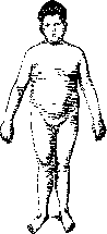
Figure 2. Pituitary obesity.
the same syndrome can be caused by damage to the diencephalon or the pituitary stalk (cerebral obesity). Biedl and Bardet described a special form of such obesity (see Bardet syndrome, Biedl syndrome). In cerebral obesity, disorders of salt and water metabolism are observed. Damage or loss of function of the sex glands is one of the most common causes of obesity, especially in women. After castration, a tendency to obesity appears, and fat accumulates on the lower abdomen, on themons Veneris, on the thighs in both men and women. The rest of the body may not become fatty. This form of obesity is called castration obesity. A similar picture is observed in patients with congenital underdevelopment of the sex glands, so-called eunuchoid obesity. The difference from pituitary obesity can be the absence of growth retardation, cerebral symptoms, and expansion of the sella turcica. However, in such cases, the differential diagnosis is often very difficult. The vast majority of cases of endogenous obesity in women are caused by weakening or cessation of ovarian function, for example, after childbirth, during lactation, to a greater degree with their inflammation, and finally at menopause, so-called hypoovarian obesity. In this form of obesity, the abdomen, thighs, buttocks, breasts, and shoulders become very fatty, i.e., those areas that are physiologically prone to obesity with overfeeding. The fat deposits produce the impression of breeches (so-called breech obesity). The extremities remain relatively thin. In many patients, the fat layer is sensitive to pressure; upon palpation, it feels like separate dense lobules embedded in loose subcutaneous tissue. The abdomen sags like an apron. Particularly characteristic are thick folds of fat on the inner surface of the thighs. The most severe forms of hypoovarian obesity occur with premature or normal menopause and are associated with a simultaneous decrease in thyroid function, as well as with overfeeding and reduced mobility. Most of these patients belong to the anemic form of obesity. The most severe cases of obesity are those caused by the increased ability of subcutaneous tissue to accumulate fat (so-called lipomatosis). There is usually a clear hereditary predisposition (with dominant transmission). Patients are obese from a very early age, sometimes already at birth; obesity relentlessly increases and reaches very large sizes (up to 180-200 kg). From the side of the endocrine glands and the central nervous system, there are usually no visible deviations. The entire body becomes fatty, especially the abdomen, thighs, legs, and shoulders (Fig. 3). Treatment is rarely successful. When food is restricted, such patients lose the body's protein and carbohydrate reserves, while fatty tissue remains untouched. In such cases, special attention should be paid to preventing fat accumulation. The mechanism of development of this form has not yet been elucidated.--The clinical picture of regional obesity and lipomatoses is very diverse. There are limited fat deposits, firm, tense, sometimes
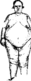
Figure 3. Constitutional obesity.
Painful, mostly on the lower extremities, in cases of nervous system lesions symmetrical. The rest of the body surface may be completely normal. There are also numerous transitional forms to general O. (see Lipodystrophia and Dercum's disease). Most cases of O. do not completely fit into the described forms, but represent transitional and mixed clinical pictures, moreover varying depending on the stage of the disease. Most often in men, O. from overfeeding on a constitutional basis is encountered, developing slowly, while in women it is associated, in addition to overfeeding, with hypofunction of the ovaries and thyroid gland. The initial stages of obesity do not particularly bother patients, except from an aesthetic point of view, but later the intensified accumulation of fat begins to disrupt the functions of other organs and systems of the body, and work capacity and general well-being sharply deteriorate. Heart and blood vessels. The cardiovascular system is affected earlier and most often. From mild subjective sensations of heart compression and palpitations, the condition can progress to the most severe forms of heart failure. Adipose tissue is extremely rich in blood vessels. Due to the sharp increase in the vascular bed and increased resistance, especially in the abdominal cavity (liver, mesentery, omentum, etc.), the work of the heart increases. Poor utilization of oxygen in the tissues of the obese (Lauter) enhances blood circulation and thereby the work of the heart. "The size of the latter corresponds not to the increased size of the body, but to the muscular system, which in the obese due to overgrowth and penetration of muscles with fat works worse than in healthy individuals, although its load is increased due to the increase in body weight. Impairment of thermoregulation and increased sweating also increase the load on the heart. The conditions of its work are significantly worse than in healthy individuals, since the diaphragm is greatly elevated, fat overgrows the pericardium, the heart muscle, penetrates between the muscle bundles, especially of the right ventricle, and purely mechanically hinders its work. Such a picture bears (Leyden) the name of O. heart, in contrast to fatty degeneration, which occurs in obesity comparatively rarely, mainly in cases of impaired nutrition of the heart muscle due to decompensation. The heart hypertrophies and dilates, especially the left ventricle. Blood pressure rises to 150-250 mm. The pulse is of good filling, slow, frequent extrasystoles. Shortness of breath, edema, sometimes anginal pains appear, which can occur due to pressure on the aorta, and sometimes due to sclerosis of the coronary vessels. With weakness of the right ventricle, stagnation in the liver, in the area of the portal vein, dropsy occurs. Such a picture is more characteristic of the plethoric form of O. In the anemic form of obesity, blood pressure is not high. The pulse is frequent. Peripheral arteries are soft. The heart is not hypertrophied. Arteriosclerosis is not observed. The work capacity of the heart is sharply reduced. Decompensation occurs earlier than in the plethoric form. Organs of respiration. In O., the function of the respiratory organs suffers. Fat deposits in the chest cavity, mediastinum, and the high position of the diaphragm compress the lungs, hinder their proper expansion, especially during physical work, which leads to insufficient saturation of the blood with oxygen and in turn increases the load on the heart due to increased blood circulation. Due to poor ventilation of the lungs, bronchitis with difficult expectoration of sputum develops. These bronchitis are persistent, pass into a chronic form and often lead to emphysema of the lungs. In the obese, catarrhal bronchopneumonia often occurs, and susceptibility to lobar pneumonia is also increased, and the prognosis is significantly more serious than in healthy individuals. Organs of digestion. Fat deposits in the abdominal cavity disrupt the proper function of the digestive tract. Appetite in severe O. decreases, although in many cases it is not large from the very beginning of the disease. However, in anemic obese individuals with poor appetite, sensations of weakness, dizziness, and a feeling of sharp hunger are often observed on an empty stomach, which forces such patients to eat frequently and prevents the vigorous implementation of dietary treatment. Disorders of gastric secretion are not frequent, mainly reduced acidity is observed. Constipation, flatulence, hemorrhoids are common. The absorptive capacity of the intestine is not impaired. Hernias are common, especially umbilical ones, and due to increased intra-abdominal pressure, the hernial ring quickly enlarges. The danger of incarceration in the obese is greater than in the thin. Therefore, the hernia should be operated on as early as possible. The liver is significantly enlarged—the fat content can reach 80% (normally 4-5%). Due to compression of the capillaries, the conditions of blood circulation worsen, and stagnation occurs in the area of the portal vein. With weakness of the right ventricle, stagnation in the inferior vena cava and in the central hepatic veins is added here, which leads to compression and atrophy of the hepatic parenchyma and to some phenomena of cirrhosis. In addition, in O., gallstones and stagnation in the gallbladder are often observed. Blood is not particularly changed. In the plethoric form, the number of erythrocytes and Hb is within normal limits. In anemic individuals, the number of erythrocytes is above normal, the percentage of Hb is lower, so the color index is significantly below unity. Kidneys. O. does not have a great influence on kidney function. In the obese, traces of protein and hyaline cylinders are often found in the urine. In arteriosclerosis of the blood vessels of the abdominal cavity, a shrunken kidney is also encountered. Impairment of regulation of salt-water exchange and water retention in tissues in thyroid and cerebral O. is expressed in the excretion of a small amount of highly concentrated urine (500-800 cm³) with a high salt content. Sexual function in O. often weakens, and not only in hypophyseal or hypogenital O., but sometimes also in exogenous obesity. Potentia coeundi is sometimes impossible due to mechanical difficulties. The skin of the obese is cold, and its thermoregulatory function is impaired, since heat loss by radiation and conduction is limited due to the powerful subcutaneous fat layer. Perspiratio insensibilis of the skin is enhanced, the skin is almost always moist and sweaty, but the sweat does not always equalize the temperature, and in the obese, hyperthermia is common with moderate physical exertion. The secretion of skin sebum is enhanced. Severe seborrhea and seborrheic eczema are common. Due to the moisture of the skin and decomposition of sweat and sebum in the obese, skin maceration is common, especially between folds; acne, furuncles, and carbuncles are also often observed. Nervous system. In anemic obese individuals, due to anemia of the brain, dizziness, nausea, general weakness, and drowsiness are common. In the plethoric type of O., phenomena of cerebral arteriosclerosis are observed. O. is often found in diabetics (lipomatous diabetes). According to Noorden, in such patients, the ability to form glycogen is impaired, but the transition of carbohydrates into fats does not suffer. Due to impaired carbohydrate metabolism, a strong appetite develops, the amount of food consumed increases, which causes O. Such patients lose sugar not with urine, but by its transition into fat. On the other hand, in O., glycosuria is often observed. Grafe explains this by increased demands on the insular apparatus, which leads to its exhaustion. The relationship of O. to gout is not yet clear. They are often found together. It is possible that excessive nutrition causes disorders not only of fat, but also of purine metabolism. Joint hereditary transmission is also possible. French authors consider O., diabetes, and gout as expressions of the so-called arthritism (see), a special pathological constitution, to which they attribute many other diseases, as a result of which this concept has become extremely vague. Prevention and treatment. Prevention of O. is no less important than treatment. This especially concerns constitutional O., where heredity and living and working conditions lead to gradually increasing obesity. In such cases, it is necessary to increase the expenditure and decrease the intake of nutrients, without waiting for the onset of O. The caloric content of the diet is reduced, alcohol is prohibited, fatty dishes are excluded, the amount of carbohydrates is reduced. Less sweets and drinks. The amount of protein should not be restricted. A diet rich in vegetables and fruits, black bread is indicated. Special attention should be paid to physical exercises. They sharply increase energy expenditure and at the same time strengthen the muscular system and heart. It is necessary to monitor the general condition of the body and not allow overexertion. In summer, volleyball, rowing, cycling, swimming are indicated, and especially country walking excursions (on free days—up to 25-30 km) or farming. In winter—skiing, skating, sawing and splitting wood. During vacation—long walking excursions, preferably mountain ones, after preliminary training. With these measures, it is possible to burn off a significant amount of fat.
Thus, according to Tsuntsu, fat loss in grams: when walking a distance of 3.6 km on a horizontal road-16 g, 6.0 km-30 g, 8.4 km-70 g, ascending 300 m on a sloping road-16-9 g, 300 m on a steep road-280 g, 3 km of road with a 10% gradient-376 g, on a bicycle 9 km-231 g, 22 km-722 g, 9 km with a 3% gradient-384 g. The same approach should be taken for beginning O. In developed disease, treatment must vary depending on age, the predominant etiological factor, the degree of O., and accompanying complications. The main form of treatment is diet, then physical exercise, hormone therapy, and balneo- and physiotherapy. Diet must be individualized depending on the degree of O. and the tastes of the patient. It is necessary to ensure that the food is tasty and contains a small number of calories with a relatively large volume, which gives the patient a feeling of satiety. Vegetables best satisfy this condition, e.g. cabbage, rutabaga, partly potatoes, as it contains only 20% carbohydrates. Sweets and flour dishes are completely forbidden. According to Hoop den, in mild obesity, the diet is limited to 4/5 of the normal requirement of the individual, calculated according to the Harris-Benedict tables, with an addition of calories for work. This diet is prescribed to robust subjects with beginning obesity, and to it are added physical exercises and walks (see prevention). In moderate obesity, the amount of calories introduced is reduced to 3/5 of the requirement. This diet is recommended by Noorden for home treatment on condition that work continues, also for patients with anemic forms of O. and weak hearts, who for this reason should avoid rapid weight loss, finally for robust obese persons going on long walking excursions. In severe obesity, the amount of calories is reduced even more, approximately to 2/5 of the requirement. This diet is prescribed to robust people under medical supervision in a medical institution. The reduction in calories comes only from fats and carbohydrates. The amount of protein should not fall below 150-120 g. Fluid intake not more than 1-1 1/2 l. Salt not more than 5-7 g. Diets I and II can be carried out for several months in succession, III-not more than 4-5 weeks. In kidney disease and in the presence of gout, it is necessary to avoid large amounts of protein and accordingly change the diet. Treatment of obese diabetics-see Diabetes mellitus. If severe obesity is accompanied by heart decompensation, the patient must be put to bed and, in addition to drug treatment, a milk diet according to Karel (see Karel's method) should be administered. It is given 5-6 times a day, 200 cm³ of milk each, which totals 650-800 cal. In addition to low calorie content, such a diet has a diuretic effect, which causes dehydration of the body; edemas disappear, the amount of water in adipose tissue decreases. Weight loss is initially very great-up to 1-1 1/2 kg per day. After removal of water, weight loss decreases significantly. Bouchard in such cases gives 1,200 cm³ of milk and 5 eggs per day. Moritz (Moritz)-as many times 25 cm³ of milk as the patient's height in cm exceeds 1 m. This diet is carried out for 10-12 days, then the patient is gradually transferred to a more caloric diet. Such milk days are recommended as an addition to diets of I and II degree 2-3 times per decade, with weight loss reaching 1 kg, of which only about 1/5 is due to fat loss, and the rest due to water loss. Milk days can be combined with fruit-vegetable days without fat and sweets-up to 1,000-1,200 g of fruits and 300-400 g of vegetables. For the treatment of O., various diets and regimens have been proposed, which were previously widely used. Thus, e.g. Rosenfeld's potato regimen. The diet consists of 800-1,200 g of potatoes and 200 g of lean meat. Calorie content approximately 1,200 cal. (90 g protein, 160-240 g carbohydrates and 10-12 g fat).-Banting's regimen allows much protein, permits alcohol, sharply restricts fats and carbohydrates. Example: 182 g protein, 8 g fat, 81 g carbohydrates = 1,112 cal.-Oertel's regimen-much protein, little fat, some carbohydrates, very little fluids (900-1,000 cm³). Example: 183 g protein, 35 g fat, 143 g carbohydrates = 1,690 cal.-Epstein's regimen-much fat, moderate amount of protein and little carbohydrates. Example: 102 g protein, 80 g fat, 48 g carbohydrates=1,401 cal.-Vegetarian regimen-vegetables, fruits, greens with fat restriction. For weak patients who cannot take walks and physical exercise, the system of dosed muscular work (so-called Terrainkur), gradually increasing, proposed by Oertel, is recommended. In addition, if possible, treatment of O. should be carried out in resort conditions, where patients more easily submit to the regimen, walk a lot, and where they use waters and baths containing Glauber's salt and CO₂, which regulates intestinal activity and beneficially affects the cardiovascular system. In the USSR, Yessentuki, Zheleznovodsk, Kislovodsk, Lipetsk, Darasun and others are recommended. Abroad-Karlovy Vary, Marienbad, Homburg, Vichy and many others. Hydrotherapeutic procedures (baths, showers) are very useful, increasing mobility and general tone in patients. In more severe cases of O., general massage should be applied, acting in the same direction, as well as enhancing blood circulation in the skin and subcutaneous adipose tissue. In many cases, good results were obtained from the application of the Bergonie chair. In addition to diet, physio- and balneotherapy, in cases of endocrine O., preparations of those glands must be applied whose loss or weakening of functions was the main factor of the disease. The most effective results are obtained in thyroid O. from the application of thyroidin or thyroxine. Initially, 0.1 thyroidin is prescribed 1-2 times a day, then the dosage is gradually increased and reaches 0.6-0.8 per day, while constantly monitoring the patient's condition. The last dosage is not carried out for more than 6-7 days. The action of thyroidin begins 3-4 days after the start of treatment and continues for several days after discontinuation. Then the doses are also gradually reduced. The entire course of treatment is 4-6 weeks. At the first signs of hyperthyroidism or increased sensitivity to thyroidin (sweating, palpitations, nervousness, diarrhea, trembling), treatment should be discontinued. If possible, the patient's condition is controlled by studying the basal metabolism. With these conditions observed, the results are very good. O. sharply decreased. General well-being improved. Treatment with thyroidin can be well combined with diet of I or II degree; it is necessary to ensure that the food contains sufficient protein. Thyroxine is taken orally in doses from 1 to 4 mg daily; 1 mg of it corresponds to 0.2 thyroidin. The results are the same as from the application of thyroidin. Besides thyroid O., thyroidin also acts well in other forms of endocrine O., where there is relative insufficiency of the thyroid gland, most often in hypogenital and climacteric O., where it should be applied together with preparations of ovaries and anterior pituitary lobe. Of preparations made in the USSR, ovariin, ovaricrin etc. are used, 1-3 ampules per day under the skin. When taken orally, there is almost no effect. Foreign preparations are also used-folliculin, progynon etc. Of pituitary preparations, prolan and prefison are used. Combined application of these preparations-thyroidin, ovariin and prolan-causes weight loss mainly of the abdomen and hips. In cases of painful adipose tissue, after treatment the pain disappeared. The course of treatment is 80-100 ampules of ovariin on average and 15-20 ampules of prolan. In cases of hyperthyroidism, the use of thyroidin can be discontinued and the others continued. The use of diet is mandatory. Milk days 2-3 times per decade are especially desirable. In cases of O. where there is no thyroid insufficiency, the use of thyroidin is not beneficial, although for the first few days thyroidin acts as a diuretic and dehydrates adipose tissue. In eunuchoid and pituitary O. in adults, pituitary preparations have no effect. In cases of pituitary O. in young age (12-17 years), the use of thyroidin can give good results, but only when there is thyroid insufficiency. This is difficult to determine clinically, so both thyroidin and pituitary preparations should be tried. Often such combined treatment gives more success. Growth accelerates, delay in sexual development disappears, O. sharply decreases. In tumors of the Turkish saddle and pituitary O., deep X-ray irradiation of the skull and in extreme cases removal of the tumor is recommended. In eunuchoid O., sometimes homo- or heterotransplantation of sex glands helps, preferably together with the pituitary and thyroid glands. In disfiguring fat accumulations on the abdomen and in the area of the greater trochanters, their surgical removal is applied. In lipomatosis, thyroidin should be applied. There are indications of good results. Tension and pain of fat accumulations decreased (Zondek).
In cases of water and salt retention, the use of novazurol or salirgan is recommended, which have a strong diuretic effect. It is best to administer them intravenously in doses of 1 cm2. Initially, the effect is very strong. The fatty tissue becomes dehydrated. It is advisable to combine the use of these drugs with thyroidin. If the first 2 injections are not effective, treatment should not be continued. Such treatment should not be used in obese patients with kidney diseases. Protein therapy (Schmidt) is also used; milk is injected in combination with thyroidin and a culture of Bacterium coli (so-called Hypertherman), but here again the main active substance is thyroidin. Prognosis. The more severe the O., the more serious the prognosis. In general, obese people live shorter than thin people. Data from American insurance societies indicate that obese people live on average 7 years less than thin people, and the difference is greater the higher the age of the compared groups. Up to 40 years of age there is no significant difference, by 60 years 60% of obese and 90% of thin people are alive, by 70 years 30% of obese and 50% of thin people are alive, by 80 years 10% of obese and 30% of thin people are alive. O. causes overload of the most important organs, especially the cardiovascular system and respiratory organs. As a result, the resistance of obese people to infections is reduced. Strokes, thromboses, and embolisms are common in obese people.
Related articles
Mentioned in
Cite this page
“OBESITY.” Soviet Medical Encyclopedia. English translation of Bolshaya Meditsinskaya Entsiklopediya, 1st ed. (Moscow, 1928–1936), ed. N. A. Semashko. https://sovietmedicalencyclopedia.pages.dev/article/obesity/