Dystrophy
Historical document, translated for reference. It reflects medical knowledge of the 1920s–30s and is not medical advice.
Summary
This article defines dystrophy as a broad category of metabolic and nutritional disorders, distinguishing between progressive and regressive types. It also provides a detailed clinical analysis of dystrophia adiposo-genitalis (Babinski-Fröhlich syndrome), exploring its symptoms, pathogenesis, and the debate regarding its pituitary versus cerebral origins.
Encyclopedia article (1928–1936)
DYSTROPHY, dystrophia (from the Greek prefix dys-, denoting poor quality, and trepho, I nourish), a disturbance in the nutrition of tissues, organs, or entire systems of the organism. It has long been customary to divide metabolic disorders in the cell into two large groups: 1) disorders characterized by the predominance of assimilation over dissimilation (disorders of a progressive nature), which include the processes of regeneration, hypertrophy, and tumor development, and 2) disorders characterized by the predominance of dissimilation over assimilation (regressive disorders), or atrophic disorders. Among deviations of the latter kind, one distinguishes in turn: simple or quantitative atrophies, characterized by a simple decrease in the volume of cells and organs, without qualitative changes in the protoplasm and tissues in general, and atrophies distinguished mainly by precisely these qualitative changes, qualitative atrophies, otherwise called degenerations. This includes degenerations of a protein, fatty, carbohydrate, or pigmentary nature. It is precisely these qualitative atrophies, or degenerations, that are called dystrophy. Some authors (Khalatov) find it possible, however, to speak of "hypertrophic dystrophy," classifying under it, among other things, so-called compensatory hypertrophies in cases where the increase in the volume of an organ occurs not so much due to the proliferation of cellular elements as due to an increase in the volume of the cells themselves; in doing so, they are guided by the consideration that there are allegedly no data supporting the possibility of an increased intake of substances into the cell during excessive nutrition, with which, however, one cannot agree. The causes of dystrophy are most often of a toxic nature, but in a number of cases, dystrophy can also be of endocrine origin, for example, the so-called mucous edema (myxedema) in cases of thyroid gland insufficiency. One also speaks of dystrophia adiposo-genitalis, in which there is actually a general disturbance of fat metabolism in the organism accompanied by atrophic phenomena in the genital apparatus. In dystrophia musculorum progressiva, atrophy of muscle fibers is observed with simultaneous hypertrophy of some of them. In a clinical sense, in relation to the human organism, dystrophy or a dystrophic state refers to those chronic nutritional disorders in which the main symptom is a change in nutrition characterized by a decrease below the norm.
G. Sakharov. Dystrophia adiposo-genitalis (adiposo-genital syndrome, Babinski-Fröhlich syndrome) is usually regarded as a syndrome of pituitary obesity in combination with pituitary eunuchoidism. It was described in 1900 by Babinski (in a 17-year-old girl) and in 1901 by Fröhlich (in a 14-year-old boy). The adiposo-genital syndrome is usually revealed in the full completeness of its clinical picture with the onset of puberty. However, it can also appear in adulthood, and sometimes it is clearly outlined in early childhood. Both girls and boys fall ill. In the latter, the clinical picture usually appears significantly more clearly and presents fewer difficulties for differential diagnosis with other forms of hypogenitalism accompanied by obesity of endocrine origin. The main place in the clinical picture is occupied by obesity and delayed sexual development. Sometimes the adiposo-genital syndrome is also accompanied by other phenomena, the pathogenesis of which is usually associated with a disturbance in the function of the pituitary gland: acromegaloidism, genu valgum, excessively tall or dwarf stature, curvature of the spine, symptoms of myxedema, or Dercum's disease. Fat deposits can reach enormous sizes. The fat is distributed unevenly and accumulates in especially large quantities on the chest, abdomen (the abdomen lies on the thighs in the form of an apron), and in the upper part of the thighs. Abundant fat deposits are usually also present in the internal organs and especially in the abdominal cavity. Sexual development is usually delayed. In girls, the onset of menstruation is delayed, often with dysmenorrhea, sometimes replaced by the complete cessation of menstruation. In young men, the sexual organs are very small, and ectopia of the testicles is often present. Pubic hair is sparse, and axillary hair is absent. The voice in young men remains childlike; a mustache and beard are absent. In men, sexual desire is absent, and impotence is usually present. In women, despite the absence of sexual desire, the ability to conceive may be preserved. Metabolism presents characteristic changes. Tolerance to carbohydrates is always increased. Their combustion is difficult. A large part of the ingested carbohydrates is converted into fats. In some cases, phenomena of diabetes insipidus have been observed. The basal metabolic rate usually does not present significant changes. Body temperature is mostly below normal. General asthenia, low blood pressure, and often melanodermia in the late period of the disease have led to the suggestion of simultaneous hypofunction of the chromaffin system. Hyposympathicotonia, whatever its origin, is observed frequently. In view of the fact that the cause of adiposo-genital dystrophy in the majority of cases is a tumor of the pituitary gland, corresponding symptoms (headaches, visual disturbances, etc.) and an enlargement of the sella turcica, detectable radiologically, are usually observed. However, the adiposo-genital syndrome can develop not only as a result of neoplasms of the pituitary gland but also with tumors originating from the third ventricle (see below). In such cases, the absence of deformation of the sella turcica, which is characteristic of neoplasms of the pituitary gland, and the presence of symptoms of a basilar nature, typical for tumors originating from the third ventricle, can to a certain extent facilitate a differential diagnosis. One must also keep in mind the possibility of the development of adiposo-genital dystrophy even without any tumors, which is often the case in early childhood after having suffered encephalitis or meningitis. The course of the disease in the presence of a tumor that is not amenable to surgical removal is always fatal. With deep X-ray therapy, it has been possible to obtain significant improvement and remissions lasting for years. Although syphilis is very rarely the cause of the adiposo-genital syndrome, specific treatment should nevertheless be tried in all cases. Patho-anatomical studies in adiposo-genital dystrophy establish that in the majority of cases of the disease, the primary change is located in the pituitary gland, with a tumor in the form of a basophilic adenoma, cancer, or cyst most often being discovered in the latter. Likewise, tumors of the pituitary duct and teratomas compressing the pituitary gland can be the basis of the disease. Less frequently, one finds a violation of the integrity of the pituitary gland as a result of past hemorrhages, a tubercular process, or syphilis. Cases of adiposo-genital dystrophy have been described with severe hydrocephalus, which compressed the pituitary gland due to the protrusion of the floor of the third ventricle, and with the destruction of the pituitary gland from a gunshot wound and other skull injuries. There are observations of adiposo-genital dystrophy on the basis of atrophy of the pituitary gland of arteriosclerotic origin. Finally, there are cases of adiposo-genital dystrophy without any changes in the pituitary gland, but in the presence of a tumor (e.g., a glioma) or (less frequently) other changes in the region of the third ventricle and the diencephalon. In the gonads in adiposo-genital dystrophy in men, one finds atrophy and a slowing down (sometimes cessation) of spermiogenesis in the seminiferous tubules, along with atrophic changes also in the interstitial cells of the testicles. In women, a decrease (up to complete disappearance) of follicles, the formation of multiple cysts, and sclerotic atrophy are observed in the ovaries. The pathogenesis of the adiposo-genital syndrome is usually associated with hypofunction of the pituitary gland, mainly its glandular part. Cushing, Aschner, and in Russia, Arkhangelsky and Karlik, experimentally produced the adiposo-genital syndrome in animals (dogs) that remained alive for more than a year after the complete removal of the pituitary gland. Despite this, the significance of disturbances in the internal secretion of the pituitary gland in the pathogenesis of the adiposo-genital syndrome remains unclear. Camus and Roussy, who also had at their disposal a large amount of experimental material, point out that during the extirpation of the pituitary gland, damage to the nuclei of the tuber cinereum is inevitable, which is the causal factor of sexual dystrophy and obesity. And if pituitary dwarfism, pituitary obesity, and pituitary infantilism can still, despite the objections of Camus and Roussy, be recognized as a regularly recurring consequence of the extirpation of the pituitary gland, these experiments in any case do not resolve the question in favor of the exclusively pituitary origin of the clinical syndrome of dystrophia adiposo-genitalis. The cerebral origin of this syndrome, alongside the pituitary one, remains highly probable. This is also supported by the variety of symptoms superimposed on the basic picture of the adiposo-genital syndrome, which sometimes leaves only obesity from it (e.g., in the presence of hypergenitalism rather than hypogenitalism and premature sexual development). The pituitary origin of Babinski-Fröhlich disease can be established only upon the confirmation of a pituitary tumor or the compression of it by a tumor of the base of the brain, and even then only with a significant degree of probability and provided that a combined disorder of the endocrine glands is excluded.
A. Bogomolets. Dystrophia musculorum progressiva (myopathia)—progressive muscular dystrophy, a disease of the muscular apparatus characterized by muscle atrophy due to damage to the muscle tissue with complete integrity of the peripheral motor neuron (cells of the anterior horns of the spinal cord, anterior roots, peripheral nerves). The name that defines the disease in essence can be considered "primary myopathy" (Oppenheim). The separation of this disease from the extensive group of myopathies into a distinct nosological unit was made in 1883 by Erb, who divided all pure cases of progressive muscular atrophy into two groups: spinal and purely muscular; Landouzy and Dejerine (1884) joined this opinion, proposing a division into myelopathic and myopathic groups. Signs uniting all varieties of primary myopathy: 1) onset of the disease at a young age (childhood, puberty); 2)
hereditary transmission, familial occurrence; 3) selectivity of muscle group involvement: a) muscles running from the pelvis to the trunk, from the pelvis to the thigh, from the thigh to the lower leg, b) muscles running from the trunk to the scapula and shoulder and from the scapula and shoulder to the forearm, c) facial musculature; 4) combination of atrophy with true and false (myosclerotic, fatty) hypertrophy; 5) qualitative decrease in electrical excitability, but without a clearly expressed reaction of degeneration; 6) preservation of sensitivity; 7) intact function of pelvic organs; 8) absence of bulbar phenomena. The basis for distinguishing typical varieties of the clinical picture is both the time of onset of the disease and the localization of atrophy; it is necessary to keep in mind that transitional forms between individual clinical types are quite common. Types of progressive muscular dystrophy. I. Childhood form of muscular dystrophy with pseudohypertrophy (lipomatosis luxurians muscularis progressiva of Heller, atrophia musculorum lipomatosa of Seidel). The first descriptions were made by Griesinger in Germany (1864) and Duchenne in France (1868). Initial manifestations are before the age of 10; inheritance is predominantly through the male line; usually, the muscles of the trunk (predominantly the extensors of the back), pelvis, and thighs atrophy first; the muscles of the upper extremities are still normal. Functional insufficiency of the diseased muscles creates peculiar conditions for the statics and movement of the body, since the corresponding movements are performed adaptively at the expense of the as yet unaffected groups. These movements are stereotypically repeated in all cases and, in developed forms, are so characteristic that they allow for an unmistakable diagnosis; but even in the very initial stages of the disease, one can notice individual typical components of adaptability, which is extremely important for early diagnosis (this fact must be kept in mind by child health care physicians, since during medical examinations, children may be encountered whose relatives do not even suspect they have muscular dystrophy). First of all, the position of the body when standing is characteristic: the back is lordotically curved, the abdomen is protruded, the upper part of the body is thrown back; such a body position
of the musculature strengthening the pelvis
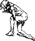
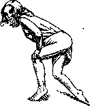
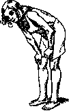
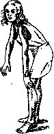
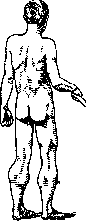
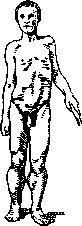
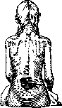
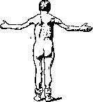
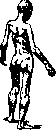
due to which the pelvis and with it the lumbar part of the spine tilt forward, while the upper part of the trunk, compensatorily, to maintain balance, tilts backward (Fig. 1). The gait acquires signs of "waddling" ("duck gait") due to weakness of the middle gluteal muscles. Especially characteristic are the movements of patients when bending the body forward and straightening it (getting up from a lying position, picking up objects from the floor): these movements are performed at the expense of the arm muscles, with which the patients lean on their thighs and knees when bending the body and push off from the thighs and knees when straightening ("climbing up their legs"; figures 2-5). A thorough examination of the musculature of the entire body reveals, along with atrophy of certain groups, an increase in the volume of individual muscles—predominantly the calf muscles, then the deltoid muscles (Fig. 6)—due to the deposition of fatty tissue in them. II. Childhood form of muscular dystrophy with involvement of the facial musculature; the first observations belong to Duchenne, a detailed description was made by Landouzy and Dejerine (type facio-scapulo-humeral). It develops in early childhood, begins with the involvement of the facial muscles (first of all, weakness of the orbicularis oculi and oris muscles appears), as a result of which closing the eyes and movements of the mouth when laughing, whistling, or talking are initially difficult. In cases where pseudohypertrophy of the orbicularis oris muscle appears, the lips become thick and protruded ("tapir lip"); subsequently, with the gradual spread of the process to the rest of the facial musculature, facial expressions become immobile, the cheeks sink in, the lower lip hangs down, and the face acquires features that greatly facilitate the diagnosis of the disease (facies myopathica). In later stages, complete immobility develops
only the supinator longus (Fig. 8). On the lower extremities, the glutei, quadriceps, peronaei, and tibialis anterior are predominantly affected. The facial muscles, with rare exceptions, remain healthy. In addition to motor disorders common to all the above-described forms
Figure 2.
Figure 3.
Figure 4.
Figures 2-5. Techniques by which the patient rises from the floor. Figure 5. face ("sphinx face"). Simultaneously or somewhat later, the muscles of the shoulder girdle and shoulder are affected, and subsequently the extensors of the back, pelvic, and thigh muscles, giving the same typical picture as in the first form (Fig. 7). III. Juvenile form. Described by Erb. Onset between 20 and 40 years of age. Inheritance is more often through the female line. Initially, the muscles of the shoulder girdle are usually affected (type scapulo-humeral of Vulpian), then the process moves to the muscles of the trunk and upper extremities. Regarding the movements (standing, gait, flexion, extension of the trunk), characteristic of the Erb type are: wing-like protrusion of the scapulae
FIG. 6.
more often through the female line. Initially, the muscles of the shoulder girdle are usually affected (type scapulo-humeral of Vulpian), then the process moves to the muscles of the trunk and upper extremities. The selective constancy of involvement, established by Erb, concerns the following muscles: pectoralis major et minor, cucullaris, latissimus dorsi, serratus anterior major, rhomboidei, lumbo-sacralis, longissimus dorsi, and later the triceps. As a rule, the sterno-cleido-mastoideus, levator anguli scapulae, coraco-brachialis, teretes, deltoideus, supra- et infraspinati, and the small muscles of the hand remain spared; of the forearm muscles, the muscle affected is
Figure 7.
Figure (paralysis of the m. serratus) and "relaxation" of the shoulders, expressed by the patient's inability to fix the shoulders downward due to weakness of the trapezius, serratus, latissimus dorsi, and pectoral muscles of the scapula (Fig. 9). In the late stages of the disease, respiratory disorders appear (serving as the cause of death) due to atrophy of the intercostal muscles and the diaphragm. The course of muscular dystrophy in all forms is chronic, slowly progressive; sometimes pauses in the process are observed. Differential diagnosis. A certain similarity of the clinical picture of d. muscularis progressiva in relation to the main external sign—muscle atrophy—exists with all forms that are closely related, such as poliomyelitis, amyotrophic lateral sclerosis, spinal muscular atrophy, neurotic muscular atrophy, myatonia congenita, etc.
Figure 9.
However, differential diagnosis does not present difficulties if the main criteria are: the typical location of lesions of muscle groups in myopathic forms, a thorough analysis of anamnestic data (heredity, familial nature, onset at a certain age), in-depth clinical examination (sensitivity, reflexes, electrical excitability), as well as microscopic examination of muscles (biopsy). -Etiology and pathogenesis. At the basis of myopathic Dystrophy lies a congenital weakness of the muscular system (abiotrophy according to Gowers). The immediate causes of the selective involvement of certain segments of the muscular system are unknown; this phenomenon belongs to the field of selective degenerations, which is difficult to study. Insufficiency of the growth energy of muscle segments apparently exists even in the embryonic formation of muscle tissue from the mesoderm. The congenital nature of myopathies is confirmed by the frequency of their combinations with other syndromes of developmental anomalies of the nervous system, as well as other systems. Research by Lenz, Siemens, and Davidenkov has established that myopathy 1) is most often transmitted in a recessive manner, 2) can often also be transmitted in a dominant manner, although this occurs for the most part in relation to mild cases, since severe cases lead to the cessation of the lineage, 3) is often transmitted in a sex-linked recessive manner. -Pathological anatomy. There is a picture of an atrophic process in various stages of its development: gradual narrowing of muscle fibers until their complete disappearance, proliferation of sarcolemma nuclei. Along with the atrophy of muscle fibers, processes of excessive growth of other tissues located in the intermuscular spaces—adipose and connective—are often observed. Macroscopically, these processes are expressed by an increase in the volume of atrophied muscles (pseudohypertrophy), whereby fatty infiltration gives the muscles a doughy softness, detectable by palpation. Sometimes, in general atrophy, a proliferation of muscle elements (true hypertrophy) may also appear in individual muscles. In the spinal cord and peripheral nerves, changes have generally not been detected to date, but in some, mainly atypical cases, changes were found in the spinal cord, specifically a decrease in the number and size of motor cells, without destructive processes in them. These observations confirm the opinion that forms of spinal, neurotic, and myopathic muscular atrophy, while being different in anatomical respects and in clinical symptoms, can be united by the essence of a congenital degenerative process. - Prognosis is always not very favorable. Myopathy has no direct effect on general life expectancy, with the exception of those cases where the respiratory musculature is affected. The general defensive forces of the organism in myopaths are weakened, and this can adversely affect the course of incidental acute diseases. -Treatment. There are no specific remedies against myopathies. The following are used: electrization, massage, and active gymnastics; however, it is necessary to avoid fatigue. Periodic use of general strengthening medicinal and nutritional preparations, and hydrotherapy is advisable.
Related articles
Mentioned in
Cite this page
“Dystrophy.” Soviet Medical Encyclopedia. English translation of Bolshaya Meditsinskaya Entsiklopediya, 1st ed. (Moscow, 1928–1936), ed. N. A. Semashko. https://sovietmedicalencyclopedia.pages.dev/article/dystrophy/